Epidemiology of Fusarium
Agave Wilt
in
Agave tequilana
Weber var. azul
María de Jesús RAMíRez-RAMíRez 1, N. Alejandra MANcillA-MARgAlli 1, lucía MezA-ÁlvARez 1, Ramón TuRiNcio-TAdeo 1, doralinda guzMÁN-de PeNA2
and Martin eduardo AvilA-MiRANdA1*
1Postgraduate Studies and Research division, Technological institute of Tlajomulco, National institute of Technology of Mexico, Tlajomulco de zúñiga, Jal, Mexico; 2department
of Biotechnology and Biochemistry, ciNveSTAv irapuato, irapuato, gto., Mexico *corresponding author: meavila@ittlajomulco.edu.mx
Abstract
Ramírez-Ramírez M.J., Mancilla-Margalli N.A., Meza-Álvarez L., Turincio-Tadeo R., Guzmán-de Pena D., Avila-Miranda M.E. (2017): Epidemiology of Fusarium agave wilt in Agave tequilana Weber var. azul. Plant Protect. Sci., 53: 144–152.
Fusarium oxysporum is reported as the principal causal agent limiting production of Agave tequilana Weber var. azul,
but frequent isolation of F. solani, and symptoms typical of F. solani as a pathogen like severe reddish coloured root rot
and loss of soil anchorage are frequently associated with diseased agaves. Inoculations of agave plantlets with F. solani
induced typical agave root rot symptoms in greenhouse trials. The incidence of both pathogens was determined molecularly with specific primers in the ITS2 sequence. Dispersion patterns of agave wilt, determined in plantations of different age, indicated a tendency to produce aggregated patterns over time as the disease spread from the initial symptomatic plant to adjacent plants. Although both fungi were isolated from agave diseased plants, and in spite of the higher percentage
of detection and root rot symptoms, it is concluded that F. solani mayhave a greater impact in agave wilt.
Keywords: Pathozone; soil-borne; plant pathogens; dispersion pattern
Agave tequilana Weber var. azul is the only raw material authorised by the Official Mexican Standard (NOM-006-SCFI-2005; www.crt.org.mx/images/ Documentos/NOM-006-SCFI-2005.pdf ) to pro-duce the alcoholic beverage tequila. This bever-age is important to the Mexican economy. In 2015, 248.3 million litres of tequila were produced and 74% was exported. To obtain this production, the Tequila Regulatory Council (CRT) reported the use of 859.2 thousand tons of agave heads (stems) (CRT 2016; www.crt.org.mx/EstadisticasCRTweb/). The agave tequilero takes from six to eight years to ma-ture sufficiently for processing. Agave wilt is the most important disease that affects the survival
of agave until it is sufficiently mature for harvest (CRT 2010). This disease has mainly been
associ-ated with Fusarium oxysporum (Luna-Hernández
1996; Aceves-Rodríguez 2002; Ortiz et al. 2011), however the disease symptoms which include severe reddish-brown necrosis in roots, crown and the lower part of the stem, and root rot (Luna-Hernández
1996; Uvalle et al. 2010; Avila-Miranda 2011),
are unusual for a vascular wilt pathogen like F. oxy- sporum (Beckman 1987, 1989; Pegg 1989; Agrios 2005; Leslie & Summerell 2006).
In previous work, F. oxysporum isolates from stems of plants with agave wilt symptoms were classified into two groups according to their genetic diversity
(determined by the DNA marker BOX-PCR) and vegetative compatibility groups (VCG). The compat-ible group included isolates with similar BOX-PCR fingerprints and the same VCG; meanwhile, the in-compatible group included isolates with high diversity in their BOX–PCR fingerprints and the VCG could not be identified (Avila-Miranda et al. 2012). Only the isolates of VCG of F. oxysporum could repro-duce symptoms of vascular wilt in inoculated agave plantlets (Avila-Miranda et al. 2010). On the other hand, Fusarium solani was frequently isolated from reddish rotted crown and stem tissue from agave plants with wilt symptoms (Avila-Miranda et al. 2010, 2012; Flores et al. 2010), but its pathogenicity was not tested. F. solani is generally associated with a strong necrotrophic process in roots, crowns, and stems in both monocot and dicot species (Graham
et al. 1985; Leslie & Summerell 2006; Koike 2011; Correia et al. 2013; Farr & Rossman 2015).
In this work, genetic diversity of Fusarium isolates from the stem or crown tissue of agave plants with agave wilt symptoms was determined, and specific primers for identification or discrimination of both pathogens in plants were designed. The distribution pattern of diseased plants with agave wilt in commer-cial agave fields was determined; the pathogenicity of F. solani on agave plants was evaluated. These analyses provide a better understanding of the spe-cific contribution of F. oxysporum and F. solani to the agave wilt disease.
MAtEriAl And MEthods
Pathogenicity testing ofFusarium solani. A patho-genicity test was performed using the FsG strain, morphologically identified as F. solani according to Booth (1971) and Leslie and Summerell (2006). Isolate was obtained from one field agave plant with severe symptoms of reddish rot on the stem. The fungus was grown on potato dextrose agar (PDA) for two weeks and then a conidial suspension was formulated and adjusted to 1.6 × 10⁴ conidia/ml.
Agave plants reproduced in vitro with 3 months of ex vitro growth were transplanted to sterile plastic pots containing a sterilised mix of peat moss, vermiculite, and sandy soil mixture (1 : 1 : 8 v/v/v) and acclimated to greenhouse conditions (28 ± 2°C), with watering and fertilisation every week. F. solani was inoculated two weeks later into the substrate introducing 3 ml of the conidial suspension; control plants were not
in-oculated. Severity of internal root rot was determined 240 days post-inoculation in 30% of the primary roots of each plant, in relation to their total length, slicing them using a scalpel, and their examination under a stereoscopic microscope. Induction of necrosis by FsG was confirmed by plating pieces of roots 1 cm in length, from areas with healthy-diseased tissue. The growth of F. solani was recorded and contrasted with the non-inoculated treatment.
Diversity on ITS1-5.8S-ITS2 of F. solaniand F. oxysporum isolates. Ten F. solani isolates (FsA, FsC, FsF, FsG, FsH, FsK, FsO, FsP, FsQ, and C7),in addition to F. oxysporum isolates, including six of the same VCG (FoxC, FoxD, FoxR, Fox24, Fox27, and Fox29) and nine non-vegetative compatibles (FoxA, FoxB, FoxCh, FoxE, FoxF, FoxQ, Fox23, Fox30, and Fox34) were plated on sterile cellophane disks overlaid on Petri dishes with PDA and grown for 10 days at 27°C. Recovered mycelia were used for total DNA extraction according to the supplier’s instructions (ZR Plant/Seed MiniPrep Kit, Zymoresearch®).
Amplifications of the ITS1-5.8S-ITS2 region of these isolates were achieved using the primers ITS1 (5'-TCCGTAGGTGAACCTGCGG-3') and ITS4 (5'-TCCTCCGCTTATTGATATGC-3') (White
et al. 1990). The PCR reaction of 50 µl contained 0.04 U/µl Taq polymerase Amplicasa® (BioTecMol),
10× PCR buffer, 1.5 mM/l MgCl2, 0.2 mM of each
dNTPs (Bio-Rad), 0.8 µM of each specific primer, and 30 ng/µl of DNA template. The amplifications were performed with initial DNA denaturation at 94°C for 2 min, followed by 35 cycles of denatura-tion at 94°C for 1 min, annealing at 55°C for 1 min, and extension at 72°C for 1 min, and final extension at 72°C for 7 minutes. PCR products were puri-fied (Gel/PCR DNA Fragments Extraction Kit, IBI Scientific) and sequenced. The obtained sequences were compared in the GenBank (http://www.ncbi. nlm.nih.gov) using the basic local alignment search tool (BLAST). Differential regions identified with the BioEdit sequence alignment editor (Hall 1999) in each group of strains were used for the design of specific primers by the Primer-BLAST program, and tested in pairs with ITS1 or ITS4 primers. Optimisa-tion of PCR amplificaOptimisa-tions was performed using a temperature gradient.
were selected, and were considered as the half of a patchy distribution point, where on one edge of the patch there was a plant with the older infection, presumably infected by the primary source of inocu-lum from the soil; meanwhile, the contiguous plants were considered infected by a secondary dispersion of the pathogen, product of the contact of healthy roots with the diseased ones (Figure 1). Plants were removed from the soil including the principal roots (minimally of 20 cm in length) and sectioned to get the internal tissue from the stems and tissue from the border between healthy and reddish necrotic areas in the crowns. Tissues were sterilised by immersion in a sodium hypochlorite, alcohol, and distilled water (1 : 1 : 8) solution for 1.5 min, and then rinsed three times in sterile distilled water, and finally they were plated on PDA media. Isolates were identified to the genus based on microbiological and morphological criteria (Leslie & Summerell 2006). The incidence of one or both Fusarium species in plants was de-termined using the designed primers.
Determination of dispersion patterns of agave wilt.Thedispersion pattern of agave wilt disease was evaluated in 2013 in sixteen commercial fields sown in 2008, 2009, 2010, and 2011, selecting groups of four fields for plants from 5 to 2 years old, respectively. The incidence of agave wilt was recorded quantifying stunted plants with dry areas in the tips of the leaves at a dif-ferent height level, corresponding to level 3 in severity according to the scale reported by Avila-Miranda et al. (2010). Four hundred agave plants were evaluated in each field, distributed in a lattice pattern of 20 rows of plants separated 3 m from each other, by 20 plants in the row, separated 1 m from each other, in a total area of 1200 m2. Join-count statistics was used to analyse the
spatial association of diseased plants. A standardised
version of join-count BWwas executed by the SAS
program in the file a:\chapter16\ex1.sas (Gumpertz 1997; Madden et al. 2007) according to the formula:
Standardised join-count =
= BW – E(BW) + 0.5 > Φ–1(0.01) = –2.326
s(BW)
where: BW – number of diseased-healthy joins in adjacent quadrants within rows, under free sampling, and expectation and variance of join-count BWwere: E(BW) = (1/2)E(r) and Var(BW) = (1/4)Var(r) (Gumpertz 1997). The value of 0.5 is used as a correction for continuity as counts are discrete
In the equation above, standardised join-count should be on the left side of the equation.
Values of standardised join-count statistics > –2.326 for BW joins were indicative of random distribution. Meanwhile, with lower values than –2.326, H0is rejected and in consequence, an aggregated pattern of diseased plants was determined in the field at a significance level α ≤ 0.01.
Statistical analyses. Data analyses were conducted using the SAS® System Version 8.0 for Microsoft®
[image:3.595.57.284.97.267.2]Windows® (SAS Institute Inc., Cary, USA).
Figure 1.Patchy distribution of diseased plants frequently observed in commercial agave fields with epidemics of agave wilt. All these plants had reddish rot symptoms on the crown
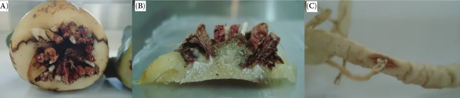
Figure 2. Rot symptoms of agave plantlets inoculated with the FsG F. solani strain: (A) rotten roots, (B) rotten tissue in the crown area, and (C) secondary rotten roots
[image:3.595.60.524.626.725.2]rEsults
Pathogenicity test of F. solani strain. The agave plants inoculated with the FsG strain averaged necro-sis on 56% of the length of their rootseight months post-inoculation. In addition, a reddish rot occurred on the crown (Figures 2A and 2B). The non-inoculated plants appeared to be healthy in the crown and root tissue. During the pathogenicity test, diseased plants generated new roots, but several of them were infected at sites where secondary or tertiary roots began to
emerge, and these new roots often presented symp-toms of root rot (Figure 2C). The aerial parts of these plants were asymptomatic. F. solani was reisolated from rotted tissues (Figure 3), demonstrating that FsG was pathogenic to A. tequilana.
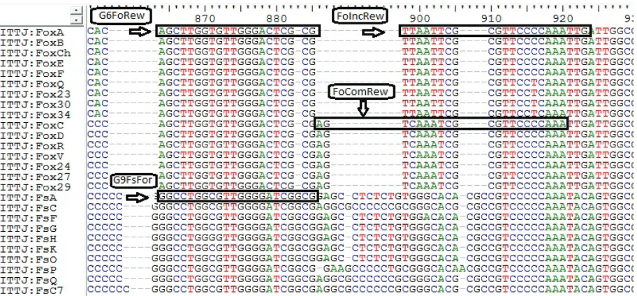
Primer design for identification of F. solaniand F. oxysporum.The more divergent region of amplified fragment with ITS1-ITS4 primers resulted in two se-quencesthat clearly differentiated between F. solani and
F. oxysporum strains (Figure 4) isolated from the tissue of agave diseased plants. Based on this zone, forward G9FsFor (5'-GGCCTGGCGTTGGGGATCG-3') and reverse G6FoRew (5'-CGCGAGTCCCAACACCAA-GC-3') primers were designed to identify F. solani and
F. oxysporum, respectively. Adjacent to this region for
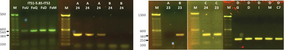
F. oxysporum, incompatible and compatible strains also showed variation (Figure 4), so it was useful to design FoIncRew (5'-CAATTTGGGGAACGCGAATTAA-3') and FoComRew (5'-TTTGGGGAACGCGATTT-GACT-3') reverse primers to identify incompatible and compatible F. oxysporum strains, respectively. PCR optimization conditions for each pair of primers are shown in Table 1. The positive reaction of this procedure amplified a band of ~380 bp for any strain of
F. oxyspo-rum as follows: combination A (ITS1-FoComRew)
identified compatible F. oxysporum strains; combina-tion B (ITS1-FoIncRew) confirmed the incompatible
[image:4.595.65.290.95.267.2]F. oxysporum lab strain; meanwhile, combination C (ITS1-G6FoRew) was useful for the identification of both groups. On the other hand, F. solani was detected
Figure 3.Long conidiophores and microconidia non-ca-tenulate characteristic of Fusarium solani, observed in isolates from the reddish rot tissue of agave plants with symptoms of agave wilt
[image:4.595.67.523.498.710.2]as a band of ~250 pb when combination D (G9FsFor-ITS4) was used (Figure 5). The primer combinations were validated in all Fusarium strains mentioned above.
Detection of frequency of F. oxysporum and F. solaniin commercial fields.The analysis of inter-nal tissues of the stem or crown from sixteen patchy groups of diseased plants indicated the presence of
F. solani in 100% of the forty-five evaluated plants. The identity of F. solani was microbiologically and molecularly corroborated in the reddish rot tissue of the crown area of the plants. Additionally, in 62.5% of the sampled plants, compatible strains of
F. oxysporum were molecularly detected; meanwhile, only in 8.3% of these plants incompatible strains
F. oxysporum were detected.
Dispersion patterns of agave wilt. In the com-mercial agave fields selected to determine dispersion patterns, the incidence of diseased plants ranged from 21.75% to 82.25%. However, the distribution of the disease had a tendency to change from a random pattern in a younger plantation like that in the 2011 IV field, to an aggregated pattern in rows like in the 2011 II field, and then to a highly aggregated distri-bution pattern in the 2008 II field (Figures 6A–C). This gradual change of the disease distribution pattern along the cycle life of agave plants was related to a
re-duction of the number of contiguous diseased-healthy plants (BW) observed in the row, in comparison with those expected (E(BW)). When the incidence of agave wilt was very high, lower BW events were observed in all the fields; however, these were not statistically dif-ferent from those expected, and a randomised pattern was determined by the join-count statistic (Table 2), such as is observed in the 2009-I field (Figure 6D).
disCussion
Agave wilt is considered the main pathological problem in A. tequilana Weber var. azul and F. oxy- sporum has been reported as its principal causal
agent (CRT 2010). Like other crops, A. tequilana
is a host to pathogenic strains of F. oxysporum that cause a vascular wilt (Avila-Miranda et al. 2010); however, in this work, it has been demonstrated that
[image:5.595.62.526.138.251.2]F. solani is pathogenic to young agave plants, caus-ing a reddish rot in roots, crown and stem tissues. This plant pathogen was detected with the use of the designed primers from the rotted tissue in roots, in-cluding plants with a range of initial symptoms, such as those with advanced foliar symptoms, and those with extreme root rot which had lost their soil anchorage
Table 1. Conditions of amplification of different primers to detect the presence of Fusarium solani, F. oxysporum (general), F. oxysporum incompatible, and F. oxysporum compatible strains in DNA from the stem or crown internal tissue of agave plants – Initial denaturation temperature (94°C) and time (2 min)
Primer pair No. of cycles Denaturation Annealing Extension Final extension
(°C) (min) (°C) (min) (°C) (min) (°C) (min)
G9FsFor-ITS4 35 94 1 58 58 72 1 72 6
ITS1-G6FoRew 35 94 1 58 58 72 1 72 6
ITS1-FoIncRew 1022 9494 11 6664 5564 7272 11 72 6
ITS1-FoComRew 1022 9494 11 6955 6966 7272 11 72 6
Figure 5. Validation of primer combinations to identify and discriminate between Fusarium solani and compatible and incompatible F. oxysporum strains (see the text): combination A confirmed compatible F. oxysporum; combina-tion B corroborated incompatible F. oxysporum strains; combinacombina-tion C identified the strains of F. oxysporum, and combination D detected F. solani strains
1500
600 540 380 100
1500
[image:5.595.61.530.616.699.2]Figure 6. Examples of spatial patterns of agave wilt epide-mics in areas of 20 plant rows by 20 contiguous plants: (A) Random pattern in the 2011 IV field, (B) Low contagious pa-ttern in the sense of the row of plants in the 2011 II field, (C) High contagious pattern in the 2008 III field, and (d) A field with high incidence of agave wilt but with random pattern reported by join-count statistics in the 2009 I field Solid squares represent diseased plants and open squares healthy plants
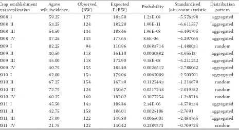
Table 2. Spatial association of diseased plants within a row in the field determined by the join-count statistics. High values of standardised join-count statistics (> –2.326) are indicative of positive autocorrelation (P ≤ 0.01) and aggre-gated pattern of diseased plant and lower values indicate random distribution
Crop establishment
year/replication wilt incidenceAgave Observed (BW) Expected E (BW) Probability join-count statisticStandardised Distributionpattern
2008 I 59.25 127 183.50 1.23E-08 –5.576398 aggregated
2008 II 53.25 124 182.20 1.90E-11 –6.611557 aggregated
2008 III 54.50 134 188.46 1.96E-08 –5.494795 aggregated
2008 IV 37.25 133 177.65 8.6E-06 –4.297065 aggregated
2009 I 82.25 94 110.96 0.0683714 –1.488031 random
2009 II 30.50 118 161.10 0.0000382 –3.95513 aggregated
2009 III 35.00 118 172.90 9.38E-08 –5.211232 aggregated
2009 IV 40.75 155 183.49 0.0026512 –2.788062 aggregated
2010 I 62.00 153 179.06 0.0062009 –2.500501 aggregated
2010 II 67.25 154 167.39 0.1122443 –1.214679 random
2010 III 72.75 128 150.67 0.0217238 –2.019382 random
2010 IV 60.25 169 182.02 0.1077254 –1.238716 random
2011 I 45.50 143 188.46 2.34E-06 –4.578334 aggregated
2011 II 42.75 158 186.01 0.0028106 –2.7691 aggregated
2011 III 27.00 122 149.80 0.0065001 –2.483765 aggregated
2011 IV 21.75 122 130.42 0.2389373 –0.709725 random
Q
uadra
t in r
ow
(A) (B)
Q
uadra
t in r
ow
Q
uadra
t in r
ow
Q
uadra
t in r
ow
(C) (d)
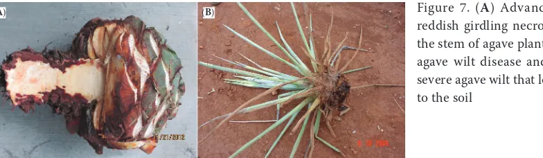
[image:6.595.67.532.505.759.2](Figure 7). These severe symptoms are similar to those described by Luna-Hernández (1996) for the stem rot of agave, attributed to F. oxysporum. However, these symptoms are different from those reported in vascular wilt diseases caused by some F. oxysporum
f.sp. strains in different host crops, where the princi-pal symptoms are injury to xylem vessels (Beckman 1987, 1989; Pegg 1989), or those symptoms caused by F. oxysporum f.spp. indicated as ‘radicis-’ in their names that induce combined vascular injury and limited destruction of the parenchyma of young roots and crown tissues (Charest et al. 1984; Benhamou
et al. 1989; Koyyappuratha et al. 2016). The severe rot symptoms in agave wilt are similar to those in the root and crown cortical tissue induced by necrotrophic soil-borne plant pathogens (MacHardy & Beckman 1981; Raaijmakers et al. 2009).
The changes observed in a distribution pattern of agave wilt from random to aggregated as plantations become older could be caused by both F. oxysporum
and F. solani agave plant pathogens, but the ability to disperse from plant to plant of the first one is prob-ably minor, if we consider that F. oxysporum induces a vascular wilt, with a specialised pathogenic process that is characterised by its penetration through the endodermis into the vascular tissue and moves up through the xylem to aboveground parts of the diseased plant, impeding the flow of water (Beckman 1987, 1989). In addition, Fusarium wilts are reported to show low transmission from plant to plant (Raaijmakers
et al. 2009). However, this kind of pattern forming a patchy distribution of diseased plants (Figure 1) could be satisfactorily explained by the necrotrophic soil-borne plant pathogen fungi such as F. solani (Rupe
et al. 2001), which cause cortical root rot, attacking preferably young roots as opposed to those lignified, and the fungus ramifies and continues to spread up the root, internally or externally, or can infect other roots in close proximity (MacHardy & Beckman 1981; Gilligan & Bailey 1997; Raaijmakers et al. 2009). This fact is in agreement with reports by
Aceves-Rodríguez (2003), who concluded that the incidence and severity of agave wilt increased in the field when the distance between plants was decreased.
F. oxysporum strains from VCG are able to colo-nise the xylem vascular system and cause vascular wilt (Avila-Miranda et al. 2010). In this work, we present evidence that F. solani is very much related to agave wilt, with its independent and specific way of pathogenicity, which adds information to explain the high incidence and severity of this disease that is frequently observed in commercial agave fields. This knowledge is necessary to design better strategies of management of this disease.
Acknowledgements. The authors gratefully acknowl-edgement to Casa Cuervo S.A. de C.V. for the facilities for agave plants and field sampling. M.J. Ramírez-Ramírez thanks Tecnológico Nacional de México for the fellowship for her doctoral studies.
references
Aceves-Rodríguez J.J. (2002): Aislamiento, identificación, incremento e inoculación de patógenos asociados a la marchitez del agave. In: Flores-López H. (ed.): Análisis
Agroecológico del Agave tequilana Weber var. azul con
Énfasis en Problemas Fitosanitarios en Jalisco. INIFAP-CIRPAC, Campo Experimental Altos de Jalisco. México, Conexión Gráfica S.A. de C.V: 33–44.
Aceves-Rodríguez J.J. (2003): Prevención y manejo integral
de la marchitez del Agave tequilana Weber var. azul en
Jalisco. Folleto técnico Núm. 1. Campo Experimental Altos de Jalisco. CIRPAC. INIFAP. Jalisco, México.
Agrios G.N. (2005): Plant Pathology. 5th Ed. Burlington,
Mass, Elsevier-Academic Press.
Avila-Miranda M.E. (2011): Enfermedades del agave y su manejo. In: Rendón-Salcido L.A., Avila-Miranda M.E., Rodríguez-Garay B., Del Real-Laborde J.I. (eds): Manual Técnico para el Establecimiento de Huertas Madre de Agave Azul, C.R.T. México, Prometeo editores SA de CV: 115–127.
Figure 7. (A) Advanced stage of the reddish girdling necrosis symptom on the stem of agave plant associated with agave wilt disease and (B) plant with severe agave wilt that lost its anchorage to the soil
Avila-Miranda M.E., Zazueta-López J.G., Arias-Castro C., Peña-Cabriales J.J. (2010): Vascular wilt caused by Fusarium oxysporum in agave (Agave tequilana Weber var. azul). Journal of the Professional Association for Cactus Development, 12: 166–180.
Avila-Miranda M.E., Campos-León C., Peña-Cabriales J.J., Rodríguez-Mendiola M.A., Mancilla-Margalli N.A., Pérez-González F., Arias-Castro C. (2012): Genetic
di-versity and vegetative compatibility groups in Fusarium
oxysporum cause of wilt symptoms in agave (Agave te-quilana Weber var. azul). Gayana Botanica, 69: 40–48. Beckman C.H. (1987): The Nature of Wilt Diseases of
Plants. St. Paul, APS Press.
Beckman C.H. (1989): Colonization of the vascular system of plants by fungal wilt pathogens: A basis for modeling the interactions between host and parasite in time and space. In: Tjamos E.C., Beckman C.H. (eds): Vascular Wilt Diseases of Plants Basic Studies and Control. Berlin-Heidelberg, Springer-Verlag: 19–32.
Benhamou N., Charest P.M., Jarvis W.R. (1989): Biology
and host-parasite relations of Fusarium oxysporum f.sp.
radicis-lycopersici. In: Tjamos E.C., Beckman C.H. (eds): Vascular Wilt Diseases of Plants Basic Studies and Con-trol. Berlin-Heidelberg, Springer-Verlag: 95–105.
Booth C. (1971): The genus Fusarium. Kew, Commonwealth
Mycological Institute.
Charest P.T., Oullette G.B., Pauzé F.J. (1984):
Cytologi-cal observations of early infection process by Fusarium
oxysporum f.sp. radicis-lycopersici in tomato plants. Ca-nadian Journal of Botany, 62: 1232–1244.
Correia K.C., Souza B.O., Câmara M.P.S., Michereff S.J. (2013): First report of stem rot of papaya caused by Fusarium solani species complex in Brazil. Plant Dis-ease, 97:140–141.
CRT (2010): Actualización de la base de datos y diagnóstico
fitosanitario: Agave tequilana Weber var. azul. Comité
Técnico Agronómico-Subcomité de Fitosanidad. Avail-able at https://www.crt.org.mx/images/documentos/ inventarioagave2010b.pdf (accessed June 10, 2015). CRT (2016): Información Estadística. Available at www.
crt.org.mx/EstadisticasCRTweb/ (accessed Mar 9, 2016). Farr D.F., Rossman A.Y. (2015): Fungal databases, System-atic Mycology and Microbiology Laboratory, ARS, USDA. Available at http://nt.ars-grin.gov/fungaldatabases/ (ac-cessed Apr 15, 2015).
Flores L.H.E., Ireta M.J., Ruíz C.J.A. (2010): Tecnología para la prevención y/o control de la marchitez del agave
tequilero en Jalisco, 1st Ed. Guadalajara, Jal. México,
Prometeo Editores: 1–5.
Gilligan C.A., Bailey D.J. (1997): Components of pathozone behavior. New Phytologist, 135: 475–490.
Graham J.H., Brlansky R.H., Timmer L.W., Lee R.F., Marais L.J., Bender G.S. (1985): Comparison of citrus tree de-clines with necrosis of major roots and their association
with Fusarium solani. Plant Disease, 69: 1055–1058.
Gumpertz M.L. (1997): Testing binary response variables for spatial autocorrelation. In: Francland L.J., Neher D.A. (eds): Exercises in Plant Disease Epidemiology. St Paul, The American Phytopathological Society: 78–84. Hall T.A. (1999): BioEdita user-friendly biological sequence
alignment editor and analysis program for windows 95/98/INT. In: Nucleic Acid Symposium Series No. 41: 95–98.
Ireta-Moreno J., Rodríguez-González P., Flores-López H.E., Flores-Mendoza J. (2002): Epidemiología de la
“marchitez” del agave azul Agave tequilana Weber
var-iedad azul. In: Flores-López H.E. (ed.): Análisis
agro-ecológico del Agave tequilana Weber variedad azul con
énfasis en problemas fitosanitarios en Jalisco. Jalisco,
México, EditorialConexión Gráfica, S.A. de C.V: 51–62.
Koike S.T. (2011): Fusarium crown and root rot of tarragon
in California caused by Fusarium solani. Plant Disease,
95: 768.
Koyyappuratha S., Atuahivab T., Le Guena R., Batinacd H., Le Squina S., Gautherone N., EdelHermanne V., Peribef J., Jahielg M., Steinberge C., Liewh E.C.Y., Alabouvette C., Bessej P., Dronk M., Sachecd I., Lavalcd V., Grisonia
M. (2016): Fusarium oxysporum f.sp. radicis-vanillae is
the causal agent of root and stem rot of vanilla. Plant Pathology, 65: 612–625.
Leslie J.F., Summerell B.A. (2006): The Fusarium Laboratory
Manual. Hoboken, Blackwell Publishing.
Luna-Hernández G. (1996): Pudrición del tallo de Agave
tequilana Weber en el estado de Jalisco. [Bachelor Thesis.] Texcoco, México, Universidad Autónoma de Chapingo. MacHardy W.E., Beckman C.H. (1981): Vascular wilt Fusaria:
infection and pathogenesis. In: Nelson P.E., Toussoun T.A., Cook. R.J. (eds): Fusarium: Diseases, Biology, and Taxonomy. Pennsylvania State University Press: 365–390. Madden L.V., Hughes G., van den Bosch F. (2007): The Study
of Plant Disease Epidemics. St. Paul, APS Press: 262. Ortiz P.G., Sánchez A.A., Virgen-Calleros G.,
Carvajal-Ca-zola C.R., Padrón-Corral E. (2011): Incidencia y severidad
de la marchitez del Agave tequilana Weber var. azul en la
zona sur del estado de Nayarit, México. Revista
Agraria-Nueva Época,8: 21–25.
Pegg G.F. (1989): Pathogenesis in vascular diseases of plants. In: Tjamos E.C., Beckman C.H. (eds): Vascular Wilt Diseases of Plants Basic Studies and Control. Berlin-Heidelberg, Springer-Verlag: 51–94.
and battlefield for soilborne pathogens and beneficial microorganisms. Plant Soil, 321: 341–361.
Rupe J.C., Correll J.C., Guerber J.C., Becton C.M. Jr., Gbur E.E., Cummings M.S., Yount P.A. (2001): Differentiation of the sudden death syndrome pathogen of soybean, Fusarium solani f.sp. glycines, from other isolates of F. solani based on cultural morphology, pathogenicity, and mitochondrial DNA restriction fragment length polymorph. Canadian Journal of Botany, 79: 829–835. Uvalle B.J.X., Angelina B.R., Apodaca S.M.A. (2010):
Mane-jo integral de enfermedades. In: División Agrícola (eds):
Manejo integral del cultivo de Agave tequilana Weber,
cv. azul. Orgánica Diseño Editorial: 99–133.
White T.J., Bruns T., Lee S., Taylor J. (1990): Amplification and direct sequencing of fungal ribosomal RNA genes for phylogenetics. In: Innis M.A., Gelfand D.H., Sninsky J.J., White T.J. (eds): PCR Protocols: a Guide to Methods and Applications. New York, Academic Press: 315–322.