Original Article
MiR-184 has prognostic implication and
is down-regulated during the malignant
progression in human astrocytoma
Ling Chen1, Yanlei Guan2, Yijun Bao2, Guangyu Li2, Run Cui2, Zhen Zhang3, Yunjie Wang2
Departments of 1Geriatric Medicine, 2Neurosurgery, 3Ultrasound, First Affiliated Hospital of China Medical
University, Shenyang, Liaoning, China
Received February 22, 2016; Accepted July 12, 2016; Epub September 1, 2016; Published September 15, 2016
Abstract: Recent studies have indicated that miR-184 is deysregulated and function both as oncogene and tumor suppressor in several types of cancer. However, the role of miR-184 in astrocytoma is still not clearly understood.
Therefore, we in the present study aimed to investigate the clinical significance of miR-184 expression in human
astrocytomas. We examined the expression level of miR-184 in 76 astrocytoma tissues and 5 cell lines by qRT-PCR and found that miR-184 expression was markedly reduced in the tumor tissues and cell lines, compared with that in non-neoplastic brain tissues and human astrocyte, respectively. In addition, low expression of miR-184 was
sig-nificantly associated with the aggressive clinicopathological features (advanced tumor degree, advanced patient
age, low Karnofsky performance score and high ki-67 index of tumor tissue) in astrocytoma patients. Furthmore, the correlations of miR-184 expression with prognosis of astrocytoma patients were also modeled by Kaplan-Meier
method and multivariate analysis. Our results showed that patients with low miR-184 expression had significantly
poor survival (P < 0.001, Kaplan-Meier method) and miR-184 was an independent prognostic indicator for astrocy-toma patients (P < 0.001; risk ratio = 5.7, Cox regression analysis). Moreover, we examined miR-184 expression in paired tumor tissues from seven patients with primary lower-grade astrocytomas and the spontaneously recurrent higher-grade astrocytomas. MiR-184 showed an absolute down-regulation in recurrent astrocytomas as compared with the corresponding primary tumors. In conclusion, our data suggest that down-regulation of miR-184 may have potential value for predicting clinical outcomes in astrocytoma patients, and miR-184 is an important candidate tumor suppressor, and its down-regulation may contribute to mailgnant progression of human astrocytoma.
Keywords: microRNA, miR-184, astrocytoma, down-regulation, prognosis, malignant progression
Introduction
Astrocytomas are the most frequent tumors of central nervous system, accounting for more than 60% of all primary brain tumors of human adults [1]. They are aggressive, highly invasive and neurological destructive tumors consid-ered being among the deadliest of human can-cers. Based on histomorphological criteria, dif-fuse infiltrating astrocytomas are classified into three ascending grades of malignancy includ-ing well-differentiated diffuse astrocytoma (DA, grade II), anaplastic astrocytoma (AA, grade III), and glioblastoma (GBM, grade IV), according to the World Health Organization (WHO) grading system [1]. In addition, GBMs can be further divided into two subtypes that have distinct
identify new biomarkers for prognostic predic-tions and treatment oppredic-tions and explore novel therapeutic targets for astocytoma.
MicroRNAs (miRNAs) are recently discovered small, non-coding endogenous RNA molecules of about 18-25 nucleotides. They are consid-ered to play important roles in a variety of biological processes including cell prolifera- tion, apoptosis, migration and differentiation, through post-translationally regulating expres-sion of their target genes [4, 5]. These short RNA molecules have been reported to be aber-rantly expressed in various human cancers and act as important regulators of tumor biologic behaviors in tumorigenesis and aggressive pro-gression, by targeting oncogenes or tumor sup-pressors [6, 7]. Accumulating evidences indi-cate that identification of specific miRNAs in cancer cells has substantial value for diagnos-tic and prognosdiagnos-tic determinations as well as for eventual therapeutic interventions [8, 9]. In the last decade, a number of specific miR-NAs have been identified to be abnormally in human astrocytomas. They are involved in tu- morigenesis and malignant progression of as- tocytoma by functioning as oncogenes or tumor suppressors [10-13]. Most of these miRNAs have also been demonstrated to significant- ly correlate with patients’ survival and could function as prognostic and predictive indica-tors in human cancers, such as miR-21 [14], miR-155 [15], miR-196 [16] and miR-326 [17]. MiR-184 is located in region 25.1 on the long arm of chromosome 15 and is particularly en- riched in human brain. This miRNA has been reported to be deysregulated and function both as oncogene and tumor suppressor in numerous human cancers [18-21]. However, the functions and the exact mechanisms of miR-184 in tumorigenesis and progression of human astrocytoma remain controversial. Se- veral recent studies showed that miR-184 was significantly down-regulated in astocytoma tis-sues compared with normal brains and acted as a potential tumor suppressor by inhibiting cell proliferation and invasion [22-24]. In con-trary, other groups found that miR-184 could enhance the aggressive biological behaviors of glioma cell lines [25, 26]. In addition, the clinical significances of miR-184 in astrocyto-mas are still poorly understood.
We in the present study examined expression level of miR-184 in a large panel of astrocyto-mas and cell lines and statistically evaluated the correlations between miR-184 expression and clinicalpathological factors in these pa- tients. As our results, miR-184 was remarkably dwon-regulated in astrocytoma tissues and tumor cell lines as compared with non-neoplas-tic brain tissues and low miR-184 expression was significantly associated with aggressive clinicalpathological features of astrocytoma. In addition, miR-184 expression statistically cor-related with patients’ survival and was an in- dependent prognostic indicator. Furthermore, significant lower expression of miR-184 was observed in recurrent higher-grade tumor com-pared with corresponding primary lower-grade tumor in a series of paired astrocytomas. Our observations suggest that miR-184 might fun- ction as a tumor suppressor and could be a potential biomarker for prognosis and aggre- ssive progression in human astrocytoma. Materials and methods
Astrocytoma specimens and patients
the alteration of miR-184 expression during the malignant progression of astrocytoma, expres-sion level of miR-184 was determined in prima-ry lower-grade (grade II or III) and recurrent higher-grade (grade III or IV) tumor pairs derived from seven independent astrocytoma patients. In addition, a validation step involved analysis of 9 DAs, 8 AAs and 8 secondary GBM from 25 independent patients. All astorcytoma patients were well followed up, and overall survival time was calculated from the date of the initial surgi-cal operation to death. Patients, who died of diseases not directly related to their astrocyto-mas or due to unexpected events, were exclud-ed from this study. The present study was approved by the Ethics Committee of China Medical University.
Glioma cell lines and human astrocyte
The astrocytoma cell lines U87, U251, U373, T98G and SF295 were obtained from the American Type Culture Collection (ATCC, Ma- nassas, VA, USA) and maintained in Dulbe- cco’s modiied eagle’s medium (DMEM) (Gibco, USA) supplemented with 10% fetal bovine serum (FBS) (Gibco) and penicillin/streptomy-cin (100 U/mL). The human astrocyte was a gift from Dr. T. Sasaki (Graduate School of Medical Sciences, Kyushu University, Fukuoka, Japan) and maintained in DMEM supplement- ed with 2% FBS and 1% N-2 supplement (Gibco).
tion Kit (Applied Biosystems) and individual TaqMan miRNA assay (Applied Biosystems), and Applied Biosystems 7500HT Fast Real-Time PCR System (Applied Biosystems), as previously described [27]. RNU6B were used as endogenous controls, and non-neoplastic brain tissues and human astrocyte were used for calibrations. Relative quantification of miR-184 expression was calculated with the 2-ΔΔCt
method.
Statistical analysis
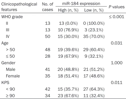
All computations were carried out using the software of SPSS version19.0 for Windows (SPSS Inc., Chicago, IL, USA). Data were expre- ssed as mean ± standard deviation (SD). Stu- dent’s t-test was used to compare the expres-sion levels of miRNAs between different sub-types of astrocytoma, as well as between as- trocytomas and non-neoplastic brains. Associ- ations of miR-184 expression with clinicopa- thological features and the ki-67 index were estimated using a Pearson’s chi-square test and Pearson’s correlation analysis, respective-ly. A life table was calculated according to the Kaplan-Meier method. Risk ratios for the time-to-event endpoint were estimated using the multivariate Cox regression analysis in a for-ward stepwise method to evaluate the effect of multiple independent prognostic factors on overall survival outcome. Differences were con-Table 1. Correlation of miR-184 relative expression level
with clinicopathological factors of astrocytoma patients
Clinicopathological features No. of cases
miR-184 expression P values High (n, %) Low (n, %)
WHO grade ≤ 0.001
II 13 13 (0.0%) 0 (100.0%) III 13 10 (76.9%) 3 (23.1%) IV 50 15 (30.0%) 35 (70.0%)
Age 0.031
> 50 48 19 (39.6%) 29 (60.4%)
≤ 50 28 19 (67.9%) 9 (32.1%)
Gender 1.000
Male 41 20 (48.8%) 21 (51.2%) Female 35 18 (51.4%) 17 (48.6%)
KPS 0.011
< 90 42 15 (35.7%) 27 (64.3%)
≥ 90 34 23 (67.6%) 11 (32.4%)
Abbreviations: KPS, Karnofsky performance scale.
RNA extraction, reverse transcription and real-time PCR quantification for miRNA detection
[image:3.612.90.341.97.295.2]sidered statistically significant when P was less than 0.05.
[image:4.612.91.374.74.312.2]184 expression of each malignancy grade of astocytomas with that of non-neoplastic brains. We found that grade II DAs had an about 1.80-fold higher expression of miR-184 relative to brain tissues (P = 0.020, Figure 1A). However, no significant difference was observed between miR-184 expression of grade III AAs and normal brain tissues (P = 0.214, Figure 1A). In con-trast, expression level of miR-184 was remark-ably reduced in grade IV pGBMs as compared with that in brain tissues (fold change = 0.35; P ≤ 0.001, Figure 1A). Furthermore, miR-184 showed a decreased expression with the in- creasing degree of malignancy of astrocyto- mas (P values in grade II vs. III, grade II vs. IV and grade III vs. IV were 0.003, ≤ 0.001 and 0.048, respectively, Figure 1A). Moreover, miR-184 expression was examined in five com- monly used model cell lines (U87, U251, U373, T98G, and SF295, Figure 1B) derived from human malignant astrocytomas. As demon-strated in Figure 1B, we found a profoundly decreased expression of miR-184 in these tu- mor cells. The expression level of miR-184 was about 0.05- to 0.17-fold lower in tumor cell Figure 1. MiR-184 expression in 76 astrocytoma tissues, 5 cell lines
com-pared with 10 non- neoplastic brain tissues and human astrocyte,
respec-tively, detected by qRT-PCR analysis. A. MiR-184 was significantly down-regu -lated in astrocytoma tissues compared with non-neoplastic brains tissues (P
= 0.041). Its expression level was decreased with the increasing malignancy degree of the tumor. B. Astrocytoma cell lines showed remarkably lower ex-pression of miR-184 in comparison with human astrocyte (HA).
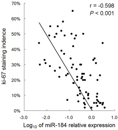
Figure 2. Pearson’s correlation analysis to evaluate the correlation of miR-184 expression with the ki-67 labeling index in 76 astrocytoma tissues.
Results
MiR-184 was down-regulated in astrocytoma tissues and cell lines
[image:4.612.92.285.416.629.2]miR-Figure 3. Prognostic performance of miR-184 for astrocytoma patients. A. Atrocytoma patients with low miR-184
expression (left, solid line, n = 38) had significantly shorter overall survival time than did patients with high miR-184
expression (right, dotted line, n = 38; P ≤ 0.001, log-rank test). B. Among the 63 high-grade astrocytoma patients (grade III AAs and grade IV pGBMs), those with low miR-184 expression (left, solid line, n = 38) had significantly
shorter survival periods than did patients with high miR-184 expression (right, dotted line, n = 25; P ≤ 0.001).
lines relative to a human astrocyte. These re- sults suggested that miR-184 might act as a tumor suppressor in astrocytoma tumorigene-sis and its down-regulation might be involved in aggressive progression of astocytoma. Low miR-184 expression was associated with aggressive clinicalpathological features of as-trocytoma
Subsequently, correlations of miR-184 expres-sion with several clinicopathological features (tumor grade, patients’ age at diagnosis, gen-der and pre-operative Karnofsky performance scale (KPS)) of these 76 patients mentioned above were statistically evaluated by Χ2 test
as demonstrated in Table 1. Patients were assigned to high-miR-184 group (n = 38) and low-miR-184 group (n = 38) that were tumors with miR-184 expression above and under the median value of miR-184 expression in all of the 76 astrocytomas, respectively. As summarized in Table 1, low miR-184 expres-sion was significantly associated with advanced malignancy degree of tumor (P ≤ 0.001, Χ2
test), advanced patient’s age (P = 0.031) and low KPS (P = 0.011). However, there was no sta-tistically significant correlation between miR-184 expression and patient’s gender. In addi-tion, we statistically analyzed the correlation
between miR-504 expression and ki-67 la- beling index in these astrocytoma tissues. As shown in Figure 2, the ki-67 index was nega-tively associated with log10 of the relative miR-504 expression (P < 0.001, r = -0.598, Pear- son’s correlation analysis).
Expression level of miR-184 had prognostic implication in astrocytoma patients
[image:5.612.95.517.74.280.2]tumor tissue (P ≤ 0.001; risk ratio 5.7) and advanced malignancy degree (P ≤ 0.001; risk ratio 8.6) were independent predictors of poor prognosis in glioma patients (Table 2). Moreover, we also performed Kaplan-Meier analysis to evaluate the prognostic perfor-mance of miR-184 expression in high-patholog-ical grade astrocytoma patients (grade III AAs and grade IV pGBMs). Similarly, we observed that low-miR-184 expression showed a statis- tically significant correlation with poor clinical outcome in patients with these malignant as- trocytomas (P ≤ 0.001, Figure 3B).
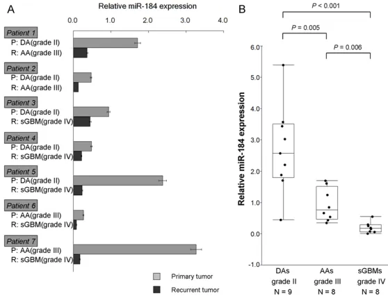
MiR-184 was down-regulated in malignant pro-gression of astrocytomas
Moreover, we discovered the dynamic altera-tion of miR-184 expression during the malig-nant progression of astrocytoma. Paired tumor tissues from seven patients with primary lower-grade astrocytomas (lower-grade II DAs or lower-grade III AAs) and the spontaneously recurrent higher-grade astrocytomas (higher-grade III AAs or higher-grade IV sGBMs) were collected for detection of
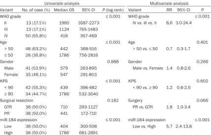
[image:6.612.93.521.97.374.2]miR-184 expression by qRT-PCR. These seven as- trocytoma patients displayed three different progression patterns of this tumor: two of them were with primary grade II DA that spon- taneously progressed to grade III AA; three of them suffered from primary grade II DA that recurred as grade IV sGBM; whereas, other two had primary grade III AA and recurrent grade IV sGBM. As shown in Figure 4A, we observed that miR-184 was absolutely down-regulated during each of these three malignant progres-sion patterns. It showed an about 1/2- to 1/20-fold significantly lower expression in the re- current higher-grade astrocytomas relative to the corresponding primary lower-grade tumors (Figure 4A). To validate the progression-asso- ciated down-regulation of miR-184, we ana-lyzed expression of miR-184 in an independ- ent series of 9 grade II DAs, 8 grade III AAs and 8 grade IV sGBMs from another panel of 25 patients. As shown in Figure 3B, miR-184 showed an obvious decrease along with the increasing malignant grade of the tumor (mean ± SD of relative miR-184 expression for grade II, III and IV astrocytomas were 2.70 ± 1.40, Table 2. Univariate and multivariate Cox regression analysis for overall survival in astrocytoma pa-tients
Univariate analysis Multivariate analysis Variant No. of case (%) Median OS 95% CI P (log-rank) Variant RR 95% CI P
WHO grade ≤ 0.001 WHO grade ≤ 0.001
II 13 (17.1%) 1980 1687-2273 IV vs. III vs. II 8.6 3.0-24.4 III 13 (17.1%) 1124 765-1483
IV 50 (65.8%) 418 367-469
Age ≤ 0.001 Age 0.401
> 50 48 (63.2%) 442 368-516 > 50 vs. ≤ 50 0.7 0.3-1.7
≤ 50 28 (36.8%) 1786 756-2816
Gender 0.888 Gender 0.266
Male 41 (53.9%) 579 263-895 Male vs. Female 1.4 0.8-2.6 Female 35 (46.1%) 547 291-803
KPS ≤ 0.001 KPS 0.602
< 90 42 (55.3%) 439 396-482 < 90 vs. ≥ 90 1.2 0.6-2.5
≥ 90 34 (44.7%) 1786 532-3040
Surgical resection 0.182 Surgery 0.066 GTR 38 (50.0%) 710 293-1127 PR vs. GTR 1.8 1.0-3.4
PR 38 (50.0%) 441 172-710
miR-184 expression ≤ 0.001 miR-184 expression ≤ 0.001
Low 38 (50.0%) 404 300-508 Low vs. High 5.7 2.4-13.8 High 38 (50.0%) 1786 681-2891
0.92 ± 0.53 and 0.20 ± 0.17, respectively). The differences were statistically significant between grade II and III (P = 0.015), grade III and IV (P = 0.016), as well as grade II and IV (P ≤ 0.001) tumors (Figure 4B).
Discussion
In recent years, accumulating evidences have indicated that miRNAs play important roles in tumorigenesis and aggressive progression by regulating multiple oncogenes and tumor sup-pressors, in various human cancers. Previous studies have identified a number of dysregulat-ed miRNAs including 21, 155, miR-196 and miR-326 in astrocytomas, the most frequent and aggressive tumors of human cen-tral nervous system. These miRNAs were dem-onstrated to play important roles in crucial biological processes such as cell proliferation, apoptosis and invasion, in tumorigenesis of astrocytoma [10-13]. In addition, their expres-sion signatures have also been proved to have
[image:7.612.91.370.74.287.2]sues had significantly poorer overall survival and low miR-184 expression was a statistically significant risk factor of poor survival for astro-cytoma patients. Moreover, miR-184 was sig-nificantly down-regulated in the recurrent high-er-grade astrocytomas compared with the cor-responding primary lower-grade tumors. To our knowledge, this is the first study to analyze the expression profile and clinical significance of miR-184 in large panel of astrocytomas. MiR-184, which is located in region 25.1 of chromosome 15q, is particularly enriched in human brain and testes. Its corresponding transcript is comparatively small (84 bp) and is not encoded other clustered miRNAs [28, 29]. Previous studies have indicated that miR-184 is dysregulated in a variety of human can-ers and acts as a tumor promoter or suppres-sor in an organ-specific fashion [18-21]. Func- tioning as a tumor suppressor, miR-184 was originally reported to be aberrantly expressed in neuroblastomas and involved in tumorige- Figure 4. Alteration of miR-184 expression during the malignant progression
of astrocytoma. A. Expression levels of miR-184 in seven astrocytoma pa-tients with parimary lower-grade tumors that recurred as higher-grade
tu-mors. MiR-184 showed an about 1/2- to 1/20-fold significantly lower expres -sion in the recurrent higher-grade astrocytomas relative to the corresponding primary lower-grade tumors. Patient numbers 1-7 encode the individual patient; Gray and black columns indicate expression levels of miR-184 in primary and recurrent tumors, respectively. P, primary tumor; R, recurrent tumor. B. Validation experiment for miR-184 expression in an indepentdent
series of 9 DAs, 8 AAs and 8 secondary-GBMs. miR-184 showed a significant
progression-associated down-regulation in gliomas with different WHO grade (P = 0.005, 0.006 and ≤ 0.001 for grade III vs. II, grade IV vs. III and grade
IV vs. II, respectively).
substantial value of diagno- sitc and prognositic deter- minations for patients with these malignant brain tumors [14-17].
tis-nesis of these aggressive pediatric tumors, in a global miRNA expression profiling study, by Chen et al. [18]. They found that miR-184 was significantly down-regulated in MYCN-amplified tumors that have poor prognosis as compared with other types of neuroblastoma, and their function analyses clearly showed that miR-184 overexpression induces apoptosis and cell cycle arrest in neuroblastoma cells [18]. The investigation by Foley et al. confirmed the tumor suppressive role of miR-184 and revealed that this miRNA inhibits cell survival by targeting AKT2 kinase in neuroblastoma [19]. Conversely, other researchers have revealed the potential oncogenic role of this miRNA. Wong et al. found that the plasma expression levels of miR-184 were significantly associated with the presence of primary tumors and might be used as a novel cancer marker in tonguesquamous cell carcio-ma [20]. In addition, Wu et al. demonstrated that miR-184 promotes cell proliferation in human hepatocellular carcinoma by post-tran-scriptionally regulating SOX7 expression [21]. However, the biological functions of miR-184 in astrocytoma tumorigenesis still remain con-troversial, and miR-184’s clinical significance in astrocytoma patients is not clearly under-stood. Actually, miR-184 was first reported to fuction as a negative regulator in malignant progression of astrocytoma by Malzkorn et al. [22]. They found that miR-184 showed signifi-cantly decreased expression upon the progres-sion from low-grade to high-grade astrocyto-mas. And their function analysis revealed that overexpression of miR-184 inhibits cell prolif-eration and invasion in glioma cell lines, A172 and T98G [22]. At almost the same time, Guan et al. reported that miR-184 was significantly down-regulated in grade IV astrocytomas as compared with grade III astrocytomas, by in- vestigating the expression profiles of 365 miR-NAs in 12 high-grade astrocytomas [16]. In ad- dition, a recent study by Cheng et al. showed that miR-184 was down-regulated in astrocy- toma tissues and decreased with the increas-ing degree of malignancy, although the number of cases used was limited [23]. These observ-astions suggested the possibility that miR-184 might act as a tumor suppressor in tumorigen-esis of astrocytoma. We in the present study increased the tumor cases to further confirm the expression and clinical implication of miR-184 in astrocytomas. In accordance with the results from these previous investigations, we
found that down-regulation of miR-184 signi- ficantly correlated with aggressive progress- ion and poor survival in astrocytoma patients. Furthermore, recent studies have revealed the biological mechanisms by which miR-184 mod-ulates astrocytoma tumorigenesis through fun- ctioning as a tumor suppressive miRNA. The study by Chen et al. mentioned above demon-strated that miR-184 inhibits cell prolifera- tion and invasion by specifically targeting TNF- AIP2 in astrocytoma [23]. Meanwhile, Emdad et al. indicated that suppression of miR-184 in malignant astrocytomas down-regulates its direct target, SND1, and promotes tumor aggre- ssiveness both in vitro and in vivo [24]. These collective data provided sufficient evidence that miR-184 functions as a negative regulator in astrocytoma tumorigenesis. Conversely, sev-eral other investigations have demonstrated that miR-184 acts as a tumor promotor in as- trocytoma cells. Yuan et al. found that upre- gulation of miR-184 enhances the malignant biological behavior of human astrocytoma cell line A172 by targeting FIH-1 [25]. Similarly, Cui et al. showed that miR-184 promotes pro- liferation ability of glioma cells by regulating FOXO3 [26]. Taken togther, the detailed bio- logical mechanism(s) through which miR-504 modulates tumorigenesis of astrocytoma still remains unclear, and thus needs further in- vestigation.
glioblastomas and not contribute to the malig-nant progression from anaplastic gliomas to secondary glioblastomas [31]. In consistent with our results, Malzkorn et al. showed that expression of miR-184 was reduced upon the progression from grade II to grade IV astrocyto-mas [22]. We in the present study clearly showed that miR-184 was absolutely down-reg-ulated during each of the three patterns (from grade II to grade IV, from grade II to grade III and from grade III to grade IV tumors) of astro-cytoma progression from low-grade to high-grade tumors (Figure 4A). Our results confirm the progression-associated down-regulation of miR-184 and suggest miR-184 as an important candidate contributes to malignant progres-sion of human astrocytoma.
In summary, we in the present study show- ed that expression of miR-184 was markedly reduced in both astrocytoma tissues and cell lines. In additon, our resutls demonstrated that miR-184 down-regulation correlated with aggressive clinicopathological features and poor survival in astrocytoma patients. Further- more, we found that miR-184 was absolutely down-regulated during the malignant progres-sion from low-grade to high-grade astrocyto-mas. Our data confirm the tumor suppressive role of miR-184 in astrocytoma tumorigenesis and suggest that miR-184 might serve as a prognostic and predictive biomarker, as well as a novel therapeutic target for these malig-nant brain tumors.
Acknowledgements
This study was supported by the National Natural Science Foundation of China (Grant No: 81302190).
Disclosure of conflict of interest
None.
Address correspondence to: Dr. Yanlei Guan, De-
partment of Neurosurgery, First Affiliated Hospital
of China Medical University, 155 North Nanjing Street, Heping District, Shenyang 110001, Liao- ning, China. Tel: 83283129; Fax: +86-24-83283390; E-mail: [email protected]
References
[1] Louis DN, Ohgaki H, Wiestler OD, Cavenee WK, Burger PC, Jouvet A, Scheithauer BW, Kleihues
P. The 2007 WHO classification of tumors of
central nervous system. Acta Neuropathol 2007; 114: 97-109.
[2] Ohgaki H, Kleihues P. Genetic pathways to pri-mary and secondary glioblastoma. Am J Pathol 2007; 170: 1445-1453.
[3] Van Meir EG, Hadjipanayis CG, Norden AD, Shu HK, Wen PY, Olson JJ. Exciting new advances in neuro-oncology: the avenue to a cure for malig-nant glioma. CA Cancer J Clin 2010; 60: 166-93.
[4] Bartel DP. MicroRNAs: genomics, biogenesis, mechanism and function. Cell 2004; 116: 281-97.
[5] Ambros V. The functions of animal microRNAs. Nature 2004; 431: 350-5.
[6] Calin GA, Sevignani C, Dumitru CD, Hyslop T, Noch E, Yendamuri S, Shimizu M, Rattan S, Bullrich F, Negrini M, Croce CM. Human mi-croRNA genes are frequently located at fragile sites and genomic regions involved in cancers. Proc Natl Acad Sci U S A 2004; 101: 2999-3004.
[7] Garzon R, Fabbri M, Cimmino A, Calin GA, Croce CM. MicroRNA expression and function in cancer. Trends Mol Med 2005; 12: 580-7. [8] Lowery AJ, Miller N, McNeill NE, Kerin MJ.
MicroRNAs as prognostic indicators and thera-peutic targets: potential effect on breast can-cer management. Clin Cancan-cer Res 2008; 14: 360-5.
[9] Schetter AJ, Leung SY, Sohn JJ, Zanetti KA, Bowman ED, Yanaihara N, Yuen ST, Chan TL, Kwong DL, Au GK, Liu CG, Calin GA, Croce CM,
Harris CC. MicroRNA expression profiles asso -ciated with prognosis and therapeutic out-come in colon adenocarcinoma. JAMA 2008; 299: 25-36.
[10] Chan JA, Krichevsky AM, Kosik KS. MicroRNA- 21 is an antiapoptitic factor in human glioblas-toma cells. Cancer Res 2005; 65: 6029-33. [11] Zhou J, Wang W, Gao Z, Peng X, Chen X, Chen
W, Xu W, Xu H, Lin MC, Jiang S. MicroRNA-155 promotes glioma cell proliferation via the regu-lation of MXI1. PLoS One 2013; 8: e83055. [12] Yang G, Han D, Chen X, Zhang D, Wang L, Shi
C, Zhang W, Li C, Chen X, Liu H, Zhang D, Kang J, Peng F, Liu Z, Qi J, Gao X, Ai J, Shi C, Zhao S. MiR-196a exerts its oncogenic effect in
glio-blastoma multiforme by inhibition of IκBα both
in vitro and in vivo. Neuro Oncol 2014; 16: 652-61.
[13] Kefas B, Comeau L, Erdle N, Montgomery E, Amos S, Purow B. Pyruvate kinase M2 is a tar-get of the tumor-suppressive microRNA-326 and regulates the survival of glioma cells. Neuro Oncol 2010; 12: 1102-12.
has-miR-21, has-miR-181b and has-miR-106a as prognositic indicator of astrocytoma. Eur J Cancer 2010; 46: 1640-9.
[15] Sun J, Shi H, Lai N, Liao K, Zhang S, Lu X. Overexpression of microRNA-155 predicts poor prognosis in glioma patients. Med Oncol 2014; 31: 911.
[16] Guan Y, Mizoguchi M, Yoshimoto K, Hata N, Shono T, Suzuki SO, Araki Y, Kuga D, Naka- mizo A, Amano T, Ma X, Hayashi K, Sasaki T. MiRNA-196 is upregulated in glioblastoma but not in anaplastic astrocytoma and has
prognostic significance. Clin Cancer Res 2010;
16: 4289-97.
[17] Wang S, Lu S, Geng S, Ma S, Liang Z, Jiao B.
Expression and clinical significance of microR -NA-326 in human glioma miR-326 expression in glioma. Med Oncol 2013; 30: 373.
[18] Chen Y, Stallings RL. Differential patterns of microRNA expression in neuroblastoma are correlated with prognosis, differentiation, and apoptosis. Cancer Res 2007; 67: 976-83. [19] Foley NH, Bray IM, Tivnan A, Bryan K, Murphy
DM, Buckley PG, Ryan J, O’Meara A, O’Sullivan M, Stallings RL. MicroRNA-184 inhibits neuro-blastoma cell survival through targeting the seine/thresnine kinase AKT2. Mol Cancer 2010; 9: 83.
[20] Wong TS, Liu XB, Wong BY, Ng RW, Yuen AP, Wei WI. Mature miR-184 as potential oncogen-ic moncogen-icroRNA of squamous cell carcinoma of tongue. Clin Cancer Res 2008; 14: 2588-92. [21] Wu GG, Li WH, He WG, Jiang N, Zhang GX,
Chen W, Yang HF, Liu QL, Huang YN, Zhang T, Zeng XC. Mir-184 post-transcriptionally regu-lates SOX7 expression and promotes cell prolif-eration in human hepatocellular carcinoma. PLoS One 2014; 9: e88796.
[22] Malzkorn B, Wolter M, Liesenberg F, Grzen- dowski M, Stühler K, Meyer HE, Reifenberger
G. Identification and functional characteriza -tion of microRNAs invovled in the malignant progression of gliomas. Brain Pathol 2010; 20: 539-50.
[23] Cheng Z, Wang HZ, Li X, Wu Z, Han Y, Li Y, Chen G, Xie X, Huang Y, Du Z, Zhou Y. MicroRNA-184 inhibits cell proliferation and
invasion, and specifically targets TNFAIP2 in
Glioma. J Exp Clin Cancer Res 2015; 34: 27.
[24] Emdad L, Janjic A, Alzubi MA, Hu B, Santhe- kadur PK, Menezes ME, Shen XN, Das SK, Sarkar D, Fisher PB. Suppression of miR-184 in malignant gliomas upregulates SND1 and promotes tumor aggressiveness. Neuro Oncol 2015; 17: 419-29.
[25] Yuan Q, Gao W, Liu B, Ye W. Upregulation of miR-184 enhances the malignant biological behavior of human glioma cell line A172 by tar-geting FIH-1. Cell Physiol Biochem 2014; 34: 1125-36.
[26] Cui QK, Liu WD, Zhu JX, Wang YH, Wang ZG. MicroRNA-184 promotes proliferation ability of glioma cells by regulating FOXO3. Asian Pac J Trop Med 2014; 7: 776-9.
[27] Chen C, Ridzon DA, Broomer AJ, Zhou Z, Lee DH, Nguyen JT, Barbisin M, Xu NL, Mahuvakar VR, Andersen MR, Lao KQ, Livak KJ, Guegler KJ. Real-time PCR of microRNA by stem-loopRT-PCR. Nucleic Acids Res 2005; 33: e179. [28] Nomura T, Kimura M, Horii T, Morita S, Soejima
H, Kudo S, Hatada I. MeCP2-dependent re-pression of an imprinted miR-184 released by depolarization. Hum Mol Genet 2008; 17: 1192-9.
[29] Weitzel RP, Lesniewski ML, Greco NJ, Laughlin MJ. Reduced methyl-CpG protein binding con-tributing to miR-184 expression in umbilical cord blood CD4+ T-cells. Leukemia 2011; 25: 169-72.
[30] Riemenschneider MJ, Reifenberger G. Astro- cytic tumors. In: Gliomas, editor. Recent Re- sults in Cancer Research, Vol. 171. A von Deimling (ed.), Springer: Berlin; 2009. pp. 3-24.