Original Article
miR-132 and miR-212 cluster function as a tumor
suppressor in thyroid cancer cells by CSDE1
mediated post-transcriptional program
Tong Chen*, Mingdong Lu*, Xiang Zhou, Xiaoyu Pan, Yifang Han, Yi Zhang, Bing Ye, Jianda Dong, Pihong Li
Department of General Surgery, The Second Affiliated Hospital of Wenzhou Medical University, Wenzhou, China.
*Equal contributors.
Received September 30, 2017; Accepted December 7, 2017; Epub February 1, 2018; Published February 15, 2018
Abstract: microRNAs (miRNAs) are small non-coding RNA molecules which have been reported to be associated with the development of cancers. However, the role of miRNAs in thyroid cancer remains unclear. Here, we identified that miR-132/212 cluster as tumor suppressor in thyroid cancer. Overexpression or knockdown of miR-132/212 in thyroid cancer cells resulted in inhibited or enhanced proliferation. Furthermore, CSDE1 was identified as the direct and functional target of miR-132/212. Knockdown of CSDE1 expression upregulated PTEN expression and inhibits AKT activation. Suppressed proliferation was also observed in CSDE1 inhibition cells. Moreover, overexpression of
CSDE1 reversed miR-132/212 mediated proliferation suppression. In summary, our findings highlight the impor-tance of miR-132/212 as tumor suppressor in thyroid cancer by directly targeting CSDE1.
Keywords: CSDE1, miR-132, miR-212, proliferation, thyroid cancer
Introduction
Thyroid cancer is the most common malignant tumor of endocrine organs [1]. The worldwide incidence of thyroid cancer has been going up steadily and has almost tripled over the past 3 decades in the US and all over the world [2]. The estimated new cancer cases of the disease are 64,300 and accounts for approximately 3% of all new diagnosed cases in the US [3]. With the development and application of next gen-eration sequencing (NGS) technology, the pro-file of thyroid cancer genetic change has been discovered. Just like other cancers, thyroid can-cer initiate and progression occurs through accumulation of multiple genetic and epigene-tic alterations. The most important changes are MAPK and PI3K-AKT pathway [4].
MicroRNAs (miRNAs) are small non-coding RNAs which function in RNA silencing and post-transcriptional regulation of gene expression by binding 3’ untranslated region (3’UTR) of mRNAs. Dysregulated expression of oncomiRs and tumor suppressor miRNAs has been found to participate the process of tumor progression
[5]. Many miRNAs have been found to be dereg-ulated in thyroid cancer, such as miR-146b, miR-221 and miR-222 in papillary carcinomas, miR-197, miR-346 and miR-155 in follicular car-cinomas and miR-30d, miR-125b, miR26a and miR-30a-5p in anaplastic carcinomas [1]. However, our understanding of miRNAs in thy-roid cancer is just at beginning.
miR-132 and miR-212 are tandem miRNAs sharing close sequences highly conserved. Deregulation of miR-132/212 cluster is associ-ated with Alzheimer’s disease and tauopathies [6]. In malignant disease, miR-132/212 cluster exhibits controversial biological functions in dif-ferent genetic context. miR-132/212 promotes gastric and pancreatic cancer cells proliferation and tumor growth, while in lung and ovarian cancer, miR-132/212 suppresses tumor cells migration and invasion [7-11].
post-transcrip-tional program to promote melanoma invasion and metastasis [12]. However, the role of CSDE1 in thyroid cancer has not been ch- aracterized.
In the current study, we explored the functional role of miR-132/212 cluster in thyroid cancer cells and established a link between miR-132/212 and CSDE1 mediated PI3K-AKT path-way activation in the disease.
Materials and methods
Cell culture, transfection and reagents
Thyroid cancer cell lines were obtained from Chinese Academy of Sciences and maintained according to their recommendations. Transient transfection was performed using the Li- pofectamine 2000 (Invitrogen). Antisense oli-gonucleotides were purchased from Sigma Aldrich. ASO-miR132 (5’-AGUAACAAUCGAAA- GCCACGGU-3’), ASO-miR212 (5’-AGUAAGC- AGUCUAGAGCCAAGGU-3’), ASO-NC (5’-CAG- UACUUUUGUGUAGUACAA-3’).
miRNA target prediction
miRNA target analysis of miR-132 and miR-212 was applied using the miRanda, TargetScan and PicTar algorithms. The functions of predi-cated targets were taken into consideration.
Lentivirus infection
Lentivirus of pGLV-miR132, pGLV-miR212 and pGLV-vector was obtained from Shanghai Genepharma Co., Ltd. Lentivirus of pLKO.1 CSDE1-1, pLKO.1 CSDE1-1 and pLKO.1 scram-ble was obtained from Shanghai Genepharma Co., Ltd. The targets of pLKO.1 CSDE1-1 or pLKO.1 CSDE1-1 are 5’-CACTAATGAAGCC- CGAGAAAT-3’ and 5’-CTGTAAGTGCTCGCAACA- TTA-3’, respectively. Virus supernatant was incubated on target cells for 12 hours with 5 μg/ml polybrene, following the manufacturer’s instructions. Infected cells were selected in puromycin, as optimized for each cell line.
Luciferase reporter assay
Cells of 80% confluence in 24-well plates were transfected using Lipofectamine 2000 reagent (Invitrogen). Firefly luciferase reporter gene construct (200 ng) with wildtype or mutant 3’-UTR of CSDE1 and 1 ng of the pRL-SV40 Renilla luciferase construct (for normaliza-
tion) were cotransfected per well. Cell extra- cts were prepared 24-48 h after transfec- tion, and the luciferase activity was measur- ed using the Dual-Luciferase Reporter Ass- ay System (Promega).
CCK8 assay
Thyroid cancer cells (3,000 cells/well) were placed in 96-well plates. At 24 h following treat-ment, the cells were continually cultured for 24-72 h. At Day 0, 1, 2, 3, 4 and 5, 10 μl of CCK8 reagent was added to each well. The cells were incubated at 37°C for another 2 h, after shaking for 20 min, the absorbance was de- tected at 450 nm on a μ Quant Univers- al Microplate Spectrophotometer (Bio-Tek In- struments, Winooski, USA).
Colony formation assay
Thyroid cancer cells infected or transfected with indicated plasmid or lentivirus were tryp-sinized and 2000 cells were placed in a 6-well plate. The cells then were cultured for 10-14 days with medium replaced every 2 days. Cells were stained for 10 minutes with 0.05% crys- tal violet. Cells in five random fields of view at ×100 magnifications were counted and expressed as the average number of cells per field of view. All assays were performed in triplicate.
BrdU incorporation assay
BrdU incorporation assay were performed as an indicator of proliferation. Cell Proliferation ELISA Kit (Roche) was applied following the manufacture’s instruction. All assays were per-formed in triplicate.
mRNA extraction and RT-qPCR assay
ATCCAAACCTTCTCCACAA and CAATAACCCCA- GTTTCACGCA, GAPDH, GGAGCGAGATCCCTCC- AAAAT and GGCTGTTGTCATACTTCTCATGG, miR- 132, ACCGTGGCTTTCGATTG and GGTCCAG- TTTTTTTTTTTTTTTAGTAAC, miR212, GCAGA- CCTTGGCTCTAGAC and TCCAGTTTTTTTTTT- TTTTAGTAAGCA, U6, GTCGTATCCAGTGCAGG- GTCCGAGGTATT and CGCACTGGATACGACAA- AATATGGAAC.
Results
miR-132 suppresses thyroid cancer cells prolif-eration in vitro
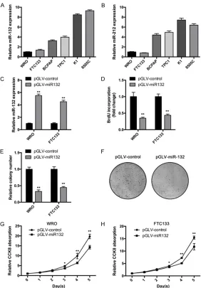
[image:3.612.92.374.70.475.2]To investigate miR-132/212 cluster’s role in thyroid cancer, we first examined their expres-sion in six thyroid cancer cell lines. As shown in Figure 1A, miR-132 showed high expression
Figure 1. miR-132 suppresses thyroid cancer cells proliferation in vitro. A and B. miR-132 and miR-212 expression in six thyroid cancer cell lines was determined by qRT-PCR assay. C. WRO and FTC133 cells were infected with expressing or control lentivirus. Then expression level of miR-132 was deter-mined by RT-qPCR assay. D. WRO and FTC133 cells overexpressing miR-132 or control cells were subjected to BrdU incorporation assay to determine the cell proliferation. E and F. WRO and FTC133 cells overexpressing miR-132 or control cells were subjected to colony formation assay. F. Representative graphs of WRO cells in colony formation assay. G and H. WRO and FTC133 cells were infected miR-132 expressing or control lentivirus. Then cell pro-liferation curve was determined using CCK8 reagent. Bars, mean of each group ± SD (n = 3). *, P < 0.05, **, P < 0.01.
Western blot assay
Whole-cell extracts were ob- tained by lysis of cells in an appropriate volume of ice-cold radioimmunoprecipitation as- say (RIPA) buffer. Nuclear pro-tein was extracted using NE-PER Nuclear and Cyto- plasmic Extraction Reagents (Thermo Scientific) following the manufacturer’s instruc-tions. Cell lysates were sepa-rated on 10% SDS denatured polyacrylamide gel electro-phoresis (PAGE) gels, trans-ferred to nitrocellulose me- mbranes and blocked in phos-phate-buffered saline/Tween-20 containing 5% nonfat milk. Membranes were probed with dilutions of primary antibodi- es followed by incubation wi- th HRP-conjugated secondary antibodies. After extensive washing, proteins were visual-ized by enhanced chemilumi-nescence and exposure to film (Fujifilm, Tokyo, Japan). Anti-CSDE1 antibody was pur-chased from Abcam, anti-PTEN, EKR1/2, p-ERK1/2 and anti-p-AKT antibodies were from Cell signaling, anti-β-ac- tin antibody was from Sigma Aldrich.
Statistical analysis
level in K1 and 8505C cells, and low expres-sion level in WRO and FTC133. Interestingly, miR-212 showed similar expression pattern in these cell lines (Figure 1B). These results indi-cate miR-132 and miR-212 could share similar effector. Then we chose WRO and FTC133 cells for further investigation because of their low level of miR-132 expression. Using lentivirus vector, we stably overexpressed miR-132 in WRO and FTC133 cells. qRT-PCR assay was performed to confirmed the expression (Figure 1C). Then we used thymidine analog BrdU (5-bromo-2’ deoxyuridine) measure cell prolif-eration. miR-132 significantly reduced BrdU incorporation of WRO cells (Figure 1D). Similar results were obtained from FTC133 cells. miR-132 reduced BrdU incorporation in FTC133 cells (Figure 1D). In consistent with these data,
WRO and FTC133 cells (Figure 2C). Cell prolif-eration was also impacted by miR-212 overex-pression. WRO and FTC133 cells infected with miR-212 expressing lentivirus showed sup-pressed CCK8 absorption at Day 4 and 5. Taken together, these results indicate that miR-212 showed similar biological function of miR-132 that inhibits thyroid cancer proliferation.
Inhibition of miR-132/212 promotes thyroid cancer cell proliferation in vitro
To validate the miR-132/212’s role in thyroid cancer cells, we inhibited their expression in miR-132/212 high expression cells, K1 and 8505C. RT-qPCR assay was performed to con-firm the downregulation (Figure 3A and 3B). In consistent with previous results, miR-132
inhi-Figure 2. miR-212 suppresses thyroid cancer cells proliferation in vitro. A. WRO and FTC133 cells was infected with miR-212 expressing or control len-tivirus. Then expression level of miR-212 was determined by RT-qPCR assay. B. WRO and FTC133 cells overexpressing miR-212 or control cells were sub-jected to BrdU incorporation assay to determine the cell proliferation. C and D. WRO and FTC133 cells overexpressing miR-212 was subjected to colony formation assay. D. Representative graphs of WRO cells in colony formation assay. E and F. WRO and FTC133 cells were infected with lentivirus express-ing miR-212. Then cell proliferation curve was determined usexpress-ing CCK8 re-agent. Bars, mean of each group ± SD (n = 3). **, P < 0.01.
miR-132 overexpressing cells produced less colonies com-pared with control cells (Figure 1E and 1F). Cell Counting Kit-8 (CCK-8) assay is a sensitive colorimetric assay for the determination of cell viability in cell proliferation and cyto-toxicity assays. CCK8 assay showed that miR-132 overex-pressing cells proliferated slower than control cells (Figure 1G and 1H). Taken together, these results indi-cate that miR-132 inhibits cell proliferation in thyroid cancer.
miR-212 suppresses thyroid cancer cells proliferation in vitro
bition significantly enhanced BrdU incorpora-tion in K1 cells. Similar results were obtained from 8505C cells (Figure 3C). miR-212 inhibi-tion also promoted BrdU incorporainhibi-tion in both K1 and 8505C cells (Figure 3C). In addition, CCK8 assay showed that downregulation of miR-132 in K1 and 8505C cells increased CCK8 absorption compared with control (Figure 3D). Inhibition of miR-212 obtained similar results (Figure 3D). Furthermore, we also
exam-nsfected ASO-NC or ASO-miR-132/212 with wildtype 3’UTR of CSDE1 mRNA reporter vector in K1 cells. Results showed that luciferase activity was significantly lower in cell transfe- cted with ASO-miR132/212 (Figure 4B). In addition, luciferase activity was also decr- eased in ASO-8505C cells transfected with ASO-miR132/212 (Figure 4B). Furthermore, RT-qPCR assay showed that miR132 or miR-212 overexpression suppressed CSDE1 mRNA
Figure 3. Inhibition of miR-132/212 promotes thyroid cancer cell prolifera-tion in vitro. A. K1 and 8505c cells were transfected with ASO-miR132 or ASO-NC. Then miR-132 expression was determined by RT-qPCR assay. B. K1 and 8505c cells were transfected with ASO-miR212 or ASO-NC. Then miR-132 expression was determined by RT-qPCR assay. C. K1 and 8505c cells were transfected with ASO-miR132/212 or ASO-NC. 48 hours later, BrdU incorporation was determined. D. K1 and 8505c cells were transfected with ASO-miR132/212 or ASO-NC. 48 hours later, CCK8 absorption was deter-mined. E. p-ERK1/2 and total ERK1/2 expression was determined by west-ern blot assay. Bars, mean of each group ± SD (n = 3). **, P < 0.01.
ined ERK activation. Result showed that inhibition of miR-132 and miR-212 enhanced ERK phosphorylation in K1 cells (Figure 3E). Taken together, these results sug-gest miR-132/212 as tumor suppressive miRNAs in thyroid cancer.
miR-132/212 co-target the 3’-UTR of CSDE1 mRNA and downregulates CSDE1 expres-sion in thyroid cancer cells
expression in both WRO and FTC133 cells (Figure 4C). Inhibition of miR-132 or miR-212 could upregulate CSDE1 mRNA in K1 and
[image:7.612.93.522.164.597.2]8505C cells (Figure 4E). In addition, we con-firmed these alterations by western blot assay (Figure 4D and 4F).
Figure 4. miR-132/212 co-target the 3’-UTR of CSDE1 mRNA and downregulates CSDE1 expression in thyroid can-cer cells. A. The sequences of the predicted miR-132/212 binding site and the CSDE1 3’ -UTR segments containing the wildtype or mutant binding site are shown. B. Relative luciferase activity was determined after 3’-UTR reporter plasmids were co-transfected indicated plasmid. C and D. WRO and FTC133 cells were infected with miR-132/212 expressing or control lentivirus. Then CSDE1 expression was determined by RT-qPCR and western blot assay. E and F. K1 and 8505C cells were transfected with ASO-miR-132/212 expressing lentivirus. Then CSDE1 expression was determined by RT-qPCR and western blot assay. Bars, mean of each group ± SD (n = 3). **, P < 0.01.
miR-132/212 and CSDE1 promotes prolifera-tion of thyroid cancer cells by activating AKT
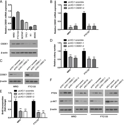
CSDE1 is a RNA binding proteins (RBPs). However, its role in thyroid cancer has not been reported. To explore the functions of CSDE1 in thyroid cancer, we first examined its expression in thyroid cancer cell lines. Results showed that CSDE1 high expresses in WRO and FTC133 cells and low expresses in K1 and 8505C cells (Figure 5A). Interestingly, these results nega-tively correlate with miR-132/212 expression (Figure 1A), which indicates the link between miR- 132/212 and CSDE1. Then we knockdowned CSDE1 expression in WRO and FTC133 cells with two different shRNA. RT-qPCR and west-ern blot assay confirmed the downregulation (Figure 5B and 5C). Colony formation was per-formed. Both two shRNA could decrease the colony number of WRO and FTC133 cells (Figure 5D). BrdU incorporation assay provides similar results. CSDE1 downregulation sup-pressed BrdU incorporation in WRO and FT- C133 cells (Figure 5E). Previous study indi-cates that CSDE1 suppress PTEN expre- ssion using a post-transcriptional program. We examined PTEN expression in CSDE1 knockdown and control cells. Upregulation of PTEN after CSDE1 suppression was observed
western blot assay confirmed the upregulation (Figure 6A and 6B). Colony formation assay showed that CSDE1 overexpression cells reversed miR-132/212 induced colony number decrease (Figure 6C). Furthermore, CSDE1 pro-moted BrdU incorporation in miR-132/212 overexpression cells (Figure 6D).
Discussion
microRNAs are critical regulators of gene expression. Dysregulation of microRNAs has been found in a variety of malignant diseases. In current study, we established a link between miR-132/212 cluster and CSDE1 mediated post-transcriptional program in thyroid cancer cells.
Although several reports indicate miR-132/212 cluster have critical roles in cancer progres-sion, the results are controversial. Furthermore, little is known about the specific function of the cluster and mechanism behind it in thyroid can-cer. Our results indicate a tumor suppressive role of miR-132/212 cluster. Colony formation assay showed that both miR-132 and miR-212 decreased colony numbers of WRO and FTC133 cells. BrdU incorporation was also suppressed by enhance overexpression of miR-132/212. In consistent with these results, inhibition of the
Figure 6. CSDE1 overexpression reversed miR-132/212 induced cell prolif-eration suppression. (A) and (B). miR-132 or miR-212 overexpression cells was transfected with CSDE1 overexpression vector or control. CSDE1 ex-pression was determined by RT-qPCR and western blot assay. Then colony formation (C) and BrdU incorporation (D) assay was performed. Bars, mean of each group ± SD (n = 3). **, P < 0.01.
(Figure 5F). Furthermore, CS- DE1 downregulation inhibit- ed AKT activation in WRO and FTC133 cells (Figure 5F). Because miR-132/212 tar-gets CSDE1, we wondered if miR-132/212 could also sup-press AKT activation. Results showed that overexpression of miR-132/212 upregulated PTEN expression and inhibited AKT activation in WRO and FTC133 cells (Figure 5F).
CSDE1 overexpression re-versed miR-132/212 induced cell proliferation suppression
expression of both miRNAs promoted K1 and 8505c cells growth.
RNA binding proteins (RBPs) are proteins that bind to the double or single stranded RNA in cells and participate in forming ribonucleopro-tein complexes. Recent studies indicate RBPs participate in tumorigenesis and progression [13]. CSDE1 is a conserved RBP which regu-lates mRNA translation and stability [14]. Recent studies indicate the oncogenic role of CSDE1 in melanoma [12]. In the current study, we confirmed oncogenic role of CSDE1 in thy-roid cancer cells. CSDE1 suppresses prolifera-tion of thyroid cancer cells. In consistent with previous study, CSDE1 negatively regulated PTEN expression and AKT activation in WRO and FTC133 cells.
In addition, we found that miR-132/212 target CSDE1 in thyroid cancer cells. CSDE1 3’- UTR segments contains both 132 and miR-212 complementary sequence. Overexpres- sion of either miR-132 or miR-212 could down-regulate CSDE1 expression. Inhibition of miR- 132/212 expression could upregulate CSDE1. Furthermore, manipulating miR-132/212 ex- pression could alter luciferase activity of CSDE1 3’UTR reporter. Forced expression of CSDE1 in miR-132/212 overexpression cells could reverse miR-132/212 induced tumor suppressive effects.
In summary, our study establishes a link between dysregulation expression of miR-132/212 cluster and CSDE1 mediated post-transcriptional program in thyroid cancer cells. PTEN and AKT activation are also regulated by the cluster. These findings provide us a new mechanism by which thyroid cancer progress and a potential therapeutic target.
Acknowledgements
This study was sponsored by a grant from Wenzhou City Science and Technology Plan Project (Y20170099).
Disclosure of conflict of interest
None.
Address correspondence to: Pihong Li and Jianda Dong, Department of General Surgery, The Second Affiliated Hospital of Wenzhou Medical University, Wenzhou 325000, China. E-mail: pihongliwz@yeah. net (PHL); [email protected] (JDD)
References
[1] Nikiforov YE and Nikiforova MN. Molecular ge-netics and diagnosis of thyroid cancer. Nat Rev Endocrinol 2011; 7: 569-580.
[2] Davies L and Welch HG. Increasing incidence of thyroid cancer in the United States, 1973-2002. JAMA 2006; 295: 2164-2167.
[3] Siegel RL, Miller KD and Jemal A. Cancer sta-tistics, 2016. CA Cancer J Clin 2016; 66: 7-30. [4] Nikiforova MN and Nikiforov YE. Molecular ge-netics of thyroid cancer: implications for diag-nosis, treatment and prognosis. Expert Rev Mol Diagn 2008; 8: 83-95.
[5] Lin S and Gregory RI. MicroRNA biogenesis pathways in cancer. Nat Rev Cancer 2015; 15: 321-333.
[6] Wanet A, Tacheny A, Arnould T and Renard P. miR-212/132 expression and functions: within and beyond the neuronal compartment. Nucle-ic Acids Res 2012; 40: 4742-4753.
[7] Park JK, Henry JC, Jiang J, Esau C, Gusev Y, Le-rner MR, Postier RG, Brackett DJ and Schmitt-gen TD. miR-132 and miR-212 are increased in pancreatic cancer and target the retinoblas-toma tumor suppressor. Biochem Biophys Res Commun 2011; 406: 518-523.
[8] Liu X, Yu H, Cai H and Wang Y. The expression and clinical significance of miR-132 in gastric cancer patients. Diagn Pathol 2014; 9: 57. [9] Li Y, Zu L, Wang Y, Wang M, Chen P and Zhou
Q. miR-132 inhibits lung cancer cell migration and invasion by targeting SOX4. J Thorac Dis 2015; 7: 1563-1569.
[10] You J, Li Y, Fang N, Liu B, Zu L, Chang R, Li X and Zhou Q. MiR-132 suppresses the migra-tion and invasion of lung cancer cells via tar-geting the EMT regulator ZEB2. PLoS One 2014; 9: e91827.
[11] Tian H, Hou L, Xiong YM, Huang JX, Zhang WH, Pan YY and Song XR. miR-132 targeting E2F5 suppresses cell proliferation, invasion, migra-tion in ovarian cancer cells. Am J Transl Res 2016; 8: 1492-1501.
[12] Wurth L, Papasaikas P, Olmeda D, Bley N, Cal-vo GT, Guerrero S, Cerezo-Wallis D, Martinez-Useros J, Garcia-Fernandez M, Huttelmaier S, Soengas MS and Gebauer F. UNR/CSDE1 drives a post-transcriptional program to pro-mote melanoma invasion and metastasis. Cancer Cell 2016; 30: 694-707.
[13] Agami R. microRNAs, RNA binding proteins and cancer. Eur J Clin Invest 2010; 40: 370-374.