Original Article
Expression of COUP-TF2 in bladder carcinoma and
effect of microenvironment of bladder
cancer on endothelial cells
Mingshan Wang1*, Chaoli Duan2*, Qiang Ren1, Peijie Chen1, Shu Lu1, Shuchao Ye1, Jiawei Wang3, Peiming
Bai1
1Department of Urology, Zhongshan Hospital Affiliated to Xiamen University, Xiamen, Fujian Province, P. R. China; 2Xiamen Diabetes Institute, 3Department of Neurology, The First Affiliated Hospital of Xiamen University, Xiamen,
Fujian Province, P. R. China. *Equal contributors and co-first authors.
Received January 18, 2017; Accepted March 24, 2017; Epub May 1, 2017; Published May 15, 2017
Abstract: Vessel plays an important role in the occurrence and development of tumors. COUP-TF2 (Chicken Oval-bumin Promoter transcription factor 2) has been found associated with tumor development, metastasis and tumor angiogenesis in a number of tumor models, such as pancreatic cancer and breast cancer. The aim of this study was to analyze the expression of TF2 in human bladder cancer to evaluate the relationship between COUP-TF2 expression and clinicopathological features, and to study the effect of tumor microenvironment on vascular endothelial cells. We investigated the expression of COUP-TF2 and microvessel density (MVD) of 35 bladder tissue specimens with immunohistochemical assay and assessed the score using the prescribed scoring method. Further-more, HUVEC was cultured with supernatant of bladder cancinoma cells and normal cells, respectively. The effect of microenvironment on HUVEC cells were evaluated using Real Time-PCR, Western blot and CCK-8. COUP-TF2 was strongly expressed in 74% of tumor tissues investigated and weakly expressed in 25% of normal bladder tissues investigated. The pathological grade and clinical stage of bladder cancer were closely related to COUP-TF2 protein expression and MVD (P<0.05). In addition, the expression of COUP-TF2 protein was closely positively correlated to MVD (P<0.05). The expression of COUP-TF2 in the bladder cancer supernatant was higher than that in normal blad-der supernatant at both RNA and protein level, and promoted the proliferation of HUVEC. This indicates that COUP-TF2 may promote the progression of bladder cancer and therefore, may serve as a molecular marker for bladder cancer progression and used a new target for medical treatment.
Keywords: Bladder neoplasms, COUP-TF2, clinicopathological data, immunohistochemical assay, vascular endo-thelial cells
Introduction
Bladder cancer is the most common malignant tumor of the urinary system, it has multi-center, easy to relapse, and invasive growth character-istics. The incidence rate of malignancy in Chinese male genitourinary system is the first, and the incidence rate of malignant tumor is the eighth. Due to the aging population, incre- ased smoking population, increased environ-mental pollution and other aspects of occupa-tional protection imperfections, China has an upward trend in its incidence [1]. At present, the standard treatment for invasive bladder cancer is radical surgery with chemotherapy
and/or radiotherapy, but the effect is not ideal. If the bladder cancer patients have bladder metastasis, the 5-year survival rate was only 6%, treatment and prognosis will be poor [2]. A better understanding of the molecular mecha-nisms underlying the development of bladder cancer may help to improve the prognosis of patients with bladder cancer. Thus, there is a strong need for new noninvasive biomarker identification for early tumor detection.
VEGFR-1 and promote endothelial cell prolifera-tion and germinaprolifera-tion. It was found that COUP-TF2 up-regulates E2F1 signaling and down-reg-ulates Notch signaling [11, 12], both of which have been shown to modulate angiogenesis through the VEGF/VEGFR axis [13, 14]. In the tumor microenvironment, there may be com-plex interaction networks between COUP-TF2 and E2F1, Notch signaling and VEGF/VEGFR signaling to coordinate angiogenesis. How- ever, the detailed mechanism by which COUP-TF2 performs these signals remains to be elucidated.
Although the expression of COUP-TF2 protein has been found in many tumors and its poten-tial clinical relevance has been studied, no studies have been done in bladder cancer. Therefore, our aim in this study is to elucidate the expression of COUP-TF2 protein in bladder cancer and its association with the clinicopath-ological features of bladder cancer and to investigate the effect of tumor microenviron-ment on endothelial cells.
Materials and methods
Bladder tissue specimens
27 cases of primary bladder transitional cell carcinoma tissues and 8 cases of normal blad-der tissue paraffin specimens as control group were collected from January 2000 to August 2016 in our hospital. All of them were fixed in formalin, Conventional paraffin embedding, conventional section thickness 3 um. The par-affin specimens come from the department of Pathology, affiliated Zhongshan Hospital, Xia- men University. Among them, 30 males and 5 females, aged between 33 to 87 years old, mean 61.6 years. According to WHO tumor grade: 1 to 2 grade, 13 cases; 3 grade, 14 cases. Clinical stage according to UICC stan-dards: Tis~T1, 12 cases; T2~T3, 15 cases, including lymphatic metastasis in 2 cases.
Immunohistochemistry
Immunohistochemical method: We analyzed the expression of COUP-TF2 protein in transi-tional cell carcinoma of urinary bladder and normal bladder tissue. COUP-TF2 monoclonal antibody was purchased from R&D Systems, mouse anti-human, monoclonal antibody CD34 and SP kit were purchased from Fuzhou Maixin growth of tumor blood vessels, and thus can
predict tumor growth, metastasis and recur-rence trend. The COUP-TF2 is a protein contain-ing 414 amino acids with a molecular weight of 47 kDa. Its gene is located on human chromo-some 15, 15q26, containing six exons, a total of 14336 bp base pairs. The COUP-TF is a member of the family of orphan receptors in the steroid hormone/thyroid hormone receptor superfamily. There are two height homologous subtypes, COUP-TF1 and COUP-TF2, possess-ing a highly conserved modular structure con-sisted of an amino-terminal DNA-binding do- main and a putative C-terminal ligand binding domain [3].
COUP-TF2 has been shown to stimulate prolif-eration and migration and induce angiogenesis in a variety of mouse tumor models including breast cancer, pancreatic cancer, and so on, and play a key role in tumor progression as a promoter of oncogenes in a variety of cancers. There is a wealth of evidence for its involve-ment in tumor infiltration and metastasis. COUP-TF2 is strongly expressed in the nuclei of neovascularization and various cancers (bre- ast, ovarian, prostate, colon, pancreas, etc.), and can participate in tumor and vascular lym-phatic vessels through multiple pathways. It is closely related to the clinical stage, pathologi-cal grade, lymph node metastasis and progno-sis of cancer [4-6]. In a number of inactivated COUP-TF2 spontaneous tumor models also showed inhibition of tumor progression and metastasis through inhibition of angiogenesis [7].
It was found that COUP-TF2 can regulate the two angiogenic signals, Ang-1/Tie2 and VEGF/ VEGFR-2. First, COUP-TF2 regulates the expres-sion of Ang-1 in the outer membrane, and Ang-1 is a paracrine ligand for the specific endothelial cell tyrosine kinase receptor Tie2 [8].
mum vascular density region, and then placed under 200 times the microscope, Microvessel counts were selected for the three highest vas-cular density regions, and the mean value was taken as the MVD value of the patient. Any stained individual cell or cell mass, whether or not it forms a bureaucratic cavity, is considered to be a countable microvessel, as long as it is clearly separated from surrounding microves-sels, tumor cells and other connective tissue components. Intratumoral sclerosis and tumor at the junction of soft tissue microvessels are not counted, with thick smooth muscle wall and bureaucratic diameter greater than 8 red blood cell diameter of the blood vessels are excluded.
Cell culture
Bladder cancer cells J82, 5637 and bladder normal epithelial cells SV-HUC-1 were pur-chased from the Chinese Academy of Sciences, Department of Culture Collection Committee cell library. Human umbilical vein endothelial cells (HUVEC) is from the Affiliated Zhongshan Hospital, Xiamen University, Gastroenterology Laboratory gift. The culture medium of each cell was composed of 10% fetal bovine serum (Bioind company), 1% double antibiotic (penicil-lin and streptomycin, Hyclone) and 500 ml of basal medium. The basic medium of J82, 5637, SV-HUC-1 and HUVEC were MEM, RMPI1640, F-12K and DMEM of Gibico respectively. The cells were cultured at 37°C in an incubator con-taining 5% CO2.
Real Time-PCR fluorescence quantitative re-verse transcription polymerase chain reaction Total RNA was extracted from the cells by using TIANGEN TRNzol total RNA extraction reagent, with 1 µg of total RNA reverse transcription in the Takara reagent kit reverse transcription into total cDNA. The COUP-TF2 expression level was amplified by using the specific COUP-TF2 prim-ers on an ABI 7500 instrument using a Takara kit. Human GAPDH internal controls were used as controls.
COUP-TF2: Forward primer sequence [5’-3’]: CACAGGCATCTGAGGTGAACAGG; COUP-TF2: Re- verse primer sequence [5’-3’]: CGCCTTTATGG- ACCACATACGG. GAPDH: Forward primer sequ- ence [5’-3’]: CAGGAGGCATTGCTGATGAT; GAP- DH: Reverse primer sequence [5’-3’]: GAAG- GCTGGGGCTCATTT.
Biotech. Co., Ltd. PBS buffer instead of primary antibody as a negative control group, the known COUP-TF2 and CD34 positive bladder tissue sections as a positive control. Samples were excised from a paraffin specimen (approxi-mately 3 µm) and placed on a poly-L-lysine coated slide. Dewaxed for 2 h in a thermostat at 60°C and then hydrated in xylene, xylene, 100%, 100%, 90%, 80%, 70% alcohol. PBS wash 3 times, 3 min/times. The antigen solu-tion of citrate antigen was used for autoclave antigen retrieval and boiled for 1-2 minutes. Then, according to the experimental method of UltraSensitiveTM SP hypersensitization kit (mouse/rabbit) of Fuzhou Maixin Biotech. Co., Ltd, incubation of A liquid blocking (peroxidase blocking solution) 10 min, PBS washed 3 times, 3 min/time, incubation of B liquid (normal non-immune animal serum) 10 min, remove B solu-tion, COUP-TF2 antibody (R&D Systems, 1:200) incubated overnight at 4°C, washed with PBS, incubated with C (Streptomycin antibiotic-per-oxidase solution) for 10 min, rinsed with PBS, then incubated with D solution (streptomycin antibiotic-peroxidase solution) for 10 min, washed with PBS, then observed under DAB staining microscope for 1-3 min, tap water rinse, Hematoxylin stained 45 s, PBS washed, and finally through the reverse concentration gradient of alcohol dehydration, xylene trans-parent, neutral gum sealing. After two days, the neutral gums were dried and the sections were fixed under microscope to observe the photo-graphs, and the results were graded.
Immunohistochemical results to judgment: COUP-TF2 staining score calculation: COUP-TF2 was uniformly distributed in the nucleus of vas-cular endothelial cells in transitional cell carci-noma of bladder, which was brown and was also expressed in vascular smooth muscle cells of pericancerous stroma. The total expression of COUP-TF2 was determined by the sum of the staining intensity and the percentage of posi-tive cells. Scoring criteria: (1) staining intensity: negative for 0; weak positive for 1; positive for 2; strong positive for 3. (2) the percentage of positive cells: positive cells 0% to 0; 25% to 1; 25% to 2; positive cells >50% to 3. (1)+(2) is 0~3 points is low expression, 4~6 for the high expression.
maxi-PVDF membrane by electrotransfer, blocked with 5% skim milk for 1 hour, washed 3 times with TBST, 10 min/time. And incubated over-night at 4°C in COUP-TF2 antibody (1:1000). The next day, the membrane was exposed to horseradish peroxidase-conjugated secondary antibody (1:5000) for 1 hour at room tempera-ture, washed 3 times with TBST, 10 min/time, and the protein was visualized by the enhanced chemiluminescence reagent ECL kit under the FluorChem HD2 multifunctional imaging system.
CCK8
[image:4.612.96.523.84.243.2]HUVEC were plated into 96-well plates at 3000 cells per well. HUVEC was cultured with 200 µl supernatant of J82, 5637 and SV-HUC-1 respectively. After 24, 48, 72, 96 and 120 hours of culture, the supernatant fluid was removed, 200 µl of the mixture of 180 µl DMEM medium and 20 µl of CCK-8 reagent solution (Kyushu, Japan) was incubated in an incubator for 2 hours. The number of viable cells was measured and the number of HUVEC cells was reflected by absorbance at 450 nm using a microplate reader.
Table 1. 27 cases of bladder cancer COUP-TF2 expression and clinicopathological factors
Clinicopathological features Classification Total number of samples COUP-TF2 P-value Positive Negative
Age (years) ≥60 17 12 5 0.678
<60 10 8 2
Tumor size (cm) ≥3 10 9 1 0.204
<3 17 11 6
Pathological grade 1-2 grade 13 7 6 0.033*
3 grade 14 13 1
Clinical stage Tis-T1 12 6 6 0.024*
T2-T4 15 14 1
Lymph node metastasis without metastasis Without metastasis 24 18 6 0.610
With metastasis 3 2 1
[image:4.612.91.333.302.369.2]*P<0.05.
Table 2. 35 cases of bladder cancer and normal tissue COUP-TF2 expression comparison
Classification COUP-TF2 Positive COUP-TF2 Negative Total P-value
Bladder cancer 20 7 27 0.032*
Normal bladder tissue 2 6 8
Total 22 13 35
*P<0.05.
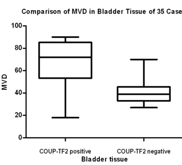
Table 3. 35 cases of bladder tissue COUP-TF2 expression and the relationship be-tween tumor microvessel count statistics COUP-TF2
expression The number of cases MVD* (X±S) P value
Positive 22 67.95±19.87 0.000
Negative 13 40.92±11.77
MVD*: Microvascular density.
Western blot
The cells were lysed on RIPA lysate and protease inhibitor (100:1) on ice fol-lowed by shaking with ultrasonic instru-ment (12,000 rpm, 15 min). The super-natant was collected and boiled at 100°C for 10 min. The extracted protein was then separated by electrophoresis in 10% SDS-PAGE and transferred to
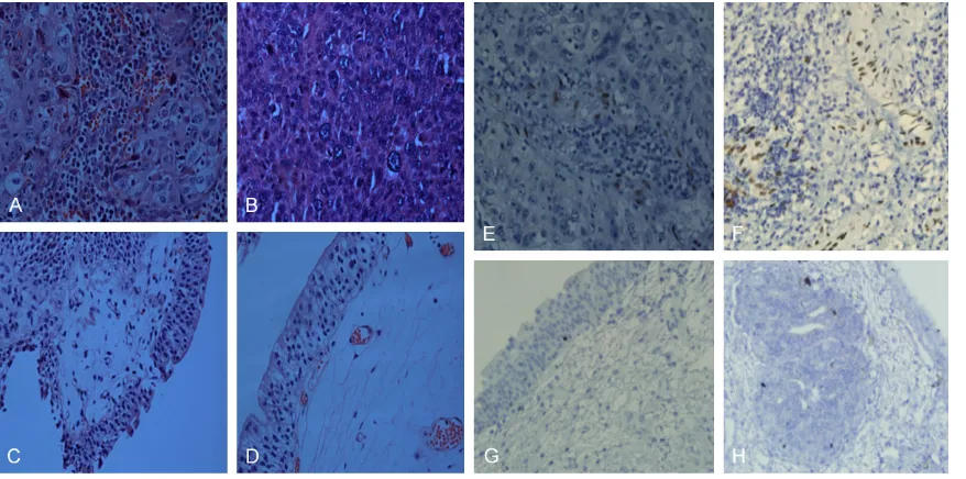
[image:4.612.91.282.436.486.2] [image:4.612.92.280.512.682.2]sue of vascular endothelial cells, vascular smooth muscle cells (Figure 2). But there were only 2 positive cases (25%) in 8 cases of nor-mal bladder tissues and weakly positive expres-sion. The distribution was mainly in the small vascular endothelial cells of stroma, and there was significant difference between the two groups (P=0.032<0.05). COUP-TF2 and CD34 negative control group were not brown particles exist. The expression of COUP-TF2 was closely related to the histological grade and clinico-pathological stage of bladder cancer (P<0.05,
Table 1), but not to the age, tumor size and lymph node metastasis (P>0.05, Table 1). The expression of COUP-TF2 in bladder cancer was significantly higher than that in normal bladder tissues (92.9% vs 53.8%, P<0.05). The expres-sion of COUP-TF2 in bladder cancer was signifi-cantly higher than that in normal bladder tis-sues (P<0.05, Table 2).
The expression of COUP-TF2 and MVD in blad-der cancer
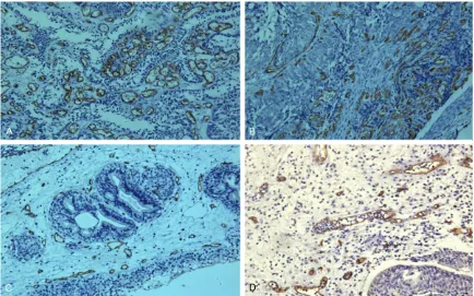
CD34, a marker of vascular endothelial cells, is used to label MVD, which is mainly expressed in the vascular endothelial cell membrane and cytoplasm. The mean MVD of COUP-TF2 posi-tive group was 67.95±19.87, which was higher than COUP-TF2 negative group (40.92±11.77). There was significant difference between the Statistical analysis
Each experiment is repeated at least three times. We used SPSS 19.0 software for statisti-cal analysis. In Table 1, the relationship bet- ween the expression of COUP-TF2 and clinico-pathological variables in bladder cancer was analyzed by chi-square test. In Table 2, the expression of COUP-TF2 in bladder cancer and bladder normal tissues was analyzed by chi-square test. In Table 3, the rank sum test was used to treat the association between MVD and clinicopathological variables in bladder cancer. The correlation analysis of two variables using rank correlation analysis; Real Time-PCR and CCK-8 were analyzed by GraphPad 5.0 soft-ware. The Real Time-PCR data were analyz- ed by multi-group independent rank analysis ANOVA. CCK-8 data were grouped by t-test; P<0.05 was considered statistically significant when the data were statistically analyzed.
Results
Expression of COUP-TF2 in bladder tissue and its relationship with clinicopathological factors COUP-TF2 was expressed on the nucleus of endothelial cells and stained into brown gran-ules. In 27 cases of bladder cancer, 20 cases of positive expression (74.1%), and all showed a positive, mainly in the surrounding cancer
tis-Figure 2. 1. Hematoxylin-eosinstaining in Bladder patient groups. (A and B) Hematoxylin-eosinstaining in Bladder
normal tissue (C and D) (original magnification ×400). 2. Immunohistochemical staining of COUP-TF2 protein in
Bladder patient groups. (E and F) Negative staining in Bladder normal tissue. (G and H) Strong staining in Bladder
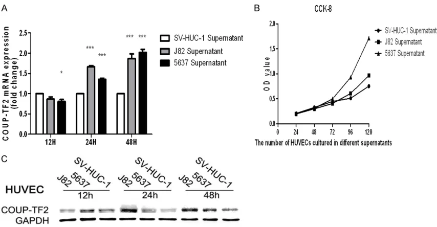
[image:5.612.91.530.71.289.2]The proliferation of HUVEC cells after different supernatant culture
The supernatants of bladder cancer cells and normal bladder epithelial cells were collected. The same amount of HUVEC cells were plated and cultured in 48-well plates for 24 h, 48 h, 72 h, 96 h and 120 h, respectively, the viable cells of HUVEC were detected by CCK-8. The results show that the number of HUVEC in the superna-tant of bladder cancer group was more than that in normal bladder epithelium group on the fourth day and the fifth day, and further pro-mote the proliferation of vascular endothelial cells, and the gap is statistically significant (P<0.05, Figure 4C).
Discussion
Bladder cancer is one of the most common neoplasms in urology in China, and is a disease that directly threatens the survival of patients. Hematuria is the most common symptoms of bladder cancer. When bladder cancer has metastasized, the 5-year survival rate was only 6%, treatment and prognosis is poor. In animal tumor models, COUP-TF2 has been shown to two groups (P<0.05, Table 3; Figures 2, 3). The
correlation between MVD and COUP-TF2 was analyzed, and there was a positive correlation between the expression of COUP-TF2 and MVD in tumor tissue (P<0.05, Figure 1). MVD increases as the expression of COUP-TF2 increased.
The expression of COUP-TF2 in HUVEC after different supernatant culture
The same number of cells was plated in 10 cm dishes, after waiting for the cells to grow over the dish, the supernatant of bladder cancer cells (J82 and 5637) and the supernatant of normal bladder epithelial cells (SV-HUC-1) were collected. HUVEC cells were cultured with these supernatant liquid for 12 h, 24 h, 48 h, respec-tively, the cells were collected and subjected to RT-PCR and Western blot. The results show that the expression level of COUP-TF2 in HUVEC cultured in supernatant of bladder cancer was significantly higher than that in normal cell cul-ture supernatant HUVEC, especially in 48 h group, and had statistical significance (P<0.05,
[image:6.612.89.523.71.342.2]Figure 4A, 4B).
Figure 3. Immunohistochemical staining of CD34 protein in Bladder patient groups (A and B). Immunohistochemical staining of CD34 protein in in Bladder normal tissue (C and D). CD34 is vascular endothelial cell markers (original
COUP-TF2, thereby promote the proliferation of vascular endothelial cells. This is consistent with the animal tumor model. But in the human body, the tumor microenvironment for COUP-TF2 regulation pathway is not yet known, the mechanism of COUP-TF2 to promote the prolif-eration of vascular endothelial cells also needs further study. For example, the relationship between COUP-TF2 and cell cycle, cell apopto-sis has not been clearly reported. However, COUP-TF2 has not been studied in bladder can-cer. The clinical significance of COUP-TF2 in bladder cancer remains to be elucidated. COUP-TF2 is expected to be a potential thera-peutic target for bladder cancer.
Conclusion
In this study, we examined the expression of COUP-TF2 protein in bladder tissues and found that COUP-TF2 was closely related to the clini-copathological features of bladder cancer. COUP-TF2 is highly expressed in bladder cancer tissue, which indicates that COUP-TF2 plays an important role in the progression of bladder cancer, especially in the late stage of the dis-ease. COUP-TF2 was positively correlated with modulate two angiogenic signals, Ang-1/Tie2
[image:7.612.92.519.71.293.2]and VEGF/VEGFR-2, playing an important role in tumor progression in animal tumor models. But the role of blood vessels in human tumor progression is still not fully understood. We found that COUP-TF2 was strongly expressed in the nucleus of vascular endothelial cells in human bladder cancer tissues, but not in blad-der cancer cells and normal bladblad-der cells, and occasionally in vascular endothelial cells of nor-mal bladder tissues. The expression of COUP-TF2 was closely correlated with the pathologi-cal grade and clinipathologi-cal stage of bladder cancer, especially in advanced bladder cancer. The positive rate of COUP-TF2 was positively corre-lated with MVD, and the number of MVD in advanced bladder cancer is the highest. It sug-gests that COUP-TF2 can promote the progres-sion of bladder cancer through promoting the proliferation of endothelial cells. In a cell exper-iment, the tumor microenvironment can prolif-erate HUVEC and result in HUVEC cells express-ing more COUP-TF2 protein, which all indicate that COUP-TF2 plays an important role in the progression of bladder cancer. These results suggest that the microenvironment of bladder cancer can lead to increased expression of
[5] Wang C, Zhou Y, Ruan R, Zheng M, Han W, Liao
L. High expression of COUP-TF II cooperated with negative Smad4 expression predicts poor prognosis in patients with colorectal cancer. Int J Clin Exp Pathol 2015; 8: 7112-21. [6] Wang L, Xu M, Qin J, Lin SC, Lee HJ, Tsai SY,
Tsai MJ. MPC1, a key gene in cancer metabo-lism, is regulated by COUPTFII in human pros-tate cancer. Oncotarget 2016; 7: 14673-83. [7] Hawkins SM, Loomans HA, Wan YW,
Ghosh-Choudhury T, Coffey D, Xiao W, Liu Z,
Sangi-Haghpeykar H, Anderson ML. Expression and functional pathway analysis of nuclear recep-tor NR2F2 in ovarian cancer. J Clin Endocrinol Metab 2013; 98: E1152-62.
[8] Qin J, Chen X, Xie X, Tsai MJ, Tsai SY. COUP-TFII
regulates tumor growth and metastasis by modulating tumor angiogenesis. Proc Natl Acad Sci U S A 2010; 107: 3687-92.
[9] Fukuhara S, Sako K, Noda K, Zhang J, Minami
M, Mochizuki N. Angiopoietin-1/Tie2 receptor signaling in vascular quiescence and angio-genesis. Histol Histopathol 2010; 25: 387-96. [10] Qin J, Chen X, Yu-Lee LY, Tsai MJ, Tsai SY. Nu -clear receptor COUPTFII controls pancreatic islet tumor angiogenesis by regulating vascu-lar endothelial growth factor/vascuvascu-lar endo-thelial growth factor receptor-2 signaling. Can-cer Res 2010; 70: 8812-21.
[11] You LR, Lin FJ, Lee CT, DeMayo FJ, Tsai MJ, Tsai SY. Suppression of Notch signalling by the COUP-TFII transcription factor regulates vein identity. Nature 2005; 435: 98-104.
[12] Chen X, Qin J, Cheng CM, Tsai MJ, Tsai SY.
COUP-TFII is amajor regulator of cell cycle and Notch signaling pathways. Mol Endocrinol 2012; 26: 1268-77.
[13] Engelmann D, Mayoli-Nüssle D, Mayrhofer C, Fürst K, Alla V, Stoll A, Spitschak A, Abshagen K, Vollmar B, Ran S, Pützer BM. E2F1 pro-motes angiogenesis through the VEGF-C/VEG-FR-3 axis in a feedback loop for cooperative induction of PDGF-B. J Mol Cell Biol 2013; 5: 391-403.
[14] Zhou W, Wang G, Guo S. Regulation of angio -genesis via Notch signaling in breast cancer and cancer stem cells. Biochim Biophys Acta 2013; 1836: 304-20.
MVD, and the tumor microenvironment could also promote the expression of COUP-TF2 in endothelial cells, suggesting that tumor micro-environment could promote the expression of COUP-TF2 in endothelial cells, further promote the proliferation of endothelial cells. This indi-cates that COUP-TF2 plays an important role in tumor progression.
Acknowledgements
The authors would like to thank the colleagues at the Zhongshan Hospital of Xiamen University for their help. This study was supported by grants from Fujian Provincial Natural Science Foundation of China (No. 2015J01527).
Disclosure of conflict of interest
None.
Address correspondence to: Dr. Peiming Bai, De-
partment of Urology, Zhongshan Hospital Affiliated to Xiamen University, 209 Hubin South Road, Xiamen, Fujian Province, P. R. China. Tel: +86
13606911821; E-mail: [email protected]
References
[1] Burger M, Catto JW, Dalbagni G, Grossman HB, Herr H, Karakiewicz P, Kassouf W, Kiemeney LA, La Vecchia C, Shariat S, Lotan Y. Epidemiology and risk factors of urothelial bladder cancer. Eur Urol 2013; 63: 234-41. [2] Xie F, Ye L, Ta M, Zhang L, Jiang WG. MTSS1: a
multifunctional protein and its role in cancer invasion and metastasis. Front Biosci (Schol Ed) 2011; 3: 621-31.
[3] Xu M, Qin J, Tsai SY, Tsai MJ. The role of the
orphan nuclear receptor COUP-TFII in tumori-genesis. Acta Pharmacol Sin 2015; 36: 32-6. [4] Wang X, Jiang R, Cui E, Feng W, Guo H, Gu D,
Tang C, Xue T, Bao Y. COUP-TFII suppresses