Original Article
Antagonism of miR-21 reverses radiation-induced EMT
in alveolar epithelial cells via PI3K/Akt pathway
Xin Li1*, Zi-jie Mei1*, Ya-Cheng Wang2, Jing Chen1, Shao-Xing Sun1, Ye Yao1, Zhen-Zhen Li1, Cong-Hua Xie1 1Department of Radiation and Medical Oncology, Hubei Cancer Clinical Study Center, Hubei Key Laboratory of Tu-mor Biological Behaviors, Zhongnan Hospital, Wuhan University, Wuhan, Hubei, China; 2Department of Oncology, The Central Hospital of Wuhan, Wuhan, Hubei, China. *Equal contributors and co-first authors.
Received November 10, 2015; Accepted January 3, 2016; Epub February 1, 2016; Published February 15, 2016
Abstract: Radiation-induced lung injury (RILI) is a common complication after thoracic radiotherapy and epithelial-to-mesenchymal transition (EMT) an important change among this process. The aim of this study was to determine the role of miR-21 in radiation-induced EMT in alveolar epithelial type II cells RLE-6TN and explored the underlying molecular mechanism. The results showed that treating RLE-6TN cells with 8 Gy of X-ray promoted EMT and re-sulted in up-regulation of miR-21. Transfection of Rle-6TN cells with miR-21 inhibitor before IR caused an increase in expression of epithelial marker e-cadherin, as well as a decrease in the mesenchymal markers α-smooth muscle actin and vimentin. It indicated that downregulation of miR-21 in RLE-6TN cells inhibited the radiation-induced EMT. Moreover, miR-21 inhibitor decreased the progress of this EMT accompanied by a decrease of phosphorylated-Akt protein level. However, PI3K activator IGF-1 reversed the suppression of phosphorylated-phosphorylated-Akt and promoted the radiation-induced EMT by miR-21 knockdown. In addition, we found a dose-dependent relationship between PI3K inhibitor LY294002 and radiation-induced EMT. In total, we concluded that antagonism of mir-21 reversed radiation-induced EMT in alveolar epithelial cells via PI3K/Akt pathway. This might be a much promising target in the cure the radiation-induced lung injury.
Keywords: RILI, EMT, miR-21, PI3K/Akt
Introduction
Radiation-induced lung injury (RILI) is the com-mon complication of thoracic radiotherapy. Because of the lung is one of the most sensi-tive tissues to ionizing radiation, the damage to normal lung tissue remains a major obstacle in the treatment of lung cancer. It contains tion pneumonia in the early period and radia-tion pulmonary fibrosis in the later period [1, 2]. Generally, we considered that pulmonary fibro-sis is characterized by alveolar epithelial cell injury, leading to the accumulation of fibro-blasts, myofibrofibro-blasts, collagen and other extracellular matrix proteins and resulting in impaired lung function [3, 4]. But recent evi-dence suggests that injured epithelial cells may directly turn to myofibroblasts by epithelial-mesenchymal transition (EMT) [5]. This transi-tion is characterized by the loss of epithelial markers such as E-cadherin and the increase of mesenchymal markers including α-SMA,
vimentin. Signaling pathway such as ERK/Snail, PI3k/Akt involved in the process [6]. Studies have confirmed that several microRNAs regu-late the process of EMT, such as miR-200 fam-ily, let-7d, miR-21 [7].
Antagonism of miR-21 reverses radiation-induced EMT
that the expression of miRNAs has changed either in peripheral blood cells of radiotherapy patients and in the cancer cells treated with ionizing radiation [13, 14]. And in an animal experiment, the miRNAs in the lung of the rat with ionizing radiation has changed, including the up-regulation of miR-21 [15].
MiR-21 is one of the miRNAs who located on chromosome 17q23.2, which controls a wide range of biological processes, including cell growth, proliferation, migration, invasion, and survival [16]. It has been reported to be overex-pressed in almost all types of human cancers [17]. Besides, miR-21 related to many other dis-eases especially fibrosis [18]. It is reported that miR-21 mediated fibrogenic activation of pul-monary fibroblasts and lung fibrosis [19]. Many studies have shown the relation between miR-21 and radiation, such as circulating mir-miR-21 upregulated in the patients who treated with radiotherapy, miR-21 may promote radioresis-tance of cancer and inhibition of miR-21 may represent effective approaches for reversing this radioresistance [20]. Therefore, in this study, we investigate the role of miR-21 in the radiation-induced EMT in alveolar epithelial cells for the sake of the therapy of radiation-induced lung injury.
Materials and methods
Cell culture
RlE-6TN cells, a rat alveolar type II epithelial cell line, were obtained from the Chinese Academy of Sciences and cultured in RPMI-1640 medium (GE Healthcare Life Scinences/ HyClone™ Laboratories, Logan, UT) containing 10% fetal bovine serum (FBS) (GE Healthcare Life Scinences/HyClone™ Laboratories, Logan, UT), 100 IU/ml penicillin and 100 μg/ml strep-tomycin at 37°C with 5% CO2 in air. Growth medium was changed every 48 hours. Cells were subcultured every 3-5 days. Suspensions of RLE-6TN cells were obtained from mostly confluent cultures (about 80-90%) using Tryp- sin/EDTA solution.
Transfection
Rno-miR-21 inhibitors, miRNA inhibitor nega-tive controls, rno-miR-21 mimics and miRNA mimic negative controls were designed and synthesized by Guangzhou RiboBio Co., Ltd.
(Guangzhou, China). The transfection of miR-NAs was performed using Lipofectamine® 2000 (Invitrogen™, Carlsbad, CA). In six-well plates (Costar™), cells were plated per well at 40-60% confluence 24 h prior to the transfec-tion. MiR-21 inhibitors, mimics, the negative controls and cy3-labeled scrambled were trans-fected into the cells at a final concentration of 100 nM with 5 μl of Lipofectamine 2000 according to the manufacturer’s protocol. The medium was replaced with new culture medi-um at 4-6 h after transfection. The initial cali-bration of transfection efficiency was done by demonstrating that, after transfection with cy3-labeledscrambled microRNA, over 80% of the cells expressed cy3.
Irradiation conditions
Linear accelerator producing 6MV X-ray beams were provided by Zhongnan Hospital of Wuhan University (Wuhan, China). The source-to-skin distance (SSD) was 100 cm and the dose rate was 200 cGy/min. Packing material 3 cm thick was placed around and underneath the culture dishes. Then the cells that after 24 h post transfection were treated with a single dose of 8 Gy. All irradiations were performed at room temperature.
RNA isolated and quantitative real-time PCR
morphol-Antagonism of miR-21 reverses radiation-induced EMT
synthesized by Guangzhou RiboBio (Guangzhou, China). The other primers were designed and synthesized by GenePharma (Shanghai, China): E-cadherin (forward primer 5’-TGACTACTAC- TTGAACGAATGGG-3’, reverse primer 5’-GGAA- GGGAGCTGAAAAACCAC-3’); α-SMA (forward primer 5’-TGACGCTGAAGTATCCGATAGA-3’, re- verse primer 5’-GTACGTCCAGAGAGGCATAGA- GG-3’); Vimentin (forward primer 5’-ATGTGGA- TGTTTCCAAGCCTGAC-3’, reverse primer 5’-GA- GTGGGTATCAACCAGAGGGAGT-3’); β-actin (for-ward primer 5’-AGAAAATCTGGCACCACACC-3’, reverse primer 5’-CCATCTCTTGCTCGAAGCTC- C-3’).
Western blots analysis
Cells were prepared at 48 h postirradiation, washed with PBS twice and lysed using RIPA buffer containing 50 mM Tris (pH 7.4), 150 mM NaCl, 1% NP-40, 0.25% sodium deoxycholate, 1% protease inhibitor cocktail (Beyotime, China), 1% Na3VO4 and 1% phosphatase inhibi-tor cocktail (Roche Diagnostics Corp., Indianapolis, IN). The proteins were separated by SDS-PAGE and then transferred using a PVDF membrane (Millipore, Billerica, MS) by Western blotting. The nonspecific sites were blocked in 5% nonfat dry milk for 1-2 h at room temperature. In this study, antibodies directed against E-cadherin, α-SMA, Vimentin, PTEN, Akt, p-Akt (each at 1:500; Proteintech Group Inc., Chicago, IL) and β-actin (1:2000; Cell Signaling Technology®, Danvers, MA) were used. The secondary antibodies were goat rabbit horseradish peroxidase or goat anti-mouse horseradish peroxidase (Bio-Rad La- boratories Inc.) used at 1:20,000 or 1:10,000, respectively.
Scratch assay
RlE-6TN cells were cultured in six-well dishes and treated or not as described above. The scratch wounds were creased using a p-10 micropipette tip into confluent cells. Two
verti-cal lines and one horizontal line were scored per well to simulate a “wound” by scratching the culture. After scratching, cells were washed twice with PBS to remove cell debris and sup-plemented with regular growth medium. Images were captured by phase-contrast microscopy at 0, 24, and 48 h after wounding.
Statistical analysis
The results were performed as the mean ± SD from at least three independent experiments. The data obtained in experiments were ana-lyzed by one-way ANOVA and unpaired two-tailed t tests. P values < 0.05 were considered statistically significant.
Results
Radiation induced EMT and up-regulated-miR-21
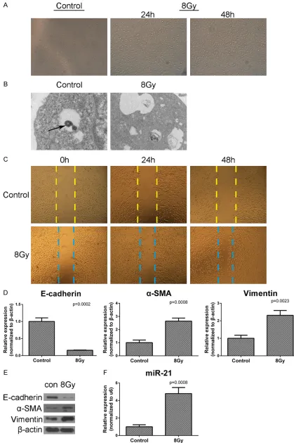
RLE-6TN cells were irradiated with a single dose of 8 Gy X-ray which is an appropriate dose to induce EMT [21]. Cell morphology was observed at 24 h and 48 h postirradiation under the optical microscopy. Cells irradiated with 8Gy lost their cuboidal appearance and showed an elongated mesenchymal-like mor-phology (Figure 1A). Then we observed the cells under the electron microscope, the osmio-philic lamellar body disappeared in the cells after irradiation (Figure 1B). In scratch assay, we found that the cells irradiated were quicker to close a gap than the cells untreated (Figure 1C). Furthermore, we harvested the cells at 24 h postirradiation and examined the mRNA expression of EMT associated proteins by real-time PCR. We found significant decrease of E-cadherin, increase of α-SMA and vimentin in mRNA levels (Figure 1D). Then we examined the protein level expression of these proteins by western blot at 48 h postirradiation (Figure 1E). Examined the expression of miR-21, we found significant increase of miR-21 after irra-diation (Figure 1F).
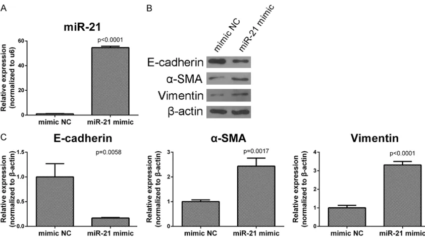
Figure 2. Overexpression of miR-21 induced EMT in RLE-6TN cells. Cells were transfected with miR-21 mimic or mimic NC. A. The transfection efficiency of miR-21 mimic in normal RLE-6TN cells was detected by real-time PCR after 24 h posttransfection. B. After 48 h posttransfection, the protein expression of E-cadherin, α-SMA and Vimen-tin were examined by western blot. C. After 24 h posttransfection, cells were collected and mRNA expression of E-cadherin, α-SMA and Vimentin were detected by real-time PCR. Data are mean ± SD, n = 3, P < 0.05.
Overexpression of miR-21 increased EMT
Transfected miR-21 mimic or mimic NC in RLE-6TN cells and examined the expression of miR-21, we confirmed the transfection efficiency (Figure 2A). Then, we harvested the cells at 48 h after transfection and examined the EMT associated proteins by Western blots. Then we examined the gene expression of these pro-teins at 24 h after transfection by real-time PCR. The results show that with transfection of miR-21 mimic the expression of α-SMA and vimentin get a significant increase and the expression of E-cadherin also had a significant decrease in both protein and mRNA levels (Figure 3C).
Inhibition of miR-21 reduced radiation-induced EMT
Transfection of miR-21 inhibitor in RLE-6TN cells resulted in a significant decrease of its expression compared with endogenous levels of this miRNA (Figure 3A). The cells transfected with either miR-21 inhibitor or inhibitor NC were irradiated with 8 Gy at 24 h after transfection. Cell morphology was observed at 48 h postir-radiation under the optical microscope. While
the cells transfected with miR-21 inhibitor NC lost their cuboidal appearance and showed an elongated mesenchymal-like morphology as same as the only irradiation group, the cells transfected with miR-21 inhibitor reversed the EMT like change (Figure 3B). Then, we harvest-ed the cells at 24 h postirradiation and exam-ined the gene expression of EMT associated proteins by real-time PCR. Then we examined the protein level expression of these proteins at 48 h postirradiation by Western blots. With transfection of miR-21 inhibitor, the expression of α-SMA and vimentin get a significant decrease and the expression of E-cadherin get a significant increase compared with the cells transfected with inhibitor NC in both protein and mRNA levels (Figure 3C-F). In scratch assay, the cells transfected with miR-21 inhibi-tor were slower to close a gap than the inhibiinhibi-tor NC group at 24 h and 48 h postirradiation (Figure 3G).
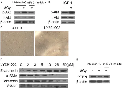
Inhibition of miR-21 attenuated PI3K/Akt path-way activation after irradiation
endogenous level of phospho-Akt expression (Ser473) in miR-21 inhibitor transfected RLE-6TN cells was downregulated compared with that in inhibitor NC transfected RLE-6TN cells after irradiation (Figure 4A). Interestingly, phos-pho-Akt (Ser473) expression was significantly increased in the case of being treated with IGF-1 [22], a PI3K activator, in miR-21 inhibitor transfected RLE-6TN cells after irradiation (Figure 4B). This suggested that activation of PI3K/Akt signaling pathway by irradiation in RLE-6TN cells was suppressed by knockdown
of miR-21, and the suppression was reversed by PI3K activator IGF-1. LY294002, a PI3K/Akt inhibitor, was used by concentration gradient before irradiation. Optical microscope showed the cells which treated with only radiation showed an elongated mesenchymal-like mor-phology and the cells treated with LY294002 keep their cuboidal appearance (Figure 4C). By western blot, we detected a dose-dependent increase of e-cadherin as well as a dose-depen-dent decrease of α-SMA and vimentin (Figure 4D). It indicated that inhibition of PI3K/Akt sig-Figure 3. The effects of miR-21 inhibitor in radiation-induced EMT in RLE-6TN cells. Cells were irradiated with 8Gy after miR-21 inhibitor or inhibitor NC was transfected. A. The transfection efficiency of miR-21 inhibitor in normal RLE-6TN cells was detected by real-time PCR after 24 h posttransfection. B. The changes in cell morphology were observed by light microscope (×100). C-E. After 24 h postirradiation, cells were collected and mRNA expression of E-cadherin, α-SMA and Vimentin were detected by real-time PCR. Data are mean ± SD, n = 3, P < 0.05. F. After 48 h postirradiation, the protein expression of E-cadherin, α-SMA and Vimentin were examined by western blot. G. The cells immigration of RLE-6TN cells were detected by Scratch Assay and observed by light microscope (×40).
[image:7.612.93.517.162.473.2]Antagonism of miR-21 reverses radiation-induced EMT
naling pathway can reduce EMT. PTEN is a known target of miR-21, and also it is an impor-tant suppressor gene of PI3K/Akt signaling pathway. We determined the expression of PTEN by western blot. As depicted in Figure 4E, the expression of PTEN decreased after irradia-tion, and the cells transfected with miR-21 inhibitor before irradiation were higher to express PTEN then the cells only treated with radiation. These results demonstrated that miR-21 inhibitor could up-regulate the expres-sion of PTEN and then affect PI3K/Akt signaling pathway further reduce EMT.
Discussion
Radiotherapy is the common treatment for the patients with lung cancer. The Radiation-induced lung injury is inevitable and currently has no effective cure. New methods are need-ed for decrease this damage. Radiation has been shown to induce the alternation of many miRNAs expression. As a tumor gene, miR-21 has been shown to be a biomarker for the detection of various carcinomas [23]. Furthermore, it is also reported that miR-21 played a role in radiation and might be a poten-tial biomarker in the breast cancer patients treated with radiotherapy [24]. In our study, we first demonstrated the potential involvement of miR-21 in radiation-induced injury in alveolar epithelial cells.
One of the major mechanisms of radiation-induced injury is epithelial-mesenchymal tran-sition (EMT). To clarify the relationship between miRNAs and EMT will be benefit to solve the problem that radiation caused. Generally, EMT happens in embryonic development, most can-cer cells and normal epithelial cells inflamma-tion and fibrosis. Different kind of EMT has the similar mechanism which is characterized by a change of cell shape, cell immigration, and the loss of epithelial characteristics and the acqui-sition of mesenchymal phenotype [25]. The cell shape changes from cuboidal to fibroblastoid can be observed by microscope, and cell immi-gration can be examined by scratch assay to detect the progress of EMT. The other vital molecular feature of EMT is down-regulation of E-cadherin, which presents in the membrane of normal epithelial cells maintaining the cell polarity and morphological structure [26]. So, loss of E-cadherin disrupts the cell junction and changes the cell phenotype [27]. Meanwhile,
vimentin and α-SMA are used to define the mesenchymal phenotype [28]. In our study, we first confirmed the radiation-induced EMT. We observed the cell shape by the optical micro-scope after treated with irradiation and found the EMT like change in phenotype. Then, we detected the expression changes of E-cadherin, α-SMA and vimentin in both gene and protein level by qPCR and Western blot analysis. And in scratch assay, we found that cells postirradia-tion were quicker to close a gap than the cells untreated. We concluded that radiation indeed induced EMT.
MiRNA played an important role in the process of EMT. Overexpression and inhibition of microRNAs have been demonstrated to be vital for cell properties and phenotypes. For exam-ple, miR-200s was explored for the cancer related EMT by targeting ZEB1 [29], and Let-7d was regarded as the member who took part in the fibrosis related EMT by targeting HMGA2 [30]. In this work, we explored the radiation-induced EMT through a single miRNA, miR-21. Following the radiation-induced EMT, the up-regulation of the miR-21 expression was found in RLE-6TN cells after irradiation. To investigate the relevance of radiation-induced EMT and miR-21, we transfected miR-21 inhibitor into suppress the miR-21 expression. Once we transfected miR-21 inhibitor before irradiation, the radiation-induced down-regulation of E-cadherin was reversed, as well as the chang-es of α-SMA and vimentin. And the same ten-dency was observed in cell type change through optical microscope and the cells immigration through scratch assay. These results revealed that miR-21 inhibitor could decrease the radia-tion-induced EMT. And in the other way round, the transfection of miR-21 mimic in normal RLE-6TN cells could enhance EMT.
activator IGF-1 was used to prove this. We added IGF-1 after transfection miR-21 inhibitor before irradiation. The result showed that the expression of p-Akt got restoration. It means that miR-21 inhibitor attenuates the activation of Akt by PI3K. Afterwards, to expatiate this fur-ther, we used PI3K inhibitor LY294002 by con-centration gradient before irradiation and detected the expression of E-cadherin, α-SMA and Vimentin. The western blot results of the tendency of these three critical proteins showed that the radiation-induced EMT recedes gradu-ally according to concentration gradient. Then we detected the expressing of the miR-21 tar-get protein PTEN. And an expected trend shows up. Taken together, miR-21 inhibitor attenuates radiation-induced EMT through PI3K/Akt by tar-geting PTEN.
In conclusion, we described the transfection of miR-21 inhibitor into RLE-6TN cells and exam-ined its ability to cause changes in their pheno-type and the expression of EMT relative protein and gene. We found transfecting miR-21 inhibi-tor into RLE-6TN cells before irradiation caused a less decrease in epithelial gene and protein expression levels, as well as a less increase in mesenchymal gene and protein levels, which accompanied changes in cellular phenotype and properties. It indicated that miR-21 inhibi-tor can attenuate the radiation-induced EMT. Furthermore, we have demonstrated the mech-anism of this process and found that the activa-tion of Akt decreased. It means that the PI3K/ Akt signaling pathway involved in this process. And we confirmed this by using PI3K inhibitor LY294002. Then we determined the predicted target gene PTEN related to this process by western blot. These results suggested the miR-21 inhibitor had profound effects on reversing the radiation-induced EMT and may serve as a promising tool in increasing the potential thera-peutic benefits of the lung cancer patient with radiation-induced lung injury.
Acknowledgements
This work was supported by the National Natural Science Foundation of Hubei Province (No. 2013CFA006) and the National Natural Science Foundation of China (No. 81572967).
Disclosure of conflict of interest
None.
Address correspondence to: Dr. Cong-Hua Xie, De- partment of Radiation and Medical Oncology, Zhong- nan Hospital, Wuhan University, 169 Donghu Road, Wuhan 430071, Hubei, China. E-mail: chxie_65@ hotmail.com
References
[1] Marks LB, Yu XL, Vujaskovic Z, Small W, Folz R and Anscher MS. Radiation-induced lung inju-ry. Semin Radiat Oncol 2003; 13: 333-345. [2] Graves PR, Siddiqui F, Anscher MS and Movsas
B. Radiation Pulmonary Toxicity: From Mecha-nisms to Management. Semin Radiat Oncol 2010; 20: 201-207.
[3] Ding NH, Li JJ and Sun LQ. Molecular mecha-nisms and treatment of radiation-induced lung fibrosis. Curr Drug Targets 2013; 14: 1347-1356.
[4] Wynn TA and Ramalingam TR. Mechanisms of fibrosis: therapeutic translation for fibrotic dis-ease. Nat Med 2012; 18: 1028-1040. [5] Kalluri R and Weinberg RA. The basics of
epi-thelial-mesenchymal transition. J Clin Invest 2009; 119: 1420-1428.
[6] Chen XF, Zhang HJ, Wang HB, Zhu J, Zhou WY, Zhang H, Zhao MC, Su JM, Gao W, Zhang L, Fei K, Zhang HT and Wang HY. Transforming growth factor-beta 1 induces epithelial-to-mes-enchymal transition in human lung cancer cells via PI3K/Akt and MEK/Erk1/2 signaling pathways. Mol Biol Rep 2012; 39: 3549-3556. [7] Zhang JS and Ma L. MicroRNA control of epi-thelial-mesenchymal transition and metasta-sis. Cancer Metastasis Rev 2012; 31: 653-662.
[8] Lee RC, Feinbaum RL and Ambros V. The C-El-egans Heterochronic Gene Lin-4 Encodes Small Rnas with Antisense Complementarity to Lin-14. Cell 1993; 75: 843-854.
[9] Ambros V. The functions of animal microRNAs. Nature 2004; 431: 350-355.
[10] Bartel DP. MicroRNAs: Genomics, biogenesis, mechanism, and function. Cell 2004; 116: 281-297.
[11] Lu M, Zhang QP, Deng M, Miao J, Guo YH, Gao W and Cui QH. An Analysis of Human MicroRNA and Disease Associations. PLoS One 2008; 3: e3420.
[12] Pandit KV, Milosevic J and Kaminski N. MicroR-NAs in idiopathic pulmonary fibrosis. Transl Res 2011; 157: 191-199.
[13] Czochor JR and Glazer PM. microRNAs in Can-cer Cell Response to Ionizing Radiation. Anti-oxid Redox Signal 2014; 21: 293-312.
Pa-Antagonism of miR-21 reverses radiation-induced EMT
tients. Int J Radiat Oncol Biol Phys 2011; 80: 549-557.
[15] Xie L, Zhou JD, Zhang SY, Chen Q, Lai RS, Ding WQ, Song CJ, Meng XJ and Wu JC. Integrating microRNA and mRNA expression profiles in re-sponse to radiation-induced injury in rat lung. Radiat Oncol 2014; 9: 111.
[16] Liu ZL, Wang H, Liu J and Wang ZX. MicroR-NA-21 (miR-21) expression promotes growth, metastasis, and chemo- or radioresistance in non-small cell lung cancer cells by targeting PTEN. Mol Cell Biochem 2013; 372: 35-45. [17] Wang Y, Gao XJ, Wei F, Zhang XW, Yu JP, Zhao
H, Sun Q, Yan F, Yan CH, Li H and Ren X. Diag-nostic and progDiag-nostic value of circulating miR-21 for cancer: A systematic review and meta-analysis. Gene 2014; 533: 389-397.
[18] Wang G, Kwan BC, Lai FM, Chow KM, Li PK and Szeto CC. Urinary 21, 29, and miR-93: Novel Biomarkers of Fibrosis. Am J Nephrol 2012; 36: 412-418.
[19] Liu G, Friggeri A, Yang YP, Milosevic J, Ding QA, Thannickal VJ, Kaminski N and Abraham E. miR-21 mediates fibrogenic activation of pul-monary fibroblasts and lung fibrosis. J Exp Med 2010; 207: 1589-1597.
[20] Liu J, Zhu HC, Yang X, Ge YY, Zhang C, Qin Q, Lu J, Zhan LL, Cheng HY and Sun XC. MicroRNA-21 is a novel promising target in cancer radiation therapy. Tumor Biol 2014; 35: 3975-3979. [21] Nagarajan D, Melo T, Deng ZY, Almeida C and
Zhao WL. ERK/GSK3 beta/Snail signaling me-diates radiation-induced alveolar epithelial-to-mesenchymal transition. Free Radic Biol Med 2012; 52: 983-992.
[22] Ma YF, Xia H, Liu Y and Li M. Silencing miR-21 Sensitizes Non-Small Cell Lung Cancer A549 Cells to Ionizing Radiation through Inhibition of PI3K/Akt. Biomed Res Int 2014; 2014: 617868.
[23] Wu KL, Li LW and Li SY. Circulating microR-NA-21 as a biomarker for the detection of vari-ous carcinomas: an updated meta-analysis based on 36 studies. Tumor Biol 2015; 36: 1973-1981.
[24] Halimi M, Parsian H, Asghari SM, Sariri R, Mos-lemi D, Yeganeh F and Zabihi E. Clinical trans-lation of human microRNA 21 as a potential biomarker for exposure to ionizing radiation. Transl Res 2014; 163: 578-584.
[25] Lamouille S, Xu J and Derynck R. Molecular mechanisms of epithelial-mesenchymal transi-tion. Nat Rev Mol Cell Biol 2014; 15: 178-196. [26] Saha B, Chaiwun B, Imam SS, Tsao-Wei DD,
Groshen S, Naritoku WY and Imam SA. Overex-pression of E-cadherin protein in metastatic breast cancer cells in bone. Anticancer Res 2007; 27: 3903-3908.
[27] Mohamet L, Hawkins K and Ward CM. Loss of function of e-cadherin in embryonic stem cells and the relevance to models of tumorigenesis. J Oncol 2011; 2011: 352616.
[28] Willis BC, Liebler JM, Luby-Phelps K, Nicholson AG, Crandall ED, du Bois RM and Borok Z. In-duction of epithelial-mesenchymal transition in alveolar epithelial cells by transforming growth factor-beta1: potential role in idiopathic pulmonary fibrosis. Am J Pathol 2005; 166: 1321-1332.
[29] Burk U, Schubert J, Wellner U, Schmalhofer O, Vincan E, Spaderna S and Brabletz T. A recipro-cal repression between ZEB1 and members of the miR-200 family promotes EMT and inva-sion in cancer cells. EMBO Rep 2008; 9: 582-589.
[30] Huleihel L, Ben-Yehudah A, Milosevic J, Yu G, Pandit K, Sakamoto K, Yousef H, LeJeune M, Coon TA, Redinger CJ, Chensny L, Manor E, Schatten G and Kaminski N. Let-7d microRNA affects mesenchymal phenotypic properties of lung fibroblasts. Am J Physiol Lung Cell Mol Physiol 2014; 306: L534-542.
[31] Bakin AV, Tomlinson AK, Bhowmick NA, Moses HL and Arteaga CL. Phosphatidylinositol 3-ki-nase function is required for transforming growth factor beta-mediated epithelial to mes-enchymal transition and cell migration. J of Biol Chem 2000; 275: 36803-36810.
[32] Meng FY, Henson R, Wehbe-Janek H, Ghoshal K, Jacob ST and Patel T. MicroRNA-21 regu-lates expression of the PTEN tumor suppressor gene in human hepatocellular cancer. Gastro-enterology 2007; 133: 647-658.