Original Article
Effects of prior treatment with simvastatin on skeletal
muscle structure and mitochondrial enzyme activities
during early phases of sepsis
Elif Ozkok1, Hatice Yorulmaz2, Gulten Ates3, Piraye Serdaroglu-Oflazer4, Ayse Sule Tamer3
1Deparment of Neuroscience, The Institute for Experimental Medicine, Istanbul University, Istanbul, Turkey; 2School of Nursing, Halic University, Istanbul, Turkey; 3Department of Physiology, Istanbul Medical Faculty,
Istanbul University, Istanbul, Turkey; 4Department of Neurology/Neuromuscular Disorders Unit, Istanbul Faculty of
Medicine, Istanbul University, Istanbul, Turkey
Received October 20, 2014; Accepted December 1, 2014; Epub December 1, 2014; Published December 15, 2014
Abstract: We investigated the effects of the early phase of sepsis and prior treatment of Simvastatin on muscle structure and mitochondrial enzymes treated with lipopolysaccharide (LPS) in rats. We divided rats into control, LPS, simvastatin, simvastatin + LPS groups. Mitochondrial citrate synthase, complex I, II, I + III, II + III, cytochrome c oxidase (COX) activities were measured. Muscle tissue was stained using modified Gomori trichrome (MGT), succinic dehydrogenase (SDH) and cytochrome oxidase (COX). In all treated groups, complex I and citrate synthase activities were higher than in the controls. In the control and LPS groups, COX activity was increased when compared with simvastatins’. Complex II, II-III activities were higher in the LPS group than in the control group. Complex I-III activities were higher in the Simvastatin and Simvastatin + LPS groups than in the control and LPS groups (P < 0.05). Myopathic changes with LPS group were observed in MGT stained sections. Our findings showed improve -ments in the alterations of enzyme activities and muscle myofibrils after treating rats with LPS that had received a prior dose of simvastatin.
Keywords: Lipopolysaccharide, mitochondrial enzyme activites, rat, simvastatin, skeletal muscle
Introduction
Sepsis is a life threatening disease which affects many organ systems, if left untreated it can cause shock, organ dysfunction and multi-organ failure [1]. Patients with sepsis treated in Intensive Care Units often develop muscle weakness, muscle fatigue, and neuropathy resulting in proteolysis [2, 3]. An incremental loss of proteins may appear in the presence of infection, which causes weight-loss and muscle fatigue and increases the risk of thromboem-bolic complications [4]. Many studies have shown that the release of cytokines such as TNF-α and IL-1 and increased ubiquitin mRNA during an infection cause loss of myofibrillary nitrogen and impairment of proteins, which activates proteolysis [5-8].
Skeletal tissue comprises 50%-60% of body cell mass and represents the largest organ
affected by systemic inflammation. Pathologies such as ultrastructural mitochondrial altera-tions and oxidative mechanisms in sepsis and septic shock have attracted attention in recent years [9].
Statins are key precursor inhibitors of HMG-CoA reductase, which synthesizes mevalonate, in the cholesterol synthesis pathway. Some stud-ies have suggested that prior statin treatment may prevent vascular hyporeactivity and inhibit the development of severe sepsis in patients with acute systemic inflammation [10, 11]. However, using statins can also have dose-dependent adverse effects. High concentra-tions of statins can cause muscle damage and myalgia [12, 13].
Simvastatin on skeletal muscle with LPS
and histologic changes in skeletal muscle of rats treated with LPS.
Materials and methods
Experimenta l groups
This study was conducted at the University of Istanbul experimental research center, (Reso- lution No: 2012/138). We used male adult Wistar albino rats weighing 200-250 g in the experiments. The animals were fed with a com-mercial diet and tap water ad libitum, were housed in cages and kept at a controlled tem-perature (22±2°C) and humidity (55 to 60%) with a 12-h light/dark cycle.
The rats were divided into four groups, each composed of eight rats: (1) control group, (2) LPS group, (3) Simvastatin group, (4) Simv- astatin + LPS group.
Experimental procedures
Lipopolysaccharide (LPS) from Escherichia coli O127: B8 (Sigma Aldrich, Product No: L5668) was dissolved in 1 ml of sterile saline solution and a single dose was injected intraperitoneally at a daily dose of 20 mg/kg. Simvastatin (20 mg/kg) (Sigma Aldrich, Product No: S0650000) were given p.o. via oral gavage for 5 days [14]. In the Simvastatin + LPS group, LPS was given 1.5 h after the fifth dose of simvastatin. Four hours after LPS treatment, animals (for both of LPS and Simvastatin + LPS groups) were sacri-fied. Gastrocnemius muscles were dissected in all groups at the end of 4th hours.
Biochemical procedures
At the end of the experimental period, blood samples were taken to determine serum con-centrations of creatinine kinase (CK), aspartate aminotransferase (GOT), alanine
aminotrans-lyzer, DPC, Diagnostic products corporation, USA).
Cytokine levels
The blood samples were centrifuged at 2500 rpm for 20 minutes and the serum stored in ep- pendorf tubes at -20°C until analysis. The cyto-kine levels were measured using the ELISA method for TNF-α (BioSource, Invitrogen, USA, Cat No, KRC3012) and IL-10 (BioSource, Invitrogen, USA, Cat No, KRC0101). The absor-bance values were measured at 450 nm using a micro-ELISA automatic analyzer. The sensitiv-ity of the ELISA kits were < 4 pg/ml for TNF-α, < 5 pg/ml for IL-10.
Neuropathological procedures
The gastrocnemius muscle was taken while the animals were under anesthesia. The muscle samples were rapidly frozen in isopentane cooled by liquid nitrogen. We took serial cross-sections 8 μm thick and stained them with modified Gomori’s trichrome (MGT) method, succinic dehydrogenase (SDH) and cytochrome oxidase (COX) using standard protocols [15, 16]. The stained sections were visualized and photographed using a Nikon microscope (ECLIPSE 80i Nikon Corporation, JAPAN).
Mitochondrial enzyme activities
Absorbances of all activities were recorded kinetically in tissue homogenate using a spec-trophotometer (Shimadzu, Japan) at 30°C. Citrate synthase
[image:2.612.91.431.85.170.2]The reaction was initiated by adding of dithio-bis 2-nitrobenzoic acid into a mixture contain-ing oxaloacetate, Acetyl-CoA, and tissue homogenate. Absorbance was measured at 412 nm [17].
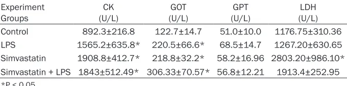
Table 1. Values of serum CK, GOT, GPT, LDH for groups Experiment
Groups (U/L)CK (U/L)GOT (U/L)GPT (U/L)LDH Control 892.3±216.8 122.7±14.7 51.0±10.0 1176.75±310.36 LPS 1565.2±635.8* 220.5±66.6* 68.5±14.7 1267.20±630.65 Simvastatin 1908.8±412.7* 218.8±32.2* 58.2±16.96 2803.20±986.10* Simvastatin + LPS 1843±512.49* 306.33±70.57* 56.8±12.21 1913.4±252.95 *P < 0.05.
autoana-Complex I (NADH dehydrogenase)
Complex I-oxidation of NAD-activity was moni-tored using first potassium phosphate, NADH, and potassium ferrisiyanide and then by adding tissue homogenate at 340 nm [18].
Complex II + III (succinate cytochrome c reductase)
Tissue homogenate was added to a mixture of fresh cytochrome c containing succinate, and potassium cyanide (KCN). Complex II + III activ-ity was observed due to the increase in rate of reduction of cytochrome c. This was measured at 550 nm [18].
Complex I + III (NADH Cytochrome c Reductase)
Complex I + III activity was observed as a result of the rate of decrease in the addition cyto-chrome c homogenate including fresh prepared cytochrome c, NADH, and KCN alterations after adding rotenone at 550 nm [19].
Complex II (succinate dehydrogenase)
Complex II activity was determined adding tis-sue homogenate by the reduction of 2,6-dichlo-rophenolindophenol (DCIP) including succinate, KCN, and DCPIP at 600 nm [20].
Complex IV (cytochrome c oxidase)
Cytochrome c oxidase (COX) activity was mea-sured as the oxidation rate of decrease in reduced cytochrome c at 550 nm at 30°C
(CYTOCOX1, Sigma Aldrich, Saint Louise, USA) [21].
Statistical analyses
Data were expressed as mean ± standard devi-ation. Overall statistical significance between the groups was tested with oneway ANOVA or Kruskal-Wallis test, depending on normality of distribution. In all cases, P < 0.05 was set as limit of significance.
Results
Biochemical findings
Serum CK and GOT values were found to be increased in experimental groups as compared to those of controls (P < 0.05).
There were no significant changes in GPT levels in any groups (P > 0.05). However, levels of GPT were lower in the control than the other groups’. In the simvastatin group, LDH levels were observed to be higher in the Simvastatin group than in the other groups (P < 0.05) (Table 1).
Cytokine findings
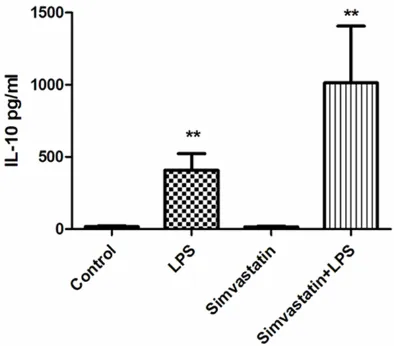
Serum TNF-α and IL-10 values were found to be significantly higher in the LPS and Simvastatin + LPS groups compared to those of the controls (P < 0.01) (Figures 1, 2).
Mitochondrial enzyme activities
[image:3.612.90.288.70.242.2]Complex I and citrate synthase activities were significantly increased in all experimental
Figure 1. Serum TNF-α values in experimental groups. **Significant differences at P < 0.01, LPS and Simvastatin + LPS vs. other groups.
[image:3.612.324.521.71.244.2]Simvastatin on skeletal muscle with LPS
groups than those of the controls. The results were as follows: Control group; complex I 20.47±14.15 µmol/min/g tissue; LPS group 72.23±21.99 µmol/min/g tissue; Simvastatin group 58.20±16.76 µmol/min/g tissue; and the Simvastatin + LPS group 59.52±17.94 µmol/min/g tissue. For citrate synthase: Control group 22.8±1.8 µmol/min/g tissue; LPS group 31.0±4.2 µmol/min/g tissue; Simvastatin group 31.29±7.63 µmol/min/g tissue; and the Simvastatin + LPS group 36.02±6.33 µmol/ min/g tissue (Figures 3 and 4).
COX activity in the control and LPS groups were observed to be significantly increased in com
-parison with the Simvastatin group: Control group 2.37±0.7 µmol/min/g tissue; LPS group 2.31±0.7 µmol/min/g tissue; Simvastatin group 1.25±0.15 µmol/min/g tissue; and the Simvastatin + LPS group 1.89±0.46 µmol/ min/g tissue (Figure 5).
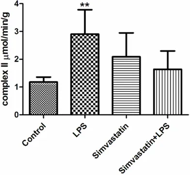
[image:4.612.90.287.71.249.2]Complex II activity was higher in the LPS and control groups than the Simvastatin + LPS groups: Control group 1.18±0.18 µmol/min/g tissue; LPS group 2.90±0.88 µmol/min/g tis-sue; Simvastatin group 2.09±0.86 µmol/min/g tissue; and Simvastatin + LPS 1.64±0.66 µmol/ min/g tissue (Figure 6).
[image:4.612.323.522.71.252.2]Figure 3. Activities of complex I in groups of muscle tissues, *: P < 0.05, Simvastatin and Simvastatin + LPS vs. control; **: P < 0.01 LPS vs. control.
Figure 4. Activities of citrate synthase in groups of muscle tissues, *: P < 0.05, experimental groups vs. control.
Figure 5. Activities of COX in groups of muscle
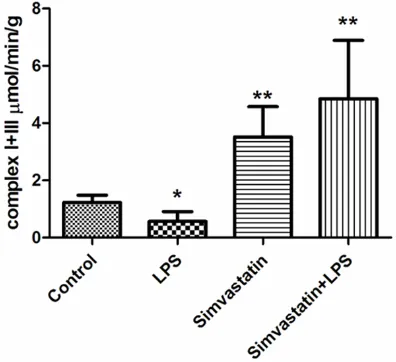
[image:4.612.90.287.313.495.2] [image:4.612.325.521.315.496.2]Complex I-III activity was higher in the Simvastatin and Simvastatin + LPS groups than the LPS and control groups: Control group 1.23±0.25 µmol/min/g tissue; LPS group 0.57±0.34 µmol/min/g tissue; Simvastatin group 3.52±1.06 µmol/min/g tissue; and the Simvastatin + LPS group 4.85±2.04 µmol/ min/g tissue (Figure 7).
Complex II-III activity was higher in the LPS and control groups than the Simvastatin and Simvastatin + LPS groups: Control group 0.75±0.09 µmol/min/g tissue; LPS group 1.18±0.35 µmol/min/g tissue; Simvastatin group 0.13±0.04 µmol/min/g tissue; and Simvastatin + LPS group 0.32±0.19 µmol/ min/g tissue (P < 0.05) (Figure 8).
Histopathological findings
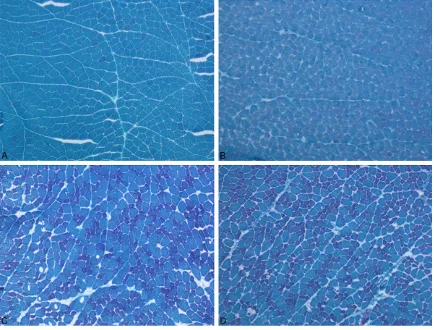
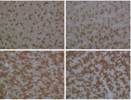
More than half of the animals that were treated with LPS showed myopathic changes charac-terized by rounding of the muscle fibers and fiber size variation. In only one animal, the fibers of some fasicles showed mitochondrial aggregates. Cytochrome c oxidase activity was found normal in the LPS group as compared to the control group.
In the Simvastatin group, myopathic changes were seen in all rats. In addition, there were no changes with COX and SDH staining in any animal.
In the Simvastatin + LPS groups, MGT and COX results were the same as those of the controls, but in less of the animals, mild myopathic changes were seen (Figures 9-11).
Discussion
In our study, we searched whether it would be possible to induce sepsis by 20 mg/kg i.p. 4 hours LPS in rats and if this effect is reduced by prior simvastatin (20 mg/kg p.o. via oral gavage) treatment. The effects were tested on muscle tissue, which represents the largest organ affected by systemic inflammation. Mitochondrial structure, changes in cytokine levels, and enzyme activities in the first hours of sepsis in all groups were evaluated.
With this method, we observed that sepsis developed in the LPS treated animals and the effect of LPS was less by prior symvastatin treatmet. The elevated CK levels in all experi-mental groups evidenced involvement of mus-cle tissue.
[image:5.612.324.522.70.249.2]We found that TNF-α and IL10 were higher than controls in the early phase of sespsis as well as in the pre-treated groups. While the increase in the proinflammatory cytokine TNF--α was more evident in the sepsis (LPS) group, increase in the anti-inflammatory cytokine IL-10 was more prominent in the pre-treated (SL) group. It has been reported that proinflammatory cytokines such as TNF-α and interleukin-6 cause loss of myofibrillary nitrogen and the destruction of
Figure 7. Activities of complex I + III in groups of muscle tissues, *: P < 0.05, LPS vs. control. **: P < 0.01, Simvastatin and Simvastatin + LPS vs. LPS and control.
[image:5.612.90.288.73.254.2]Simvastatin on skeletal muscle with LPS
proteins during sepsis [6, 8, 22]. Likewise, Almendro et al. demonstrated the increments of protein catabolism and DNA break-out that occurred in muscles of rats with inducing cecal ligation and puncture [23]. Our results were in agreement with the previous studies and showed that increase in cytokine levels were compatible with the effects of LPS and simvastatin.
In human and experimental animal models of sepsis, there are relationships between the severity of sepsis, mitochondrial damage and bioenergetic dysfunction [24]. Brealey et al. (2004) demonstrated that the analysis of mus-cle mitochondria may be an indicator of mito-chondrial changes during long-term sepsis [25]. It has been reported that using statins causes mitochondrial dysfunction and alterations in both mitochondrial swelling and membrane potential [26]. However, reports on sepsis mod-els show variable results in changes in
[image:6.612.90.524.72.402.2]mito-chondrial enzyme activities [27-29]. While Brealey et al showed that complex I activity was at higher levels, Fredriksson et al. implicated the activities of citrate synthase and complex I when they reported a 53% and 60% decrease respectively, in intercostal muscle compared with controls in patients with sepsis [27]. This variability may depend on the variability of the method in inducing sepsis, and the variability in methods of measurement of the enzyme levels. We found alterations in the activities of com-plex-I, -II, -I-III, -II-III, Citrate synthase and COX. Complex-I and citrate synthase activities were significantly higher in all experimental groups than those in the controls. As complex-I is the first enzyme in the electron transport system it may be possible that increase in its expression may be a response to the chemicals used, rath-er than being a sepsis response. This is sup-ported by the explanation of Fredriksson et al. who showed increament in complex-I levels in
muscle tissue of healthy volunteers was due to a response to acute endotoxin challenge [29]. In another study the group concluded that increased complex I activity is a compensator mechanism, which effects mitochondrial mem-brane potential [30].
In our study, COX (complex IV) activity in the control and LPS groups were significantly increased in comparison with the Simvastatin groups. Fredriksson et al. observed a decreased protein content of complex IV in patients with sepsis when compared with control subjects; this was an unexpected finding. The activity of complex IV was 30% lower in leg muscles of patients with sepsis but not in intercostal mus-cle. Morever, the activity of complex IV decreased to 30% in leg muscle [27]. In our study, we found higher activity of complex II in the LPS group than in the control and Simvastatin + LPS groups being more promi-nent in the LPS group. This suggested that the
[image:7.612.91.524.70.399.2]increase in complex II was due to sepsis. Our finding is consistent with the Larsen et al study who reported the simvastatin induced decre-ment of complex II was due to a reduction of mitochondrial volume [30]. Complex I + III activ-ity was higher in the Simvastatin and Simvastatin + LPS groups than in the control and LPS groups. It has been shown that high levels of LPS leads to a 42% lower activity of complex I + III and a 23% reduction of pyruvate oxidation in the mitochondria of skeletal mus-cle [31]. We found that complex II + III activity was higher in the LPS group than the control group, and was lower in the Simvastatin and Simvastatin + LPS groups than in the control group. In agreement with our findings, Nadanaciva et al. reported that use of simvas-tatin or lovassimvas-tatin causes mitochondrial toxicity and inhibits complex II + III activities by affect-ing uncoupled proteins [26]. It has been sug-gested that the use of statins reduces ubiqui -none, which decreases activities of certain
Simvastatin on skeletal muscle with LPS
mitochondrial enzymes along with muscle dam-age in skeletal tissue [32, 33].
Myopathic changes in muscle sections in the majority of animals in the LPS and Simvastatin groups were evident in our study. However, mitochondrial pathology in the muscle sections was very scarce. In this study we have shown that sepsis can be induced by a single dose of 20 mg/kg LPS i.p, the effects are less promi-nent with pre-treatment with 5 days oral simv-astatin at 20 mg/kg/day. The effects at early phase of sepsis were evidenced by the profiles of changes in cytokines, and in activities of electron transport chain enzymes.
Acknowledgements
Our study was granted from Istanbul University Research Projects (Project No: 42019). We would like to thank Ozgun Chemical Company and Roche Diagnostics for analyzing biochemi-cal parameters in autoanalyzer. We would like also thank Mr. David F Chapman for editing of
English and Ms. Hatice Tasli for preparing of muscle section.
Disclosure of conflict of interest
None.
Address correspondence to: Dr. Hatice Yorulmaz, School of Health Science, Halic University, Buyu- kdere Cd. No: 101 Mecidiyekoy, Sisli, Istanbul, TR 34394, Turkey. Tel: +902123430887; E-mail: [email protected]
References
[1] Balk RA. Severe sepsis and septic shock. Crit Care Clin 2000; 16: 179-92.
[2] Bolton CF. Sepsis and the systemic inflamma -tory response syndrome: neuromuscular mani-festations. Crit Care Med 1996; 24: 1408-16. [3] Larsson L, Li X, Edstrom L, Eriksson LI, Zackri-
sson H, Argentini C, Schiaffino S. Acute quad -riplegia and loss of muscle myosin in patients treated with nondepolarizing neuromuscular blocking agents and corticosteroids:
[image:8.612.90.523.71.399.2]nisms at the cellular and molecular levels. Crit Care Med 2000; 28: 34-45.
[4] Theologides A. Cancer cachexia. Cancer 1979; 43: 2004-12.
[5] Evans RD, Argilés JM, Williamson DH. Meta- bolic effects of tumour necrosis factor-alpha (cachectin) and interleukin-1. Clin Sci 1989; 77: 357.
[6] Argilés JM, García-Martínez C, Llovera M, López-Soriano FJ. The role of cytokines in mus-cle wasting: its relation with cancer cachexia. Med Res Rev 1992; 12: 637-52.
[7] Kerr JF, Wyllie AH, Currie AR. Apoptosis: a basic biological phenomenon with wide ranging im-plications in tissue kinetics. Br J Cancer 1972; 26: 239-45.
[8] Solomon V, Goldberg AL. Importance of the ATP-ubiquitin-proteasome pathway in the deg -radation of soluble and myofibrillar proteins in rabbit muscle extracts. J Biol Chem 1996; 271: 26690-97.
[9] Crouser ED, Julian MW, Blaho DV, Pfeiffer DR. Endotoxin-induced mitochondrial damage cor-relates with impaired respiratory activity. Crit Care Med 2002; 30: 276-84.
[10] Pleiner J, Schaller G, Mittermayer F, Zorn S, Marsik C, Polterauer S, Kapiotis S, Wolzt M. Simvastatin prevents vascular hyporeactivity during inflammation. Circulation 2004; 110: 3349-54.
[11] Gao F, Linhartova L, Johnston AM, Thickett DR. Statins and sepsis. Br J Anaesth 2008; 100: 288-98.
[12] Tiwari A, Bansal V, Chugh A, Mookhtiar K. Statins and myotoxicity: atherapeutic limita-tion. Expert Opin Drug Saf 2006; 5: 651-66. [13] Neuvonen PJ, Niemi M, Backman JT. Drug
in-teractions with lipid-lowering drugs: mecha-nisms and clinical relevance. Clin Pharmacol Ther 2006; 80: 565–81.
[14] Nezić L, Skrbić R, Dobrić S, Stojiljković MP, Satara SS, Milovanović ZA, Stojaković N. Effect of simvastatin on proinflammatory cytokines production during lipopolysaccharide-induced inflammation in rats. Gen Physiol Biophys 2009; 28: 119-26.
[15] Engel WK, Cunningham GG. Rapid examinaiıon of muscle tissue an improved trichrome meth-od for fresh-frozen biopsy sections. Neurology 1963; 13: 919-23.
[16] Tanji K, Bonilla E. Light microscopic methods to visualize mitochondria on tissue sections. Methods 2008; 46: 274-80.
[17] Srere PA. Citrate synthase. Methods Enzymol 1969; 13: 3-11.
[18] King TE, Howard RL. Preparation and proper-ties of soluble NADH dehydrogenase from car-diac muscle. Methods Enzymol 1967; 10: 275-94.
[19] Scottocosa GL, Kuylenstierna B, Ernster L, Bergstrand A. An electron transport system as-sociated with the outer membrane of the mito-chondria. J Cell Biol 1967; 32: 415-38. [20] King TE. Preparation of succinate
dehydroge-nase and reconstitution of succinate oxidase. Methods Enzymol 1967; 10: 322-31.
[21] Trounce IA, Kim YL, Jun AS, Wallace DC. Assessment of mitochondrial oxidative phos-phorylation in patient muscle biopsies, lym-phoblasts, and transmitochondrial cell lines. Methods Enzymol 1996; 264: 484-509. [22] Pajak B, Orzechowska S, Pijet B, Pijet M,
Pogorzelska A, Gajkowska B, Orzechowski A. Crossroads of cytokine signaling: the chase to stop muscle cachexia. J Physiol Pharmacol 2008; 59: 251-264.
[23] Almendro V, Carbó N, Busquets S, Figueras M, Tessitore L, López-Soriano FJ, Argilés JM. Sep- sis induces DNA fragmentation in rat skeletal muscle. Eur Cytokine Netw 2003; 14: 256-9. [24] Zang Q, Maass DL, Tsai SJ, Horton JW. Cardiac
mitochondrial damage and inflammation re -sponses in sepsis. Surg Infect (Larchmt) 2007; 8: 41–54.
[25] Brealey D, Karyampudi S, Jacques TS, Novelli M, Stidwill R, Taylor V, Smolenski RT, Singer M. Mitochondrial dysfunction in a long-term ro-dent model of sepsis and organ failure. Am J Physiol Regul Integr Comp Physiol 2004; 286: 491-7.
[26] Nadanaciva S, Dykens JA, Bernal A, Capaldi RA, Will Y. Mitochondrial impairment by PPAR agonists and statins identified via immunocap -tured OXPHOS complex activities and respira-tion. Toxicol Appl Pharmacol 2007; 15: 223: 277-87.
[27] Fredriksson K, Hammarqvist F, Strigard K, Hultenby K, Ljungqvist O, Wernerman J, Rooyackers O. Derangements in mitochondrial metabolism in intercostal and leg muscle of critically ill patients with sepsis induced multi-ple organ failure. Am J Physiol Endocrinol Metab 2006; 291: 1044-50.
[28] Brealey D, Brand M, Hargreaves I, Heales S, Land J, Smolenski R, Davies NA, Cooper CE, Singer M. Association between mitochondrial dysfunction and severity and outcome of sep-tic shock. Lancet 2002; 360: 219-23.
[29] Fredriksson K, Fläring U, Guillet C, Wernerman J, Rooyackers O. Muscle mitochondrial activity increases rapidly after an endotoxin challenge in human volunteers. Acta Anaesthesiol Scand 2009; 53: 299-304.
Simvastatin on skeletal muscle with LPS
[33] Sirvent P, Mercier J, Vassort G, Lacampagne A. Simvastatin triggers mitochondria- induced Ca2+ signaling alteration in skeletal muscle. Biochem Biophys Res Commun 2005; 329: 1067-75.
[31] Trumbeckaite S, Opalka JR, Neuhof C, Zierz S, Gellerich FN. Different sensitivity of rabbit heart and skeletal muscle to endotoxin-in-duced impairment of mitochondrial function. Eur J Biochem 2001; 268: 1422-29.