Int J Clin Exp Pathol 2014;7(3):1188-1192 www.ijcep.com /ISSN:1936-2625/IJCEP1401017
Case Report
Primary cardiac angiosarcoma confirmed
by multimodality imaging guided liver biopsy
Zhi-Xin Qiu1, Qing Zhang2
1Department of Respiratory Medicine, 2Department of Cardiology, West China Hospital, Sichuan University,
Chengdu, China
Received January 7, 2014; Accepted February 3, 2014; Epub February 15, 2014; Published March 1, 2014
Abstract: Primary cardiac angiosarcoma is an extremely rare malignant tumor with various clinical presentations but usually in late stage. We report a case presented with bloody pericardial effusion, in which the final diagnosis was confirmed by multiple imaging modalities such as echocardiography, computed tomography, magnetic resonance imaging and fluorine-18-fluorodeoxyglucose positron emission tomography, as well as ultrasound-guided liver bi-opsy.
Keywords: Cardiac angiosarcoma, pericardial effusion, multimodality imaging
Introduction
Cardiac angiosarcoma is an extremely rare malignant neoplasm that accounts for less than 10% of all resected primary tumors of the heart, which themselves are found in less than 0.3% of autopsies [1-3]. Since most cardiac tumors may remain clinically silent for a long time or cause a wide range of cardiac and sys-temic symptoms that mimic other diseases [4-8], it is very difficult in early diagnosis. Being a malignant type, cardiac angiosarcoma is usu-ally originated in the right atrium (RA) and asso-ciated with a poor prognosis, because distant metastasis is present in the majority of patients at the time of diagnosis [2, 9].
Two-dimensional (2D) echocardiography is the preferred initial method for detecting cardiac tumors, however, a complete assessment of the heart and surrounding tissue may some-times be difficult with this technique. Magnetic resonance imaging (MRI) and fluorine-18-fluo-rodeoxyglucose positron emission tomography (18F-FDG-PET/CT) can provide precise informa-tion on the extent of involvement of tumors and distant metastasis. Therefore, the use of these multiple imaging modalities allows simultane-ous evaluation of cardiac structures and sur-rounding tissues, as well as staging of
malig-nant tumors and treatment monitoring in some circumstances [4]. In this paper, we report a case of primary cardiac angiosarcoma con-firmed by multimodality imaging.
Case report
A 41-year-old Chinese woman was admitted to cardiac ward with exertional chest discomfort and dyspnea for 2 months. Vital signs were nor-mal. Physical examination revealed jugular vein distention, cardiac dullness enlargement and distant heart sounds. Normal cardiac enzymes and N-terminal pro-B-type natriuretic peptide. Normal blood gas analysis, liver function, renal function and thyroid function. Negative serolo-gy for viral hepatitis, cytomegalovirus, herpes, Epstein-Barr virus and human immunodeficien-cy virus. Normal values of immunoglobulin (Ig) G, IgA, IgM, IgE, and CD3, CD4, CD8 cell subsets. Serum tumor markers were normal including alpha fetal protein (AFP), carcinoembryonic antigen (CEA), carcinoma antigen (CA) 19-9 and CA-125. Negative tuberculosis skin test and serum antibody of tuberculosis. However, eryth-rocyte sedimentation rate was slightly elevated at 37 mm/hour and D-Dimer was obviously high of >38 mg/l FEU.
car-diac structures and left ventricular ejection fraction of 67%. She underwent pericardiocen-tesis and the analysis of pericardial fluid revealed red blood cells of (++++)/HP, nucleat-ed cells of 5-10/HP, albumin of 36.6 g/L,
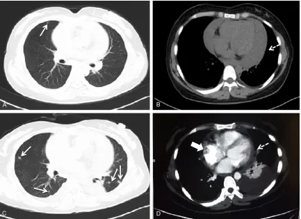
[image:2.612.91.524.71.386.2]glu-cose of 4.24 mmol/L, lactate dehydrogenase of 442 IU/L, and adenosine deaminase of 15.0 IU/L. More importantly, tumor markers were strikingly high with CEA of 7.44 ng/ml, CA19-9 of >1000.00 U/ml and CA-125 of 2689.00 U/ Figure 1. Images of the chest computed tomography (CT) on admission (A and B) and enhanced CT in a few days (C and D). (A and B) A scattered nodule in the lungs (arrow), and severe pericardial effusion (dashed arrow); (C and D) New scattered nodules in the lungs (arrows), mild pericardial effusion (dashed arrow), and a mass suspected in the right atrium (block arrow).
[image:2.612.92.525.457.615.2]Primary cardiac angiosarcoma confirmed by multimodality imaging
ml. However, neither malignant cells nor micro-organisms were identified in the pericardial fluid. Because of the quick recurrence of severe pericardial effusion, a drainage catheter was implanted. Thereafter, pericardial effusion dra- inage was sent to analysis for several times, but similar results were obtained.
Her first chest computed tomography (CT) scan on admission revealed a few scattered well-defined nodules in the lungs with very mild pleural effusion, and the following enhanced CT scan performed a few days later suspected a mass in the RA with some new scattered
[image:3.612.94.522.73.190.2]nod-ules in the lungs and moderate pleural effusion (Figure 1). Simultaneous abdominal enhanced CT scan also demonstrated a number of scat-tered low-density nodules in the liver (Figure 2). The pleural fluid analysis found red blood cells of >20000×106/L, nucleated cells of 80×106/L, albumin of 29.6 g/L, glucose of 4.47 mmol/L, lactate dehydrogenase of 225 IU/L and ade-nosine deaminase of 8.9 IU/L. Tumor markers were significantly elevated as CEA of 2.39 ng/ ml, CA19-9 of 104.60 U/ml and CA-125 of 1538.00 U/ml. In order to localize and charac-terize the cardiac tumor, cardiac MRI with gado-linium late enhancement was also arranged, Figure 3. Images of cardiac magnetic resonance imaging with gadolinium late enhancement. A: Axial view on T1 weighted turbo spin echo (TSE) image; B: Axial view on T2 weighted TSE image; C: Axial view on late gadolinium enhancement PSIR image of the right atrial mass (block arrows).
Figure 4. Images of the whole-body fluorine-18-fluorodeoxyglucose positron emission tomography (18F-FDG-PET/
[image:3.612.91.524.252.481.2]which clearly showed a huge mass with RA involvement (Figure 3).
Immediately, a whole-body 18F-FDG-PET/CT scan was performed that demonstrated intense FDG uptake with maximum standardized uptake value (SUVmax) of 8.73 in a filling defect of 86×62 mm in the RA. Intense FDG avid was also detected in the liver (SUVmax of 2.73) and in the lungs (SUVmax of 1.97) (Figure 4). The result was strongly suggestive of primary malignant tumor in the RA with distant metastasis to the liver and lungs.
As abdominal ultrasound also confirmed the nodules in the liver, ultrasound-guided biopsy was performed for pathological evidences. The microscopic findings unmasked a mesenchy-mal, poorly differentiated tumor consisting of polymorphic, predominantly spindle-shaped cells along preformed vessels, of which immu-nohistochemistry for CD31 and CD34 showed positive, that corresponding to an angiosarco-ma (Figure 5). Unfortunately, this patient refused surgical treatment where biopsy at the original site could have been performed. Discussion
Primary cardiac angiosarcoma originates in the RA and because of the involvement of the RA, it
[image:4.612.92.526.72.299.2]Primary cardiac angiosarcoma confirmed by multimodality imaging
tumor from blood stagnation or thrombus around the tumor, because old thrombus and stagnant blood may not be enhanced by Gadolinium contrast. Due to its much less avail-ability and higher cost, 18F-FDG-PET/CT has been reserved as an important non-invasive imaging modality for suspected metastasis of unknown origin and preoperative staging of various neoplasms. It offers a high sensitivity scan for metabolic activity with precise ana-tomical localization, since malignant neo-plasms and their metastases are always char-acterized by enhanced glucose utilization and therefore increased glucose uptake indicated by the 18F-FDG. Therefore, successful diagnosis in this rare case relied on the comprehensive utilization of multimodality imaging techniques, in particular when a single method was inconclusive.
Disclosure of conflict of interest
None.
Address correspondence to: Dr. Qing Zhang, Depart- ment of Cardiology, West China Hospital, Sichuan University, Chengdu, Sichuan, 610041, China. Tel: 86-28-85422607; E-mail: qzhang2000cn@yahoo. com; Zhi-Xin Qiu, Department of Respiratory Medicine, West China Hospital, Sichuan University, Chengdu, China. E-mail: [email protected]
References
[1] Dennig K, Lehmann G, Richter T. An angiosar-coma in the left atrium. N Engl J Med 2000; 342: 443-4.
[2] Putnam JB Jr, Sweeney MS, Colon R, Lanza LA, Frazier OH, Cooley DA. Primary cardiac sarco-mas. Ann Thorac Surg 1991; 51: 906-10. [3] McAllister HA Jr. Primary tumors and cysts of
the heart and pericardium. Curr Probl Cardiol 1979; 4: 1-51.
[4] Romero-Farina G, Candell-Riera J, Beltran-Ror A, Gonzalez-Moreno JB, Bigalli D, Stratta A. Pri-mary cardiac angiosarcoma: diagnostic utility of computed tomography and cardiac magnet-ic resonance. Rev Esp Cardiol 2004; 57: 1234-7.
[5] Reynen K. Frequency of primary tumors of the heart. Am J Cardiol 1996; 77: 107.
[6] Sarjeant JM, Butany J, Cusimano RJ. Cancer of the heart: epidemiology and management of primary tumors and metastases. Am J Cardio-vasc Drugs 2003; 3: 407-21.
[7] Dreon DM, John EM, DiCiccio Y, Whittemore AS. Use of NHANES data to assign nutrient densities to food groups in a multiethnic diet history questionnaire. Nutr Cancer 1993; 20: 223-30.
[8] Jimenez Mazuecos JM, Fuentes Manso R, Segovia Cubero J, Toquero Ramos J, Oteo Dominguez JF, Alonso-Pulpon Rivera L. Is heart transplantation for primary cardiac sarcoma a useful therapeutic option? Rev Esp Cardiol 2003; 56: 408-11.
[9] Janigan DT, Husain A, Robinson NA. Cardiac angiosarcomas. A review and a case report. Cancer 1986; 57: 852-9.
[10] Engelen M, Bruch C, Hoffmeier A, Kersting C, Stypmann J. Primary left atrial angiosarcoma mimicking severe mitral valve stenosis. Heart 2005; 91: e27.
[11] Loffler H, Grille W. Classification of malignant cardiac tumors with respect to oncological treatment. Thorac Cardiovasc Surg 1990; 38 Suppl 2: 173-5.
[12] Schwesinger G, Meyer B, von Suchodoletz H. Incidence of primary heart tumors. Zeitschrift fur die Gesamte Innere Medizin und Ihre Gren-zgebiete 1984; 39: 368-70.
[13] Yang HS, Sengupta S, Umland MM, Chan-drasekaran K, Mookadam F. Primary cardiac angiosarcoma evaluated with contrast two-di-mensional and real-time three-ditwo-di-mensional echocardiography. Eur J Echocardiogr 2008; 9: 733-8.
[14] Puppala S, Hoey ET, Mankad K, Wood AM. Pri-mary cardiac angiosarcoma arising from the interatrial septum: magnetic resonance imag-ing appearances. Br J Radiol 2010; 83: e230-4.