BACKGROUND AND PURPOSE: Similar to digital subtraction angiography, dynamic spin labeling angiography (DSLA) provides time-resolved measurements of the influx of blood into the cerebral vascular tree. We determined whether DSLA may help in assessing the degree of stenosis and whether it provides information about intracerebral collateralization and allows us to monitor the hemodynamic effects of vascular interventions.
METHODS:We developed a segmented DSLA sequence that allowed the formation of images representing inflow delays in 41-ms increments. Thirty patients with unilateral carotid artery stenosis and 10 control subjects underwent DSLA. Arrival times of the labeled arterial blood bolus were measured in the carotid siphon (CS) and the middle cerebral artery (MCA) on both sides, and the corresponding side-to-side arrival time differences (ATDs) were calculated. ATDs before and after carotid endarterectomy or percutaneous angioplasty were studied in 10 patients.
RESULTS: The degree of stenosis was significantly correlated with ATD in the cerebral vessels. Receiver operating characteristic analysis yielded a cutoff CS ATD of 110 ms to separate stenoses <70% from those>70%, with a sensitivity of 90% and a specificity of 67%. In one third of patients, ATD was higher in the MCA than in the CS; this finding suggested an absence of collateralization. Most patients had reduced ATD in the MCA. The degree of ATD reduction was regarded as a quantitative measure of collateralization. Successful intervention resulted in normalized ATDs.
CONCLUSION: DSLA is a promising method that allowed us to noninvasively quantify the hemodynamic effect of extracranial carotid stenosis and the resulting intracranial collateralization.
Current CT- and MR imaging-based angiographic techniques provide high spatial resolution. Combined with Doppler ultrasonography, they allow us to pre-cisely determine the degree of extracranial internal carotid artery (ICA) stenoses (1). The results are closely correlated with those of digital subtraction angiography DSA. However, both MR and CT an-giography are limited by a relatively low temporal resolution on the order of several seconds. With this
limitation, it is nearly impossible to directly assess the collateralization of an ischemic area from primarily unaffected vascular territories. However, indirect in-formation can be obtained. Supplementary to mor-phologic measurement of the site, length and lumen of a stenosis, contrast-enhanced dynamic MR or CT imaging provides physiologic and hemodynamic in-formation. Regional tissue perfusion deficits associ-ated with occlusive cerebrovascular disease can be demonstrated by using these techniques. Impaired autoregulation was found in patients with carotid ar-tery stenosis by analyzing cerebral blood flow maps before and after acetazolamide stimulation (2). An-other study showed a significant relation between hemispheric mean transit time and the degree of collateralization, as seen on DSA (3). In the circle of Willis, phase-contrast MR angiography (MRA) can be used to depict the direction of blood flow and its changes over time without the use of contrast agents (4). Nevertheless, the lack of adequate information on collateralization is an important reason why DSA is still used in patients with extracranial stenoses of
Received May 28, 2004; accepted after revision October 6. From the Departments of Radiology (C.W., M.R.) and Neurol-ogy (J.M.V., S.J.S.), Charite´–Universitary Medicine Berlin; the Department of Neuroradiology, University Hospital Leipzig (A.F., C.Z.); the Department of Neurology, Ev. Krankenhaus Ko¨nigin Elisabeth Herzberge, Berlin (H.-C.K., A.K.); and the Department of Neuroradiology, Giessen Medical School, Justus-Liebig-Univer-sita¨t Giessen (R.S.), Germany.
Supported by the German Research Foundation (Deutsche For-schungsgesellschaft, DFG), grant GRK 238/2.
Address reprint requests to Carsten Warmuth, PhD, Depart-ment of Radiology Universitary Medicine Berlin, Schumannstrae 20-21 D-10098, Berlin, Germany.
©American Society of Neuroradiology
the carotid or vertebral arteries despite the invasive-ness of this technique and the occurrence of postpro-cedural diffusion disturbances in up to 10% (5).
In the present study, we addressed whether MR spin labeling is useful for evaluating hemodynamic changes associated with stenosing cerebrovascular disease. With a number of MR techniques, spin la-beling is used to visualize blood flow noninvasively. In spin labeling, the magnetization of inflowing blood is prepared by using inversion or saturation pulses, which make it distinguishable from static tissue. The altered magnetization decays within a few seconds, but this time suffices to quantify blood flow in tissue (6). Most studies have focused on flow on the capil-lary level to assess tissue perfusion (e.g., with func-tional MR imaging) or to determine local organ per-fusion, such as that of the kidney or the heart. Various techniques are used to specifically suppress the blood signal intensity from the arteries (e.g., by applying flow-sensitive gradients or by saturating the distal end of the labeled blood bolus in combination with delayed image acquisition) (7). Although spin labeling was show to be suitable for visualizing vessels on projection angiograms by using pulsed (8, 9) or continuous inversion (10), it is rarely used for angiog-raphy because time-of-flight or contrast-enhanced MRAs are of higher quality and, more important, 3D at identical acquisition times.
We present a DSLA technique characterized by a particularly high temporal resolution. The basic ap-proach is to vary the inversion delay in pulsed spin-labeling angiography, as described in the early 1990s (11, 12). To our knowledge, no group has investigated the use of this technique for analyzing changes in cerebral blood flow dynamics in patients with ex-tracranial vascular stenoses. We optimized DSLA for clinical use by introducing segmented data acquisition to shorten the acquisition time at the expense of temporal resolution. The selection of a combination of acquired lines, bandwidth, and segmentation
al-lowed us to adjust the temporal and spatial resolu-tion, as well as the acquisition time, over wide ranges. The purpose of the present study was to determine whether this technique is suitable for determining the degree of stenosis in patients with extracranial carotid artery stenoses by precise analysis of arrival time differences (ATDs). In addition and probably more important, the investigation addressed the issue of whether DSLA provides information about intracere-bral collateralization and whether it allows us to mon-itor the hemodynamic effects of interventional vascu-lar procedures.
Methods
DSLA Technique
DSLA is based on the same principle as DSA. In DSA, contrast medium is injected directly into an artery through a catheter, and its distribution is visualized on a series of radio-graphs acquired in rapid succession. For analysis, the nonen-hanced fluoroscopic image is digitally subtracted from the sub-sequent images to eliminate the background signal intensity of static tissue. In spin labeling angiography, the contrast medium is replaced by magnetic labeling of the blood, which is done at a defined point in time (pulsed spin labeling) (13, 14). As with DSA, control images without labeling are obtained and sub-tracted, leaving only the blood signal intensity. A quasi-dy-namic study can be done by varying the delay (i.e., TI) between labeling and image acquisition (12).
We used the flow alternating inversion-recovery (FAIR) labeling scheme (15, 16). With FAIR, label and control images were acquired after global and section-selective inversion, re-spectively. This scheme had the advantage of labeling all blood in the transmitter coil and precluding subtraction errors due to magnetization transfer. We implemented hyperbolic secant in-version pulses that provided a good section profile and made the inversion almost insensitive toB1inhomogeneities. Figure 1 illustrates the technique of DSLA. Instead of acquiring data in a single temporal window with a varying postlabeling delay, multiple time steps were sampled in cine-like fashion by using gradient-echo data acquisition with a small excitation flip an-gle. This principle of simultaneously sampling multiple inver-sion times was first introduced by Look and Locker for fast T1
magnetization in the imaging plane and fully exploits the la-beling effect, but does not allow for repetitive acquisition in multiple phases. By using a smaller excitation angle, data can be acquired in multiple phases after a single blood inversion. However, with a decreased measuring signal intensity com-pared with a single phase measurement. Nevertheless, the over-all signal-to-noise ratio of such an image series is higher than that of several images acquired individually (18). Third, to achieve high temporal resolution with DSLA, only some k-space lines of each image in the series are measured after one magnetization preparation. The dynamic study thus comprises data acquired in a large number of repeated labeling experi-ments. This procedure requires blood flow dynamics to be the same after each magnetization, which is why ECG triggering should be used.
Patients and Subjects
The study was approved by the institutional review board, and informed consent was obtained from all participants. As a control group, 10 healthy subjects (three men, seven women; age range, 53– 80 years; mean age, 67 years) were examined. The absence of stenotic lesions of extracranial and intracranial brain arteries was confirmed with extracranial and transcranial ultrasonography. Thirty patients (16 men, 14 women; age range 50 – 80 years; mean age, 67 years)) with unilateral extracranial stenoses of the ICA were examined. The degree of stenosis was determined (C.W., C.Z.) on DSA in 18 patients by using the North American Symptomatic Carotid Endarterectomy Trial (NASCET) method (19). In the other 12 patients, two inde-pendent examiners (J.M.V., S.J.S.) determined the degree with extracranial duplex sonography, according to standard Euro-pean Carotid Surgery Trial (ECST) criteria (20). These results were converted into NASCET values (21). The average interval between assessment of the degree of stenosis and MR imaging was 4 days, with a maximum of 22 days.
ICA stenosis was symptomatic in 22 patients and asymptom-atic in eight. Among the patients with symptomasymptom-atic stenosis, 17 had had a cerebral infarction, and five had presented with a TIA within 4 weeks preceding the examination. In two symp-tomatic patients, cerebral infarction was older than 28 days. Ten of the patients underwent repeat imaging after either carotid endarterectomy (n⫽6) or angioplasty with stent place-ment (n⫽4). The maximum interval between carotid endar-terectomy or stent angioplasty and imaging was 11 days; the average was 8 days. Patients with additional extracranial or intracranial vascular stenoses on the contralateral side were not included in the study.
Study Protocol
All examinations were performed by using a 1.5-T whole-body MR imager (Magnetom Vision, Siemens Erlangen, Ger-many) with the transmit/receive head coil. Three-lead ECG (Bruker, Rheinstetten, Germany) with active amplification was used for triggering. Proton density- and T2-weighted axial im-ages of the entire brain were acquired by using a double-echo
Two ECG-triggered DSLA acquisitions with different sec-tion thicknesses (40 and 30 mm) were performed, one at the level of the circle of Willis and one at the level of the carotid siphon (CS). Both acquisitions were performed in the trans-verse orientation depicting the vessels in head-to-foot projec-tion. By using FAIR, blood entering the imaging section from above (e.g., in the sagittal and transverse sinuses) was likewise visualized, but this was not a problem in a transverse projec-tion. It was important for labeling to be performed distal to the stenosis, as the label was not imaged while passing through it. Consequently, possible passage delays did not contribute to the arrival time, and instead slower flow or late filling through the circle of Willis was solely responsible.
Analysis
Movie sequences showing cerebral blood arrival were gen-erated from each examination for a qualitative analysis of cerebral blood flow and visual assessment of collateralization, which was rated in the following way: High collateral flow was assigned to patients with bright connecting vessels and a dis-tinct directionality of blood flow to the affected hemisphere. Low collateral flow meant less prominent collaterals. Assess-ment was rated inconclusive in case of low vessel signal inten-sity or if the temporal resolution was insufficient to determine the flow direction.
For quantitative analysis, arrival of magnetized blood was measured in control subjects and in patients on both sides at two sites: proximal to the origin of the ophthalmic artery in the DSLA obtained at the level of the CS and at the bifurcation of the MCA in the DSLA obtained at the level of the circle of Willis. Figure 2 depicts such signal intensity time courses mea-sured in the MCA of a patient. They were normalized to their respective maximum for comparison. For precise determina-tion of arterial arrival, the time at which the signal intensity reached 50% of the maximal amplitude was measured on both sides. The temporal resolution was virtually increased by linear interpolation between points, thus signal intensities were used to a certain extent to improve ATD accuracy. Arrival times were determined on a 10-ms time scale instead of the 41 ms originating from the acquisition. In the group of patients, ab-solute arrival times and ipsilateral and contralateral ATDs were determined. In the control group, right-side measure-ments were subtracted from left-side measuremeasure-ments, yielding the ATD.
Results
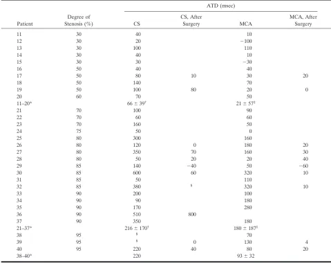
In contrast to the analysis of ATDs, evaluation of absolute arrival times yielded no meaningful results. Tables 1 and 2 summarize the data on control subjects and patients, respectively. Listed are the degree of stenosis determined on DSA or duplex ultrasonogra-phy and ATDs of arterial blood in the CS and the MCA. In two patients with 95% stenosis, no blood signal intensity could be detected in the CS.
Control group
The average ATD between the right and left side in the control group was 16 ms at the level of the CS and 19 ms at the level of the MCA. The greatest differ-ences between the right and left side were 30 and 40 ms, respectively.
Patients
Qualitative analysis of DSLA.—DSLA, in particu-lar, allowed for good qualitative assessment of eralization via the circle of Willis, i.e., whether collat-eral flow occurs mainly through the anterior communicating artery from the contralateral hemi-sphere or rather from the ipsilateral posterior
terri-tory via the posterior communicating artery. Figure 3 shows images from a patient with pronounced collat-eral flow via the circle of Willis. In one patient, ret-rograde flow in the ophthalmic artery and leptomen-ingeal collaterals were clearly depicted in the DSLA image series.
Quantitative Analysis of ATDs.—The degree of ste-nosis was clearly related to the ATDs calculated for the CS and for the MCA (Fig 4). The correlation is statistically significant for both sites of measurement (P ⬍ .01). For further evaluation, the subjects were subdivided into three groups, according to their de-gree of stenosis: 0% (controls), ⬍70% and ⱖ70%. ATDs in the CS and MCA were significantly different between the control group and patients with stenoses
ⱖ70%, as well as between patients with ⱖ70% and those with ⬍70% stenosis. The only factor that sig-nificantly differed between control subjects and pa-tients with stenoses ⬍70% was ATD in the CS. By using ROC analysis, cutoff values were determined to predict the classification of each patient into either of the groups with highest specificity and sensitivity. Be-cause the ATD values at the CS level were of highest significance regarding the degree of stenosis, only these were used in the ROC analysis. Figure 5 depicts the ROC curves. The optimal cutoff value separating controls and stenoses ⬍70% was 35 ms (sensitivity, 80%; specificity, 100%). The cutoff separating steno-ses⬍70% from thoseⱖ70% was 110 ms (sensitivity, 67%; specificity, 90%). Transit time delays of more than 150 ms in the CS are indicative for high-grade stenoses (sensitivity, 56%; specificity, 100%).
Figure 6 shows a scatterplot of the individual ATDs proximal and distal to the circle of Willis of all patients, grouping them according to the visually rated poststenotic collateral flow. The degree of collateralization visible in the circle of Willis was correlated with the ATD CS/ATD MCA ratio, with a higher degree of collateralization associated with reduced distal ATDs. No interrelation could be found between the patients’ clinical picture (asymp-tomatic, TIA, or infarction) and the degree of collateralization.
[image:4.585.60.533.55.214.2]FIG 2. Determination of ATDs.Left, Original signal intensity–time courses measured in the left and right MCAs.Right, Normalized curves (divided by the respective maximum value). By using linear interpolation between the measured points, the intersection at which the signals reached 50% was determined. Difference was referred to as the ATD (arrows).
TABLE 1: Quantitative results in the control group
Subject
ATD (msec)
CS MCA
1 30 10
2 10 20
3 20 20
4 10 20
5 30 ⫺20
6 0 40
7 20 30
8 20 30
9 20 30
10 0 0
All* 16⫾11 19⫾17
[image:4.585.52.283.272.422.2]DSLA Before and After Treatment of Carotid Stenosis
Ten patients with high-grade extracranial carotid stenoses underwent DSLA before and after treatment (carotid endarterectomy in six, stent angioplasty in fur). Figure 7 presents the image series and signal intensity-time courses in the CS a patient before and after endarterectomy. One patient (patient 32) had occlusion after the intervention. All other patients had dramatically reduced delays after treatment, but three had a CS ATD of more than 60 ms. Postinter-ventional ATDs were still significantly elevated com-pared with those of controls.
Discussion
The phenomenon of delayed arterial arrival asso-ciated with vascular stenoses was first described in 1960s, initially in studies by using conventional an-giography (22) or scintigraphy (23, 24), whereas most
later studies used contrast-enhanced dynamic CT (25), echo-enhanced transcranial Doppler sonogra-phy (26), or perfusion MR imaging (3, 27), to deter-mine tissue perfusion in the presence of stenosis. All these studies did, however, require the administration of a contrast medium. This method might alter phys-iologic blood flow conditions and cannot be repeated arbitrarily. DSLA avoids these problems because it does not need contrast agents. Conventional CT an-giography or MRA provide little information about the dynamics of poststenotic blood flow. The tempo-ral resolution achieved with two-dimensional projec-tion techniques is on the order of 0.5–1 second and 2– 6 seconds per volume dataset when 3D acquisition with temporal and spatial interpolation is used (28, 29). Both techniques incorporate a compromise be-tween temporal and spatial resolution, but the image frame rate still does not suffice for precise depiction of the hemodynamic situation in the circle of Willis or in the deeper CS. DSLA requires markedly longer
20 60 70 50
11–20* 66⫾39† 21⫾57‡
21 70 100 90
22 70 60 60
23 70 160 50
24 75 50 0
25 80 300 160
26 80 120 0 180 20
27 80 350 70 160 30
28 80 50 20 20 40
29 85 140 ⫺40 50 ⫺60
30 85 600 60 320 10
31 85 50 110
32 85 380 § 320 10
33 90 200 100
34 90 90 180
35 90 170 280
36 90 510 800
37 90 350 180
21–37* 216⫾170† 180⫾187‡
38 95 § 70
39 95 § 0 130 4
40 95 220 40 80 20
38–40* 220 93⫾32
*Data are the mean or the mean⫾standard deviation.
†
P⬍.05.
‡
P⬍.05.
§
[image:5.585.52.531.71.452.2]acquisition times, but it provides 10 –20-fold higher temporal resolution. As opposed to DSA, the method allows for arbitrary projection directions. DSLA is not limited to being performed as a projection tech-nique but also allows for acquiring 3D datasets. How-ever, the increased acquisition time is no longer ac-ceptable in clinical examinations; this limitation suggests that temporal or spatial resolution must be reduced here as well.
This study showed that the high temporal resolu-tion of the spin labeling method makes the technique especially suitable for monitoring hemodynamic
ef-fects of stenosing cerebrovascular disease. To our knowledge, the DSLA technique presented herein is the first fully noninvasive MR imaging procedure that allows quantification of ATDs in patients with ex-tracranial carotid stenosis. Moreover, DSLA enabled us to show the compensation of such ATDs by ade-quate distal collateral flow and thus provided impor-tant supplementary information on hemodynamic changes secondary to vascular stenoses.
Our results demonstrated that the degree of steno-sis was best correlated with ATD in the CS. Although the proportion of stenoses ⬍70% was small, the re-FIG 3. Patient 30, with an 85% stenosis of the left ICA. Dynamic angiograms of the circle of Willis in foot-to-head projections at 60, 100, 140, 220, 300, and 580 ms after labeling ina-f. Ina, the right ICA (upper arrow) and basilar artery (lower arrow) fill first. Inb, Collateral flow into the left MCA via the left posterior communicating artery and the anterior communicating artery is shown. Ind, Left ICA fills. In f, because of the finite length of the labeled bolus, all vessels but the left ICA and MCA contain unlabeled blood at this late phase.
sults suggested that an ATD exceeding 110 ms was indicative of a stenosis of at least 70%, with a sensi-tivity of 67% and a specificity of 90%, or even higher specificity if control subjects and subjects with low-grade stenoses are merged in one group. All stenoses with ATD of at least 150 ms were of high grade. This additional possibility for grading vascular stenoses by using MR imaging is clinically relevant. On the basis of MRA alone, even contrast-enhanced MRA, the assessment of the degree of high-grade carotid steno-ses is sometimes difficult and may lead to a misclas-sification of patients (30 –32). Combining MRA with DSLA might significantly improve the diagnostic ac-curacy in such patients and furthermore reduce the necessity for conventional invasive angiography. More-over, the technique can be used to assess the effect of treatment in patients after surgery or stent placement for ICA stenosis and to identify recurrent stenosis.
In two patients with 95% stenosis, no blood signal intensity was detected in the siphon. In both, the amount of blood was so small or it arrived so late that
its signal intensity could not be distinguished from background noise; this finding suggested near-occlu-sion of the ICA. Large ATD variations were observed within the groups (e.g., in patient 28 who had a 30% stenosis and ATDs of more than 100 ms in the MCA and CS). This patient had no anatomic abnormalities (e.g., vessel loop) at time-of-flight MRA. However, he underwent duplex ultrasonography more than 2 weeks before DSLA. Further narrowing of the vascu-lar lumen in the meantime to a high-degree stenosis is improbable, unless a nonsymptomatic thromboem-bolic event happened.
In the patients who underwent treatment, we still observed a significant transit time delay after the intervention. This result was consistent with others’ observations of a considerable variability in flow ve-locity and its changes in the cerebral arteries of treated patients (33–35).
In addition to information on the degree of steno-sis, DLSA provides information on hemodynamic changes in the brain. In 33% of patients, ATDs in the MCA are equal to or greater than those in the CS, suggesting an absence of collateralization. However, most patients had a reduced ATD distal to the circle of Willis, most probably due to collateral flow. Whether these patients have a decreased risk of ce-rebral ischemia remains to be determined in further studies. Of interest, patients with stenosesⱖ95% had relatively early MCA filling and thus small ATDs comparable to those of patients with intermediate-grade ICA stenosis. This observation was attributed to a well-developed collateralization and may explain why patients with near-occlusion hardly benefit from intervention (36). Although thromboembolic events secondary to atherosclerotic wall processes play a role in most patients with stroke, a hemodynamic infarct pattern can be detected in up to 50% of patients with high-grade ICA stenosis (37). For this subset of pa-tients, it seems to be important to have methods that allow us to quantify the hemodynamic effect of ICA stenosis. In acute ischemia, this could be of great diagnostic value. DSLA can potentially help in visu-alizing the complex postischemic vascular reaction. FIG 6. Scatterplot of the ATDs calculated for the MCA versus
A significant limitation of our method was the inability to assess the degree of bilateral carotid ste-noses, which occurred in more than 30% of patients. Determination of absolute blood arrival times in these patients is equivalent to measuring flow velocity in the vessels. However, the analysis of these absolute values gave no statistically significant results in our study. This shows that a differential method is more sensitive, as it is less dependent on section position-ing. Nevertheless DSLA depicts collateralization in patients with bilateral disease as well. Having said that, in practice, patients with unilateral carotid artery stenoses can be identified with a high level of cer-tainty by using conventional MRA, which has a high
negative predictive value (1, 38, 39). However, a re-striction is that DSLA cannot be performed after the administration of contrast agent.
stenoses, to monitor patients after intervention, and to provide additional functional information.
References
1. Nederkoorn PJ, van der Graaf Y, Hunink MG, et al. Duplex
ultrasound and magnetic resonance angiography compared with digital subtraction angiography in carotid artery stenosis: a sys-tematic review.Stroke2003;34:1324 –1332
2. Guckel FJ, Brix G, Schmiedek P, et al.Cerebrovascular reserve
ca-pacity in patients with occlusive cerebrovascular disease: assessment with dynamic susceptibility contrast-enhanced MR imaging and the acetazolamide stimulation test.Radiology1996;201:405– 412 3. Reith W, Heiland S, Erb G, Benner T, Forsting M, Sartor K.
Dynamic contrast-enhanced T2*-weighted MRI in patients with cerebrovascular disease.Neuroradiology1997;39:250 –257 4. Rutgers DR, Klijn CJ, Kappelle LJ, van Huffelen AC, van der
Grond J.A longitudinal study of collateral flow patterns in the
circle of Willis and the ophthalmic artery in patients with a symp-tomatic internal carotid artery occlusion.Stroke2000;31:1913–1920 5. Bendszus M, Koltzenburg M, Burger R, Warmuth-Metz M,
Hoff-mann E, Solymosi L.Silent embolism in diagnostic cerebral
an-giography and neurointerventional procedures: a prospective study.Lancet1999;354:1594 –1597
6. Williams DS, Detre JA, Leigh JS, Koretsky AP.Magnetic
reso-nance imaging of perfusion using spin inversion of arterial water.
Proc Natl Acad Sci U S A1992;89:212–216
7. Wong EC, Buxton RB, Frank LR.Quantitative Imaging of
perfu-sion using a single substraction (QUIPSS and QUIPSS II).Magn Reson Med1998;39:702–708
8. Dixon WT, Du LN, Faul DD, Gado M, Rossnick S.Projection
angiograms of blood labeled by adiabatic fast passage.Magn Reson
Med1986;3:454 – 462
9. Nishimura DG, Macovski A, Pauly JM, Conolly SM.MR angiography
by selective inversion recovery.Magn Reson Med1987;4:193–202 10. Roberts DA, Bolinger L, Detre JA, Insko EK, Bergey P, Leigh JS Jr.
Continuous inversion angiography.Magn Reson Med1993;29:631– 636
11. Wang SJ, Nishimura DG, Macovski A. Fast angiography using
selective inversion recovery.Magn Reson Med1990;23:109 –121
12. Wang SJ, Nishimura DG, Macovski a.Multiple-readout selective
inversion recovery angiography.Magn Reson Med1991;17:244 –251
13. Edelman RR, Siewert B, Darby DG, et al.Qualitative mapping of
cerebral blood flow and functional localization with echo-planar MR imaging and signal targeting with alternating radio frequency.
Radiology1994;192:513–520
14. Detre JA, Leigh JS, Williams DS, Koretsky AP.Perfusion imaging.
Magn Reson Med1992;23:37– 45
15. Kim SG.Quantification of relative cerebral blood flow change by
flow-sensitive alternating inversion recovery (FAIR) technique: ap-plication to functional mapping.Magn Reson Med1995;34: 293–301
16. Kwong KK, Chesler DA, Weisskoff RM, et al. MR Perfusion
Studies with T1-Weighted Echo Planar Imaging.Magn Reson Med
1995;34:878 – 887
17. Look D, Locker D.Time saving in measurement of NMR and EPR
relaxation times.Rev Sci Instrum1970;41:250 –251
18. Gunther M, Bock M, Schad LR.Arterial spin labeling in
combi-nation with a look-locker sampling strategy: inflow turbo-sampling EPI-FAIR (ITS-FAIR).Magn Reson Med2001;46:974 –984 19.Clinical alert: benefit of carotid endarterectomy for patients with
high-grade stenosis of the internal carotid artery. Stroke
1991;22:816 – 817
20.MRC European Carotid Surgery Trial: interim results for
symp-25. Davis SM, Tress BM, Hopper JL, Kaye AH, Rossiter SC.Dynamic
CT brain scanning in the haemodynamic evaluation of cerebral arterial occlusive disease.Neuroradiology1987;29:259 –265
26. Schreiber S, Weih M, Hofmann O, Ru¨ckert R, Einha¨upl KM,
Valdueza JM.Delayed transcranial echo-contrast bolus arrival in
carotid artery disease.In: Proceedings of the 4th Meeting of the European Society of Neurosonology and Cerebral Hemodynamics. 1999:48
27. Nighoghossian N, Berthezene Y, Philippon B, Adeleine P, Froment
JC, Trouillas P.Hemodynamic parameter assessment with dynamic
susceptibility contrast magnetic resonance imaging in unilateral symptomatic internal carotid artery occlusion. Stroke 1996;27: 474 – 479
28. Wang Y, Johnston DL, Breen JF, et al.Dynamic MR digital
subtrac-tion angiography using contrast enhancement, fast data acquisisubtrac-tion, and complex subtraction.Magn Reson Med1996;36:551–556
29. Korosec FR, Frayne R, Grist TM, Mistretta CA.Time-resolved
contrast-enhanced 3D MR angiography.Magn Reson Med1996;36: 345–351
30. Johnston DCC, Eastwood JD, Nguyen T, Goldstein LB.
Contrast-enhanced magnetic resonance angiography of carotid arteries: util-ity in routine clinical practice.Stroke2002;33:2834 –2838
31. Patel SG, Collie DA, Wardlaw JM, et al.Outcome, observer
reli-ability, and patient preferences if CTA, MRA, or Doppler ultra-sound were used, individually or together, instead of digital sub-traction angiography before carotid endarterectomy. J Neurol Neurosurg Psychiatry2002;73:21–28
32. Wardlaw JM, Lewis SC, Humphrey P, Young G, Collie D, Warlow CP.How does the degree of carotid stenosis affect the accuracy and interobserver variability of magnetic resonance angiography?
J Neurol Neurosurg Psychiatry2001;71:155–160
33. Araki CT, Babikian VL, Cantelmo NL, Johnson WC.
Cerebrovas-cular hemodynamic changes associated with carotid endarterec-tomy.J Vasc Surg1991;13: 854 – 859
34. Vriens EM, Wieneke GH, Hillen B, Eikelboom BC, Van Huffelen
AC, Visser GH.Flow redistribution in the major cerebral arteries
after carotid endarterectomy: a study with transcranial Doppler scan.J Vasc Surg2001;33:139 –147
35. Blohme L, Pagani M, Parra-Hoyos H, Olofsson P, Takolander R,
Swedenborg J.Changes in middle cerebral artery flow velocity and
pulsatility index after carotid endarterectomy. Eur J Vasc Surg
1991;5:659 – 663
36. Rothwell PM, Eliasziw M, Gutnikov SA, et al.Analysis of pooled
data from the randomised controlled trials of endarterectomy for symptomatic carotid stenosis.Lancet2003;361:107–116
37. Szabo K, Kern R, Gass A, Hirsch J, Hennerici M.Acute stroke
patterns in patients with internal carotid artery disease: a diffu-sion-weighted magnetic resonance imaging study.Stroke2001;32: 1323–1329
38. Wolfle KD, Schnur C, Pfadenhauer K, Bruijnen H, Bohndorf K,
Loeprecht H.MR-angiography and duplex-ultrasonography:
pre-dictive reliability for angiographically determined internal carotid artery stenosis >/ⴝ70 –99%.Zentralbl Chir2002;127:81– 88
39. Tiev KP, Sevestre MA, Reix T, et al.Quantitative assessment of
carotid stenosis: comparison between Doppler ultrasound, spiral computed tomography angiography, magnetic resonance angiogra-phy and digital angiograangiogra-phy.J Mal Vasc2000;25:325–331
40. Amann M, Warmuth C, Gu¨nther M, Zimmer C, Schad LR.