www.impactjournals.com/oncotarget/
Oncotarget, Vol. 7, No. 29
Tumor refractoriness to anti-VEGF therapy
Domenico Ribatti
1,21 Department of Basic Medical Sciences, Neurosciences and Sensory Organs, University of Bari Medical School, Bari, Italy 2 National Cancer Institute “Giovanni Paolo II”, Bari, Italy
Correspondence to: Domenico Ribatti, email: domenico.ribatti@uniba.it Keywords: angiogenesis; anti-angiogenesis; resistance; tumor growth; VEGF
Received: March 03, 2016 Accepted: April 02, 2016 Published: April 11, 2016
ABSTRACT
Vascular endothelial growth factor (VEGF) has been identified as the most potent cytokine involved in tumor angiogenesis and metastasis formation. Clinical results of anti-angiogenic therapies targeting VEGF and its receptors are very modest, resulting in a moderate improvement of overall survival. The clinical outcome is associated with the development of resistance and the increased risk of invasion and metastasis. In this article, I have analyzed the principal mechanisms of resistance to VEGF pathway inhibitors, including normalization of tumor blood vessels, hypoxia, recruitment of inflammatory cells and immature myeloid cells, alternative mechanisms of tumor vessel formation, genomic instability of tumor endothelial cells. In this context, the concept and strategies of anti-angiogenic therapies should be extensively re-considered and re-evaluated. In particular, rational combinations of anti-angiogenic agents based on pharmacokinetic and pharmacodynamics data are needed to overcome resistance and it is extremely important to determine the optimal duration and scheduling of anti-VEGF agents.
INTRODUCTION
The introduction of chemotherapy in 1950-60
resulted in the development of curative therapeutic
interventions for patients with solid and hematologic
tumors. While chemotherapy in the neo-adjuvant setting
has typically resulted in improved survival following
surgical intervention, similar benefits with anti-angiogenic
therapy remain largely untested.
Tumor cells and tumor-associated stroma are
sources of vascular endothelial growth factor (VEGF),
which is responsible of vascular proliferation and altered
permeability of newly formed vessels [1]. Several
strategies to inhibit VEGF-VEGF receptors (VEGFRs)
signaling pathway for the treatment of cancer have been
explored. In addition to monoclonal antibodies, alternative
approaches of inhibiting VEGFRs by using anti-VEGFRs
small receptor tyrosine kinase inhibitors (TKIs) have been
explored (Table 1).
Even if the majority of pre-clinical studies have
shown that the growth of all experimental tumors can be
effectively inhibited by various anti-angiogenic agents, the
clinical benefits of these treatments are relatively modest,
because the drugs merely slow down tumor progression
and prolong survival by only a few more months [2-4]. In
multiple randomized phase III clinical trials, bevacizumab
conferred a survival benefit only when administered in
combination with chemotherapy. Examples of metastatic
cancers where anti-angiogenic therapy failed to make a
significant impact on overall survival include breast,
melanoma, pancreatic and prostate.
Moreover, as it has been demonstrated in animal
models, anti-angiogenic therapy caused marked regression
of normal microvessels in endocrine glands (thyroid,
adrenal glands, pancreatic islets) and in the liver, kidney
and gastrointestinal wall [5, 6], and angiogenesis inhibitors
can decrease the delivery of cytotoxic drugs [7].
It is important to note that when VEGF-targeted
therapies are discontinued, the tumor vasculature ca
become rapidly re-established [8]. These data suggest
that prolonged use of VEGF-targeted therapy is necessary
to achieve maximal therapeutic effect. An observational
study has shown that continuation of bevacizumab
treatment beyond progression was associated with greater
benefit in terms of overall survival [9].
Intrinsic resistance is characterized by inefficacy
of tumor treatment with anti-angiogenic anti-VEGF,
fusion proteins that trap VEGF [10], and anti-VEGFRs
small receptor TKIs [11-13]. TKIs target VEGFR-1,
-2, -3 signaling pathways and other members of the
platelet-derived growth factor (PDGF) receptor and
fibroblast growth factor (FGF) receptor families. In this
context, TKIs inhibitors would be more effective than
antibody-based therapy that solely target the VEGF
pathway. Nevertheless, in acquired resistance, alternative
mechanisms lead to activation of angiogenesis even when
the target of the drug remains inhibited [14, 15].
Trials that have combined monoclonal antibodies
and TKIs have given rise to an increase in the side
effects profile, including hypertension, gastrointestinal
perforation, hemorrhages, proteinuria, anemia, leucopenia,
and thrombocytopenia.
NORMALIZATION OF TUMOR BLOOD
VESSELS
In 2001, Rakesh Jain introduced the concept of
“normalization” of tumor blood vessels by anti-angiogenic
molecules [16]. VEGF inhibition could temporarily restore
or normalize the function of tumor-associated vasculature,
decreasing vascular permeability in conjunction with
restoration of sustained pressure gradients, thereby
enhancing systemic delivery of oxygen or perfusion
of cytotoxic agents to intratumoral sites . Moreover,
abrogation of VEGF signaling increases collagenase
IV activity, leading to restoration of normal basement
membrane , which generally in tumors has an abnormally
thickness .
Moreover, tumor vascular normalization is
accompanied by increased pericyte coverage. Pericyte
deficiency could be partly responsible for vessel
abnormalities in tumor blood vessels [17] and partial
dissociation of pericytes [18, 19] contribute to increased
tumor vascular permeability.
Anti-angiogenic refractory tumors contained blood
vessels with a investment of pericytes expressing alpha
smooth muscle actin (α-SMA) [20]. Pericyte coverage
promote resistance through direct support or paracrine
interactions with endothelial cells and tumor vessels
covered by pericytes are less sensitive to VEGF blockade
[21]. Pericytes can activate compensatory
PDGFR-mediated pro-angiogenic signaling in anti-VEGF therapy
[22]. Bergers et al. showed that combined treatment or
pre-treatment with anti-PDGF-B/PDGFBR-β reducing
pericyte coverage increases the success of anti-VEGF
treatment in the mouse RIP1-TAG2 model [23]. However,
extensive regression of endothelial cells was not observed
in tumors after inhibition of PDGFR-β signaling [24].
After treatment of RIP1-TAG-2 tumors and Lewis lung
carcinomas with VEGF-Trap, surviving pericytes may
become more tightly associated with endothelial cells
or have no apparent association with tumor vessels [25].
Treatment of RIP1-TAG2 tumors with anti-PDGFR-β
antibody for three weeks reduces pericytes, increases
endothelial cell apoptosis but does not seem to reduce
tumor vascular density [26]. Treatment with a novel DNA
oligonucleotide aptamer (AX102) that selectively binds
PDGF-B led to progressive reduction of pericytes in Lewis
lung carcinomas [27].
[image:2.612.58.459.61.194.2]VEGFR-2 blockade can lead to the up-regulation of
angiopoietin-1 (Ang-1) that increases pericyte coverage
of the vessels [28]. Ang-2 is responsible for blood vessel
destabilization in vasculature surrounding tumors. In
glioblastoma patients, the Ang-1/Ang-2 ratio correlates
Table 1: Principal anti-VEGF molecules approved by FDA for anti-angiogenic cancer treatment
Name
Target
Bevacizumab
VEGF-A
Aflibercept
VEGF-A
Axitinib
VEGFRs
Cabozantinib
VEGFRs
Regorafenib
VEGFRs
Sorafenib
VEGFRs
Sunitinib
VEGFRs
Vandetanib
VEGFRs
Pazopanib
VEGFRs
Table 2: Biomarkers to predict response to anti-VEGF inhibitors
Functional imaging
Hypertension
Circulating proteins (baseline plasma VEGF concentration and soluble VEGFR-2)
Circulating endothelial or tumor cells
Single nucleotide polymorphisms (SNPs)
with survival [29] and vascular normalization, whereas
high Ang-2 levels correlate with resistance to anti-VEGF
therapy [30]. Blockade of VEGF signaling with the TKI
cediranib significantly reduced levels of Ang-2 in some
patients, even if the decrease was transient and modest
[30]. Chae et al. expressed Ang-2 in an orthotopic glioma
model and demonstrated that ectopic expression of Ang-2
had no effect on vascular permeability, tumor growth, or
survival, but it resulted in higher vascular density, with
dilated vessels and reduced mural cell coverage [31].
When combined with anti-VEGFR-2 treatment,
Ang-2 destabilized vessels and compromised the survival
benefit of VEGFR-2 inhibition by increasing vascular
permeability, suggesting that VEGFR-2 inhibition
normalized tumor vasculature, whereas ectopic expression
of Ang-2 diminished the beneficial effects of VEGFR-2
blockade by inhibiting vessel normalization.
The inhibitors of VEGF in the therapy of
central nervous system malignancy normalize tumor
vasculature and decrease tumor interstitial pressure,
leading to an improved access of cyto-reductive drugs
and radiotherapy efficacy, due to an increased oxygen
delivery [32]. However, these agents may also restore
the low permeability characteristics of normal brain
microvasculature, counteracting beneficial effects.
HYPOXIA
Hypoxia mediates immune cells recruitment and
these cells concentrate at the tumor periphery, while in
the tumor core hypoxia contributes to cancer cell escape
by providing an aggressive selection for stem-like tumor
cells. Most cancers are hypoxic at the beginning of therapy
[33] and hypoxic areas are refractory to chemotherapy and
radiotherapy and contribute to select tumor populations
able to survive in poorly oxygenated niches and escape
to metastatic sites and pro-angiogenic cancer stem cells
(CSCs) [34].
VEGF blockade aggravates hypoxia, which
up-regulates the production of other angiogenic factors or
increases tumor cell invasiveness [14, 35]. Tumor cells
respond to hypoxia by becoming tolerant and modifying
their metabolic characteristics to resist to low oxygenation
[36]. Increased intratumor hypoxia induces the selection
of more invasive metastatic clones of the cancer cells that
are resistant to anti-angiogenic agents [37], through the
production of pro-migratory proteins, such as stromal
cell derived factor 1 alpha (SDF1-α) and hepatocyte
growth factor- scatter factor (HGF-SF) and pro-invasive
extracellular matrix proteins [38, 39]. Hypoxia generated
by angiogenesis inhibition triggers pathways that make
tumors more aggressive and metastatic and less sensitive
to anti-angiogenic treatment, as demonstrated by Paez et
al. [35], who used blocking VEGFR-2 antibodies to mouse
models of pancreatic neuroendocrine carcinoma and
glioblastoma, and found that cancers showed heightened
invasiveness or metastasis.
RECRUITMENT OF INFLAMMATORY
CELLS AND IMMATURE MYELOID
CELLS
Inflammatory cells act in concert with tumor
cells, stromal cells, and endothelial cells to create
a microenvironment that is critical for the survival,
development, and diffusion of the neoplastic mass. These
synergies represent important mechanisms for tumor
development and metastasis by providing an efficient
vascular supply and an easy escape pathway. The most
aggressive human cancers, such as malignant melanoma,
breast carcinoma, and colorectal adenocarcinoma, are
associated with a dramatic host response composed of
various inflammatory cells, including macrophages, mast
cells and lymphocytes.
Therapy resistance may be mediated by the
recruitment by tumor cells of tumor associated
macrophages and mast cells [40], pro-angiogenic bone
marrow-derived cells including CD11b
+Gr1
+myeloid
cells [41] and Tie2
+monocytes [42], tumor associated
fibroblasts (TAFs) [43] and production of alternative
pro-angiogenic factors [44], including FGF-2 [45],
interleukin-8 (IL-8) [46], IL-17 [47], and ANG-2 [48].
TAFs generated PDGF-C, which is involved
in tumor refractoriness to anti-angiogenic therapy, as
demonstrated by the use of neutralizing antibodies
anti-PDGF-C ameliorate TAF-resistant induced angiogenesis
[49]. Moreover, cancer-associated inflammatory cells
trigger a catabolic pathway that causes severe adipose and
muscular atrophy [50].
A clear involvement of IL-8 has been reported in
anti-VEGF tumor resistance in sunitinib-treated renal
carcinoma [46]. IL-17 mobilizes the granulocyte-colony
stimulating factor (G-CSF)-dependent recruitment of
CD11b
+Gr1
+immature myeloid cells, involved in
anti-VEGF tumor refractoriness [47]. Human late-stage breast
cancers expressed several angiogenic cytokines in contrast
to earlier stage lesions, which preferentially expressed
only VEGF [51].
ALTERNATIVE MECHANISMS OF
TUMOR VESSEL FORMATION
Other modes of tumor vascularization, including
intussusceptive microvascular growth (IMG),
vasculogenic mimicry, vascular co-option, differentiation
of CSCs into endothelial cells, and vasculogenic vessel
growth, might be less sensitive to VEGF blockade.
proliferation is required for IMG: endothelial cells
only increase their volume and IMG occurs through
the splitting of the existing vasculature by transluminal
pillars or transendothelial bridges (Figure 1) [52]. Tumors
might prefer IMG during anti-angiogenic therapy as it is
faster and metabolically more feasible as compared with
sprouting angiogenesis [8]. IMG occurs in several tumors,
including colon and mammary carcinomas, melanoma,
B-cell non Hodgkin’s lymphoma, and glioma [53-57].
Treatment of mammary carcinoma allografts with a TKI
results in transient reduction in tumor growth rate with
decreased tumor vascularization followed by post-therapy
relapse with extensive IMG, and the switch to IMG
improves the perfusion of the tumor mass [58].
Maniotis et al. [59] described for the first time a
new model of formation of vascular channels by human
melanoma cells and called it “vasculogenic mimicry” to
emphasize the
de novo
generation of blood vessels without
the participation of endothelial cells and independent of
classical angiogenic factors, including FGF-2 and VEGF
[59].
In vitro
stimulation with VEGF do not enhance
vasculogenic mimicry [60] and it has been proposed that
vasculogenic mimicry might be dependent by CSCs [61].
In vascular co-option, tumor cells have immediate
access to blood vessels, as it occurs in in site of metastases
or in densely vascularized organs, including brain, lung,
liver, and initiate blood-vessel-dependent tumor growth as
opposed to classical angiogenesis. Tumor cells co-opt and
growth as cuffs around adjacent vessels [62]. The co-opted
vessels initiate an apoptotic cascade mediated by Ang-2
followed by regression of the co-opted vessels. Shortly
after regression, hypoxic tumor cells expressing VEGF
up-regulate the angiogenic response [62]. Treatment
of glioma with a monoclonal antibody anti-VEGFR-2
induces co-option of quiescent cerebral vessels [63] and
treatment of cerebral melanoma metastasis with the TKI
ZD6474 is associated with increase in vessel co-option
[64].
CSCs reside in a vascular niche in close proximity
to blood vessels named as CSC niche [65], and generate
angiogenic factors to stimulate tumor angiogenesis;
tumor vasculature, in turn, supports CSC self-renewal
and maintaining. CSCs produce high levels of VEGF in
both normal and hypoxic conditions [66]. Moreover, CSCs
recruit endothelial precursors for revascularization and
tumor re-growth [67, 68].
[image:4.612.76.549.387.659.2]Ricci-Vitiani et al. demonstrated that
in vitro
culture
of glioblastoma stem-like cells in generated a progeny
with phenotypic and functional features of endothelial
cells [69]. Moreover, orthotopic or subcutaneous injection
of glioblastoma stem-like cells in immunocompromised
mice generated large anaplastic tumor xenografts, showing
a vessel wall formed by human endothelial cells derived
from glioblastoma stem-like cells whereas tumor derived
endothelial cells formed large anaplastic tumors in
secondary recipients [69].
Postnatal vasculogenesis may contribute to tumor
vascular supply throughout endothelial precursor cells
(EPCs), which circulate from bone marrow, migrate
and differentiate in the stromal environment of tumors
[70]. High levels of VEGF produced by tumors result
in the mobilization of bone marrow-derived EPCs in the
peripheral circulation and enhance their recruitment into
the tumor vasculature [70].
GENOMIC INSTABILITY OF
TUMOR ENDOTHELIAL CELLS AND
REVERSIBILITY OF RESISTANCE
Comprehensive genomic analysis of tumors
demonstrates significant genetic intra- and inter-tumor
heterogeneity [71]. St Croix et al. [72], were the first to
show that colorectal cancer endothelial cells overexpress
specific transcripts as a result of qualitative differences
in gene profiling compared with endothelial cells of the
normal colorectal mucosa. Further studied in glioma [73]
and in invasive breast carcinoma [74] demonstrated a
distinct gene expression pattern related to extracellular
matrix and surface proteins characteristic of proliferating
and migrating endothelial cells, and pointed to specific
roles for genes in driving tumor angiogenesis and
progression of tumor cells. Moreover, endothelial
cells isolated from various tumors acquired genotype
alterations, leading to altered anti-angiogenic targets
and resistance [75], and proximity of tumor cells and
endothelial cells within the tumor microenvironment may
be responsible for the genotype alterations [76].
Development of a resistance-like phenotype to
sorafenib by human hepatocellular carcinoma cells
is reversible and can be delayed by metronomic UFT
chemotherapy [77]. The continued administration of
bevacizumab beyond progression still results in a small
significant overall survival [78], suggesting that the
resistance if reversible and raising the possibility of
re-treating with the same of an alternative VEGF-A inhibitor.
PREDICTIVE MARKERS
Predictive markers of angiogenesis or
anti-angiogenesis are needed to demonstrate the activity and
efficacy of anti-angiogenic agents in clinical trials and
for the future monitoring of anti-angiogenic treatments in
clinics. There are currently no validated biomarkers for
selecting patients that benefit from the treatment with
anti-angiogenic agents from those patients that will not.
VEGF and VEGF isoforms expression levels
serve as a predictive marker for selecting cancer patients
who are likely to benefit from anti-VEGF therapy [79].
[image:5.612.56.560.445.687.2]Elevated levels of sVEGFR-1 prior to treatment were
associated with a poor outcome from bevacizumab in
rectal carcinoma, hepatocellular carcinoma, and metastatic
colorectal carcinoma patients [80, 81]. Increased
VEGFR-1 levels may induce increased pro-angiogenic
signaling by placental growth factor (PlGF) when VEGF
is blocked [79]. Circulating levels of the chemokine
SDF1α rise in patients who evade various anti-VEGF
therapies including rectal carcinoma with bevacizumab,
glioblastoma multiforme with cediraninb, hepatocellular
carcinoma with sunitinib, and soft tissue sarcoma with
sorafenib [82]. However, measurement of circulating
markers is difficult to standardize across different centers
due to technical issues associated with sample handling
[83].
Medical imaging techniques play an important role
in the evaluation of anti-angiogenic treatment efficacy.
Dynamic contrast-enhanced perfusion magnetic resonance
imaging (MRI), perfusion computed tomography (CT)
give only an indirect estimation of angiogenesis. New
molecular imaging techniques can give an overall
estimation of angiogenesis and anti-angiogenic therapy
effects. These include nuclear imaging techniques
such as positron emission tomography (PET), that
uses paramagnetic nanoparticles to track angiogenesis
by targeting avb3 integrin and sonography with novel
contrast agents such as gas-filled microbubbles directed
against specific target endothelial cell receptors and optical
techniques.
INCREASE OF METASTATIC
POTENTIAL
VEGF pathway inhibition may change the natural
history of tumor progression after anti-angiogenic therapy
and include potential metastasis promoting effects.
Short-term treatment with sunitinib prior to intravenous
inoculation of breast and melanoma cells could accelerate
metastasis and short survival, despite cessation of
treatment [84]. Moreover, sunitinib increases metastasis
in orthotopic mouse models of breast and colon cancer,
whereas it does not promote metastasis in lung cancer
[85]. Increased invasiveness might result from enhanced
expression of various angiogenic cytokines induced by
the treatment, such as VEGF and PlGF, or recruitment
of EPCs that promote the formation of a pre-metastatic
niche [86]. Hypoxia-driven effects may be also involved,
because hypoxia generated by angiogenesis inhibition
triggers pathways that make tumors more aggressive and
metastatic and less sensitive to anti-angiogenic treatment
[35, 84]. Finally, VEGF-targeted therapy can allow an
epithelial-mesenchymal transition, which could in turn
promote increased invasion and metastasis [87].
CONCLUSIONS AND PERSPECTIVES
Mechanisms of resistance can be divided into
non-oncogenic and non-oncogenic, and these latter are associated
with highly aggressive cancer phenotype [88].
Anti-angiogenic treatment induces a reactive resistance which
is mediated by the HIF/VEGF pathway, allowing both
endothelial and cancer cells to resist to therapy [89].
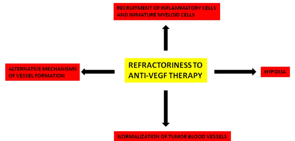
Resistance to VEGF pathway inhibitors involves different
mechanisms that are summarized in Figure 2. All of these
mechanisms deserve further investigation in both animal
models and in humans to clarify their significance and
importance.
The successful development of anti-VEGF targeted
therapy will require a greater understanding of how tumors
become vascularized and how they evade the effects of
anti-angiogenic therapy. VEGF blockade aggravates
tumor hypoxia, which up-regulates the production of
other angiogenic factors in the tumor microenvironment.
In this context, targeting VEGF and other pathways
implicated in angiogenesis should result in more effective
tumor growth inhibition. Moreover, rational combinations
of anti-angiogenic agents based on pharmacokinetic
and pharmacodynamics data are needed to overcome
resistance and it is extremely important to determine the
optimal duration and scheduling of anti-VEGF agents. It
has been underlined the importance of the time interval
of the normalization effects of anti-angiogenesis, the
so-called “window of normalization”[90]. Recently,
Paez-Ribes et al. [91] have demonstrated that metastatic effects
of preclinical anti-angiogenic therapy with an antibody
targeting mouse VEGFR-2 are prevented by concurrent
chemotherapy.
The identification of specific predictive biomarkers
(Table 2) remains an important end-point even if
biomarkers that are predictive of anti-VEGF therapy may
be specific to different tissues and tumor subtypes.
ACKNOWLEDGMENTS
This work was supported by European Union
Seventh Framework Programme (FP7/2007-2013) under
grant agreement n.278570 to DR.
CONFLICTS OF INTEREST
There is no conflict of interest.
REFERENCES
1. Senger DR, Brown LF, Claffey KP and Dvorak HF. Vascular permeability factor, tumor angiogenesis and stroma generation. Invasion Metastasis. 1994; 14:385-394. 2. Hurwitz H, Fehrenbacher L, Novotny W, Cartwright T,
3. Miller K, Wang M, Gralow J, Dickler M, Cobleigh M, Perez EA, Shenkier T, Cella D and Davidson NE. Paclitaxel plus bevacizumab versus paclitaxel alone for metastatic breast cancer. N Engl J Med. 2007; 357:2666-2676.
4. Sandler A, Gray R, Perry MC, Brahmer J, Schiller JH, Dowlati A, Lilenbaum R and Johnson DH. Paclitaxel-carboplatin alone or with bevacizumab for non-small-cell lung cancer. N Engl J Med. 2006; 355:2542-2550.
5. Kamba T, Tam BY, Hashizume H, Haskell A, Sennino B, Mancuso MR, Norberg SM, O’Brien SM, Davis RB, Gowen LC, Anderson KD, Thurston G, Joho S, Springer ML, Kuo CJ and McDonald DM. VEGF-dependent plasticity of fenestrated capillaries in the normal adult microvasculature. Am J Physiol Heart Circ Physiol. 2006; 290:H560-576.
6. Yang Y, Zhang Y, Cao Z, Ji H, Yang X, Iwamoto H, Wahlberg E, Lanne T, Sun B and Cao Y. Anti-VEGF- and anti-VEGF receptor-induced vascular alteration in mouse healthy tissues. Proc Natl Acad Sci U S A. 2013; 110:12018-12023.
7. Van der Veldt AA, Lubberink M, Bahce I, Walraven M, de Boer MP, Greuter HN, Hendrikse NH, Eriksson J, Windhorst AD, Postmus PE, Verheul HM, Serne EH, Lammertsma AA and Smit EF. Rapid decrease in delivery of chemotherapy to tumors after anti-VEGF therapy: implications for scheduling of anti-angiogenic drugs. Cancer Cell. 2012; 21:82-91.
8. Mancuso MR, Davis R, Norberg SM, O’Brien S, Sennino B, Nakahara T, Yao VJ, Inai T, Brooks P, Freimark B, Shalinsky DR, Hu-Lowe DD and McDonald DM. Rapid vascular regrowth in tumors after reversal of VEGF inhibition. J Clin Invest. 2006; 116:2610-2621.
9. Grothey A, Sugrue MM, Purdie DM, Dong W, Sargent D, Hedrick E and Kozloff M. Bevacizumab beyond first progression is associated with prolonged overall survival in metastatic colorectal cancer: results from a large observational cohort study (BRiTE). J Clin Oncol. 2008; 26:5326-5334.
10. Lockhart AC, Rothenberg ML, Dupont J, Cooper W, Chevalier P, Sternas L, Buzenet G, Koehler E, Sosman JA, Schwartz LH, Gultekin DH, Koutcher JA, Donnelly EF, et al. Phase I study of intravenous vascular endothelial growth factor trap, aflibercept, in patients with advanced solid tumors. J Clin Oncol. 2010; 28:207-214.
11. Batchelor TT, Sorensen AG, di Tomaso E, Zhang WT, Duda DG, Cohen KS, Kozak KR, Cahill DP, Chen PJ, Zhu M, Ancukiewicz M, Mrugala MM, Plotkin S, et al. AZD2171, a pan-VEGF receptor tyrosine kinase inhibitor, normalizes tumor vasculature and alleviates edema in glioblastoma patients. Cancer Cell. 2007; 11:83-95. 12. Gotink KJ and Verheul HM. Anti-angiogenic tyrosine
kinase inhibitors: what is their mechanism of action? Angiogenesis. 2010; 13:1-14.
13. Kindler HL. Pancreatic cancer: an update. Curr Oncol Rep. 2007; 9:170-176.
14. Bergers G and Hanahan D. Modes of resistance to anti-angiogenic therapy. Nat Rev Cancer. 2008; 8:592-603. 15. Kerbel RS. Molecular and physiologic mechanisms of drug
resistance in cancer: an overview. Cancer Metastasis Rev. 2001; 20:1-2.
16. Jain RK. Normalizing tumor vasculature with anti-angiogenic therapy: a new paradigm for combination therapy. Nat Med. 2001; 7:987-989.
17. Gerhardt H and Semb H. Pericytes: gatekeepers in tumour cell metastasis? J Mol Med (Berl). 2008; 86:135-144. 18. Hashizume H, Baluk P, Morikawa S, McLean JW, Thurston
G, Roberge S, Jain RK and McDonald DM. Openings between defective endothelial cells explain tumor vessel leakiness. Am J Pathol. 2000; 156:1363-1380.
19. Hobbs SK, Monsky WL, Yuan F, Roberts WG, Griffith L, Torchilin VP and Jain RK. Regulation of transport pathways in tumor vessels: role of tumor type and microenvironment. Proc Natl Acad Sci U S A. 1998; 95:4607-4612.
20. Franco M, Paez-Ribes M, Cortez E, Casanovas O and Pietras K. Use of a mouse model of pancreatic neuroendocrine tumors to find pericyte biomarkers of resistance to anti-angiogenic therapy. Horm Metab Res. 2011; 43:884-889.
21. Ribatti D, Nico B and Crivellato E. The role of pericytes in angiogenesis. Int J Dev Biol. 2011; 55:261-268.
22. Song N, Huang Y, Shi H, Yuan S, Ding Y, Song X, Fu Y and Luo Y. Overexpression of platelet-derived growth factor-BB increases tumor pericyte content via stromal-derived factor-1alpha/CXCR4 axis. Cancer Res. 2009; 69:6057-6064.
23. Bergers G, Song S, Meyer-Morse N, Bergsland E and Hanahan D. Benefits of targeting both pericytes and endothelial cells in the tumor vasculature with kinase inhibitors. J Clin Invest. 2003; 111:1287-1295.
24. Abramsson A, Lindblom P and Betsholtz C. Endothelial and nonendothelial sources of PDGF-B regulate pericyte recruitment and influence vascular pattern formation in tumors. J Clin Invest. 2003; 112:1142-1151.
25. Inai T, Mancuso M, Hashizume H, Baffert F, Haskell A, Baluk P, Hu-Lowe DD, Shalinsky DR, Thurston G, Yancopoulos GD and McDonald DM. Inhibition of vascular endothelial growth factor (VEGF) signaling in cancer causes loss of endothelial fenestrations, regression of tumor vessels, and appearance of basement membrane ghosts. Am J Pathol. 2004; 165:35-52.
26. Song S, Ewald AJ, Stallcup W, Werb Z and Bergers G. PDGFRbeta+ perivascular progenitor cells in tumours regulate pericyte differentiation and vascular survival. Nat Cell Biol. 2005; 7:870-879.
28. Winkler F, Kozin SV, Tong RT, Chae SS, Booth MF, Garkavtsev I, Xu L, Hicklin DJ, Fukumura D, di Tomaso E, Munn LL and Jain RK. Kinetics of vascular normalization by VEGFR2 blockade governs brain tumor response to radiation: role of oxygenation, angiopoietin-1, and matrix metalloproteinases. Cancer Cell. 2004; 6:553-563. 29. Sie M, Wagemakers M, Molema G, Mooij JJ, de Bont ES
and den Dunnen WF. The angiopoietin 1/angiopoietin 2 balance as a prognostic marker in primary glioblastoma multiforme. J Neurosurg. 2009; 110:147-155.
30. Batchelor TT, Duda DG, di Tomaso E, Ancukiewicz M, Plotkin SR, Gerstner E, Eichler AF, Drappatz J, Hochberg FH, Benner T, Louis DN, Cohen KS, Chea H, et al. Phase II study of cediranib, an oral pan-vascular endothelial growth factor receptor tyrosine kinase inhibitor, in patients with recurrent glioblastoma. J Clin Oncol. 2010; 28:2817-2823. 31. Chae SS, Kamoun WS, Farrar CT, Kirkpatrick ND,
Niemeyer E, de Graaf AM, Sorensen AG, Munn LL, Jain RK and Fukumura D. Angiopoietin-2 interferes with anti-VEGFR2-induced vessel normalization and survival benefit in mice bearing gliomas. Clin Cancer Res. 2010; 16:3618-3627.
32. McGee MC, Hamner JB, Williams RF, Rosati SF, Sims TL, Ng CY, Gaber MW, Calabrese C, Wu J, Nathwani AC, Duntsch C, Merchant TE and Davidoff AM. Improved intratumoral oxygenation through vascular normalization increases glioma sensitivity to ionizing radiation. Int J Radiat Oncol Biol Phys. 2010; 76:1537-1545.
33. Blagosklonny MV. Antiangiogenic therapy and tumor progression. Cancer Cell. 2004; 5: 13-17.
34. Blagosklonny MV. Hypoxia-inducible factor: Achille’s heel of antiangiogenic cancer therapy (review). Int. J. Oncol. 2001; 19: 2567-262.
35. Paez-Ribes M, Allen E, Hudock J, Takeda T, Okuyama H, Vinals F, Inoue M, Bergers G, Hanahan D and Casanovas O. Antiangiogenic therapy elicits malignant progression of tumors to increased local invasion and distant metastasis. Cancer Cell. 2009; 15:220-231.
36. Rapisarda A and Melillo G. Role of the hypoxic tumor microenvironment in the resistance to anti-angiogenic therapies. Drug Resist Updat. 2009; 12:74-80.
37. Semenza GL. Hypoxia-inducible factors: mediators of cancer progression and targets for cancer therapy. Trends Pharmacol Sci. 2012; 33:207-214.
38. Finger EC and Giaccia AJ. Hypoxia, inflammation, and the tumor microenvironment in metastatic disease. Cancer Metastasis Rev. 2010; 29:285-293.
39. Semenza GL. Oxygen sensing, hypoxia-inducible factors, and disease pathophysiology. Annu Rev Pathol. 2014; 9:47-71.
40. Ribatti D. Mast cells and macrophages exert beneficial and detrimental effects on tumor progression and angiogenesis. Immunol Lett. 2013; 152:83-88.
41. Shojaei F, Wu X, Malik AK, Zhong C, Baldwin ME, Schanz
S, Fuh G, Gerber HP and Ferrara N. Tumor refractoriness to anti-VEGF treatment is mediated by CD11b+Gr1+ myeloid cells. Nat Biotechnol. 2007; 25:911-920.
42. De Palma M, Venneri MA, Galli R, Sergi Sergi L, Politi LS, Sampaolesi M and Naldini L. Tie2 identifies a hematopoietic lineage of proangiogenic monocytes required for tumor vessel formation and a mesenchymal population of pericyte progenitors. Cancer Cell. 2005; 8:211-226. 43. Raffaghello L, Vacca A, Pistoia V and Ribatti D. Cancer
associated fibroblasts in hematological malignancies. Oncotarget. 2015; 6:2589-2603. doi: 10.18632/ oncotarget.2661.
44. Azam F, Mehta S and Harris AL. Mechanisms of resistance to antiangiogenesis therapy. Eur J Cancer. 2010; 46:1323-1332.
45. Casanovas O, Hicklin DJ, Bergers G and Hanahan D. Drug resistance by evasion of antiangiogenic targeting of VEGF signaling in late-stage pancreatic islet tumors. Cancer Cell. 2005; 8:299-309.
46. Huang D, Ding Y, Zhou M, Rini BI, Petillo D, Qian CN, Kahnoski R, Futreal PA, Furge KA and Teh BT. Interleukin-8 mediates resistance to antiangiogenic agent sunitinib in renal cell carcinoma. Cancer Res. 2010; 70:1063-1071.
47. Chung AS, Wu X, Zhuang G, Ngu H, Kasman I, Zhang J, Vernes JM, Jiang Z, Meng YG, Peale FV, Ouyang W and Ferrara N. An interleukin-17-mediated paracrine network promotes tumor resistance to anti-angiogenic therapy. Nat Med. 2013; 19:1114-1123.
48. Rigamonti N, Kadioglu E, Keklikoglou I, Wyser Rmili C, Leow CC and De Palma M. Role of angiopoietin-2 in adaptive tumor resistance to VEGF signaling blockade. Cell Rep. 2014; 8:696-706.
49. Crawford Y, Kasman I, Yu L, Zhong C, Wu X, Modrusan Z, Kaminker J and Ferrara N. PDGF-C mediates the angiogenic and tumorigenic properties of fibroblasts associated with tumors refractory to anti-VEGF treatment. Cancer Cell. 2009; 15:21-34.
50. Argiles JM, Busquets S and Lopez-Soriano FJ. Anti-inflammatory therapies in cancer cachexia. Eur J Pharmacol. 2011; 668 Suppl 1:S81-86.
51. Relf M, LeJeune S, Scott PA, Fox S, Smith K, Leek R, Moghaddam A, Whitehouse R, Bicknell R and Harris AL. Expression of the angiogenic factors vascular endothelial cell growth factor, acidic and basic fibroblast growth factor, transforming growth factor beta-1, platelet-derived endothelial cell growth factor, placenta growth factor, and pleiotrophin in human primary breast cancer and its relation to angiogenesis. Cancer Res. 1997; 57:963-969.
52. Ribatti D, Djonov V. Intussusceptive microvascular growth in tumors. Cancer Lett 2012; 316: 126-131.
54. Djonov V, Hogger K, Sedlacek R, Laissue J and Draeger A. MMP-19: cellular localization of a novel metalloproteinase within normal breast tissue and mammary gland tumours. J Pathol. 2001; 195:147-155.
55. Nico B, Crivellato E, Guidolin D, Annese T, Longo V, Finato N, Vacca A and Ribatti D. Intussusceptive microvascular growth in human glioma. Clin Exp Med. 2010; 10:93-98.
56. Patan S, Munn LL and Jain RK. Intussusceptive microvascular growth in a human colon adenocarcinoma xenograft: a novel mechanism of tumor angiogenesis. Microvasc Res. 1996; 51:260-272.
57. Ribatti D, Nico B, Floris C, Mangieri D, Piras F, Ennas MG, Vacca A and Sirigu P. Microvascular density, vascular endothelial growth factor immunoreactivity in tumor cells, vessel diameter and intussusceptive microvascular growth in primary melanoma. Oncol Rep. 2005; 14:81-84. 58. Hlushchuk R, Riesterer O, Baum O, Wood J, Gruber G,
Pruschy M and Djonov V. Tumor recovery by angiogenic switch from sprouting to intussusceptive angiogenesis after treatment with PTK787/ZK222584 or ionizing radiation. Am J Pathol. 2008; 173:1173-1185.
59. Maniotis AJ, Folberg R, Hess A, Seftor EA, Gardner LM, Pe’er J, Trent JM, Meltzer PS and Hendrix MJ. Vascular channel formation by human melanoma cells in vivo and
in
vitro
: vasculogenic mimicry. Am J Pathol. 1999; 155:739-752.60. van der Schaft DW, Hillen F, Pauwels P, Kirschmann DA, Castermans K, Egbrink MG, Tran MG,Sciot R, Hauben E, Hogendoorn PC, Delattre O, Maxwell PH, Hendrix MJ, Griffioen AW. Tumor cell plasticity in Ewing sarcoma, an alternative circulatory system stimulated by hypoxia. CancerRes. 2005; 11520-11528.
61. El Hallani S, Boisselier B, Peglion F, Rousseau A, Colin C, Idbaih A, Marie Y, Mokhtari K, Thomas JL, Eichmann A, Delattre JY, Maniotis AJ, Sanson M. A new alternative mechanism in glioblastoma vascularization: tubular vasculogenic mimicry. Brain. 2010; 133: 973-982.
62. Holash J, Maisonpierre PC, Compton D, Boland P, Alexander CR, Zagzag D, Yancopoulos GD and Wiegand SJ. Vessel cooption, regression, and growth in tumors mediated by angiopoietins and VEGF. Science. 1999; 284:1994-1998.
63. Kunkel P, Ulbricht U, Bohlen P, Brockmann MA, Fillbrandt R, Stavrou D, Westphal M and Lamszus K. Inhibition of glioma angiogenesis and growth in vivo by systemic treatment with a monoclonal antibody against vascular endothelial growth factor receptor-2. Cancer Res. 2001; 61:6624-6628.
64. Leenders WP, Kusters B, Verrijp K, Maass C, Wesseling P, Heerschap A, Ruiter D, Ryan A and de Waal R. Antiangiogenic therapy of cerebral melanoma metastases results in sustained tumor progression via vessel co-option. Clin Cancer Res. 2004; 10:6222-6230.
65. Calabrese C, Poppleton H, Kocak M, Hogg TL, Fuller C, Hamner B, Oh EY, Gaber MW,Finklestein D, Allen M, Frank A, Bayazitov IT, Zakharenko SS, Gajjar A, Davidoff A,Gilbertson RJ. A perivascular niche for brain tumor stem cells. Cancer Cell 2007; 11: 9-82.
66. Alvero AB, Fu HH, Holmberg J, Visintin I, Mor L, Marquina CC, Oidtman J, Silasi DA and Mor G. Stem-like ovarian cancer cells can serve as tumor vascular progenitors. Stem Cells. 2009; 27:2405-2413.
67. Folkins C, Shaked Y, Man S, Tang T, Lee CR, Zhu Z, Hoffman RM and Kerbel RS. Glioma tumor stem-like cells promote tumor angiogenesis and vasculogenesis via vascular endothelial growth factor and stromal-derived factor 1. Cancer Res. 2009; 69:7243-7251.
68. Ribatti D. Cancer stem cells and tumor angiogenesis. Cancer Lett. 2012; 321:13-17.
69. Ricci-Vitiani L, Pollini R, Biffoni M, Todato M, Invernici G, Cenci T, Maira G,Parati EA,Stassi G, Larocca LM, De Maria R, Tumour vascularization viae ndothelial differentiation of glioblastoma stem-like cells. Nature. 2010; 468: 824-828.
70. Asahara T, Takahashi T, Masuda H, Kalka C, Chen D, Iwaguro H, Inai Y, Silver M, Isner JM,. VEGF contributes to postnatal neovascularization by mobilizing bone marrow-derived endothelial progenitor cells. EMBO J. 1999; 18: 3964-3972.
71. Yap TA, Gerlinger M, Futreal PA, Pusztai L and Swanton C. Intratumor heterogeneity: seeing the wood for the trees. Sci Transl Med. 2012; 4:127ps110.
72. St Croix B, Rago C, Velculescu V, Traverso G, Romans KE, Montgomery E, Lal A, Riggins GJ, Lengauer C, Vogelstein B and Kinzler KW. Genes expressed in human tumor endothelium. Science. 2000; 289:1197-1202. 73. Madden SL, Cook BP, Nacht M, Weber WD, Callahan MR,
Jiang Y, Dufault MR, Zhang X, Zhang W, Walter-Yohrling J, Rouleau C, Akmaev VR, Wang CJ, et al. Vascular gene expression in nonneoplastic and malignant brain. Am J Pathol. 2004; 165:601-608.
74. Parker BS, Argani P, Cook BP, Liangfeng H, Chartrand SD, Zhang M, Saha S, Bardelli A, Jiang Y, St Martin TB, Nacht M, Teicher BA, Klinger KW, Sukumar S and Madden SL. Alterations in vascular gene expression in invasive breast carcinoma. Cancer Res. 2004; 64:7857-7866.
75. Hida K, Hida Y, Amin DN, Flint AF, Panigrahy D, Morton CC and Klagsbrun M. Tumor-associated endothelial cells with cytogenetic abnormalities. Cancer Res. 2004; 64:8249-8255.
76. Hida K and Klagsbrun M. A new perspective on tumor endothelial cells: unexpected chromosome and centrosome abnormalities. Cancer Res. 2005; 65:2507-2510.
delayed by metronomic UFT chemotherapy. Neoplasia. 2010; 12:928-940.
78. Bennouna J, Sastre J, Arnold D, Osterlund P, Greil R, Van Cutsem E, von Moos R, Vieitez JM, Bouche O, Borg C, Steffens CC, Alonso-Orduna V, Schlichting C, et al. Continuation of bevacizumab after first progression in metastatic colorectal cancer (ML18147): a randomised phase 3 trial. Lancet Oncol. 2013; 14:29-37.
79. Lambrechts D, Lenz HJ, de Haas S, Carmeliet P and Scherer SJ. Markers of response for the antiangiogenic agent bevacizumab. J Clin Oncol. 2013; 31:1219-1230. 80. Willett CG, Duda DG, di Tomaso E, Boucher Y,
Ancukiewicz M, Sahani DV, Lahdenranta J, Chung DC, Fischman AJ, Lauwers GY, Shellito P, Czito BG, Wong TZ, et al. Efficacy, safety, and biomarkers of neoadjuvant bevacizumab, radiation therapy, and fluorouracil in rectal cancer: a multidisciplinary phase II study. J Clin Oncol. 2009; 27:3020-3026.
81. Zhu AX, Ancukiewicz M, Supko JG, Sahani DV, Blaszkowsky LS, Meyerhardt JA, Abrams TA, McCleary NJ, Bhargava P, Muzikansky A, Sheehan S, Regan E, Vasudev E, et al. Efficacy, safety, pharmacokinetics, and biomarkers of cediranib monotherapy in advanced hepatocellular carcinoma: a phase II study. Clin Cancer Res. 2013; 19:1557-1566.
82. Duda DG, Kozin SV, Kirkpatrick ND, Xu L, Fukumura D and Jain RK. CXCL12 (SDF1alpha)-CXCR4/CXCR7 pathway inhibition: an emerging sensitizer for anticancer therapies? Clin Cancer Res. 2011; 17:2074-2080.
83. Maru D, Venook AP and Ellis LM. Predictive biomarkers for bevacizumab: are we there yet? Clin Cancer Res. 2013; 19:2824-2827.
84. Ebos JM, Lee CR, Cruz-Munoz W, Bjarnason GA, Christensen JG and Kerbel RS. Accelerated metastasis after short-term treatment with a potent inhibitor of tumor angiogenesis. Cancer Cell. 2009; 15:232-239.
85. Shojaei F, Simmons BH, Lee JH, Lappin PB and Christensen JG. HGF/c-Met pathway is one of the mediators of sunitinib-induced tumor cell type-dependent metastasis. Cancer Lett. 2012; 320:48-55.
86. Ebos JM, Lee CR, Christensen JG, Mutsaers AJ, Kerbel RS. Multiple circulating proangiogenic factors induced by sunitinib malate are tumor-independent and correlate with antitumor efficacy. Proc Natl Acad Sci USA 2007;104:17069-74.
87. Lu KV, Chang JP, Parachoniak CA, Pandika MM, Aghi MK, Meyronet D, Isachenko N, Fouse SD, Phillips JJ, Cheresh DA, Park M and Bergers G. VEGF inhibits tumor cell invasion and mesenchymal transition through a MET/ VEGFR2 complex. Cancer Cell. 2012; 22:21-35.
88. Blagosklonny MV. Why therapeutic response may not prolong the life of a cancer patient:selection for oncogenic resistance. Cell Cycle 2005; 4: 1693-1698.
89. Blagosklonny MV. How Avastin potentiates
chemotherapeutic drugs: action and reaction in antiangiogenic therapy. Cencer Biol Ther. 2005; 4: 1307-310.
90. Weissleder R. Scaling down imaging: molecular mapping of cancer in mice. Nat Rev Cancer 2002; 2: 11-18.