© Indian Journal of Medical Research and Pharmaceutical Sciences http://www.ijmprs.com/
[11]
PREVALENCE OF
CRYPTOSPORIDIUM PARVUM
IN DUNG
COLLECTED FROM CATTLE IN BAMENDA, NORTHWEST REGION OF
CAMEROON
PN Chia ⃰, CN Ukaga, KA yongabi, BEB Nwoke, PM Tih
*Department of Animal and Environmental Biology, Imo State University Owerri, Nigeria. *Department of Biochemistry Catholic University of Cameroon (CATUC) Bamenda - Cameroon. Department of Animal and Environmental Biology, Imo State University Nigeria.
Phyto-Biotechnology Research Foundation Institute,/Catholic University of Cameroon CATUC), Bamenda, Cameroon.
Department of Animal and Environmental Biology, Imo State University Nigeria. Cameroon Baptist Convention Health Board, Bamenda Cameroon.
Abstract
Keywords:
Diarrhoeic; non dirrhoiec; Zoonotic, Modified Kinyoun’s acid-fast stain; Oocysts; Zoonotic.
The dynamic of Cryptosporidia parvum and its zoonotic potentials have not been established in the Northwest Region of Cameroon, which is an important watershed in Cameroon with a lot of cattle grazing at free range.
The aim of this study was to identify Cryptosporidia oocysts and other intestinal parasites in cattle from the 6 out of the 7 divisions that make up the Northwest Region of Cameroon. Single stool samples were randomly collected from 60 cattle (30 diarrhoeic and 30 non-diarrhoeic) immediately the cattle passed dung. Dungs were examined for intestinal parasites using standard techniques (Formalin-ether sedimentation method and Modified Kinyoun’s acid-fast stain).
Out of the 60 dung samples examined, 34 were found to be positive with one or more parasites, giving an overall prevalence of 56.67%. Fasciola species recorded the highest prevalence 10(16.67%), followed by Cryptosporidium parvum 07(11.7 Fasciola spp has the highest prevalence 7(23.33%), followed by Cryptosporidium parvum 5(16.67%), in diarrhoeic dung. For non-diarrhoeic dung Fasciola spp 2(6.67% and Cryptosporidium parvum is 2(6.67) that were the same.Cryptosporidium parvum is common in the North West Region of Cameroon and can easily contaminate the environment leading to human infections.
Introduction
© Indian Journal of Medical Research and Pharmaceutical Sciences http://www.ijmprs.com/
[12]
Cryptosporidium parvum is major a species having zoonotic potential. However, other species including Cryptosporidium bovis, Cryptosporidium andersoni and Cryptosporidium ryanae have been isolated from infected cattle [10, 11].
Cryptosporidiosis is an infection caused by an apicomplexan protozoan known as Cryptosporidia. Cryptosporidia spp common parasites of vertebrates have recently attracted increasing interest due to several serious waterborne outbreaks, and the life-threatening nature of infection in immunocompromised patients, children, the elderly, and patients on chemotherapy, pregnant women; and also the realization of economic losses caused by these pathogens in livestock. It is a common enteric pathogen in humans and domestic animals worldwide with a very low infective dose of one to ten ooysts(Pereira[12]. The sporulated ooysts are immediately infectious when excreted in faeces as there is no intermediate host.
Cattle are reared throughout Cameroon but the major production areas are in the West and North West Regions and from the Adamawa Province [13]. The cattle are transported on foot to the cattle market and the dung they pass along the road is likely to contaminate the environment and the oocysts possibly end up in streams after torrential rains. The aim of this study was to identify Cryptosporidiosis parvum and other intestinal parasites in cattle being transported to the main cattle market in Bamenda, the Northwest Regional headquarter
Materials and methods
Study area
Bamenda, the North West Regional capital: The Western High Plateau or Western Highlands, or Bamenda Grassfields is a region of Cameroon characterized by high relief, cool temperatures, heavy torrential rainfall, and savanna vegetation.
The area experiences two major seasons: A long, wet season of nine months, and a short, dry season of three months. Average rainfall per year ranges from 1,000 mm to 2,000 mm.Sudan savanna forms the dominant vegetation. This consists of grassfields—leading to the name Bamenda grassfields around the city of Bamenda—and short shrubs and trees that shed their foliage during the dry season as a defence against brush fires and dry weather.
With this climate, topography and vegetation, the North West Region is an excellent zone for animal husbandry. [13]There exist the extensive, semi-intensive and intensive types of animal husbandry which involve large ruminants (cattle), small ruminants (goats and sheep), pigs, poultry and non-conventional livestock. A lot of the extensive cattle production by free ranging is practiced in Donga Mantung, Menchum, Bui, Boyo, Momo and Mezam Divisions [14]. The Ndop plain in Ngoketunjia Division serves as an important transhumance zone for the cattle population from neighbouring Divisions and the West Region.
Farming is done along the streams or swamps rice in the plains and gardens, irrigated with the water from the streams [15].
Bamenda is situated between longitude 100.08̍ to 100.12̍ E and latitude 50.55̍ to 6000̍ N. The city covers a surface area
© Indian Journal of Medical Research and Pharmaceutical Sciences http://www.ijmprs.com/
[image:3.612.79.535.117.407.2][13] Figure. 1. Cameroon Map showing the study area, Bamenda. Source: North West Regional master plan for sustainable
development 2001[19]
River Mezam is the main river which drains Bamenda. The hydrography presents a characteristic dendritic drainage pattern. This study will provide adequate information that the government should use to protect and advice the inhabitants on water use and prevention of waterborne infections. Overgrazing, the seasonality of streams and scarcity of potable water in the dry season are just part of the chain of problems originating from the degradation of the watershed due to population pressure. Watersheds are generally considered as points of development especially in countries which rely on water dependent activities [16].
Method of sample (Dung) collection:
A month before sample collection was to commence a visit was paid to the abattoir to sensitize the director and the cattle farmers about the importance of the study. The dung was collected from cattle being transported from the divisions of the Northwest Region to the main cattle market, near the abattoir in Bamenda metropolis.
Fresh dung was collected from cattle being transported to Bamenda Central cattle market in the month of December 2014. The samples were randomly collected from the anal region of the cattle with the assistance of the herdsmenof which a total of 60 samples were collected for analysis. 60 samples (30 diarrhoeic and 30 non diarrhoiec) were collected from each of the 6 routes that the cattlemen used in transporting the cattle to the market. Dung was collected by scooping with plastiques spoons into a well labeled dry sterile universal plastic bottle following defaecation. The collected samples were immediately transported to PRF for analysis or refrigeration.
Sample processing and staining
© Indian Journal of Medical Research and Pharmaceutical Sciences http://www.ijmprs.com/
[14]
Identification of the Parasites
This was based on the microscopic and morphological appearance of the eggs encountered during examination of each sample under magnification 10X and 40X objectives. Microscopic appearance of the eggs was then carefully compared with those in standard texts, literature and micrographs for proper identification by two Laboratorians. Prevalence was expressed as the percentage of cattle infected. Stained slides were compared with positive oocysts and identified on the basis of microscopic morphological features [17]. Photographs of oocyst and /or other parasites seen under the Microscope were provided by the Phyto-Biotechnology Research Foundation.
Statistical analysis
Data was entered into Ms Excel® 2003 (Microsoft corporation, USA) and analysis were conducted using SPSS for
Windows version 12.0. Prevalence was calculated as a percentage of d/n where d is the number of animals infected and n = Total number of animals examined.
Results
Of the 60 dung samples examined, 34(56.67%) had intestinal parasites detected. Table 1 shows the overall
prevalence of different parasites. Fasciola spp was the most predominant10 (16.67%), followed by Cryptosporidium parvum 7(11.67%).
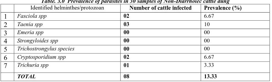
Fasciola spp has the highest prevalence 7(23.33%), followed by Cryptosporidium parvum 5(16.67%), in diarrhoeic dung Table 2. For non-diarrhoeic dung Fasciola spp 2(6.67% and Cryptosporidium parvum is 2(6.67) that were the same Table. 3.
The prevalence of the Cryptosporidium spp determined by microscopy, showedthe distribution in non-diarrhoea and diarrhoea dung samples as 1:2.5 ratio.
[image:4.612.74.557.403.699.2]Comparing the prevalence of all microbes present in the overall sample, cryptosporidium is second in prevalence in all samples.
Table 1: Overall prevalence of parasites in 60 cattle in Bamenda Central cattle market
Identified helminthes/protozoan Number of cattle infected Prevalence (%)
1
Fasciola spp 10 16.672
Taenia spp 06 10.03
Emeria spp 05 8.334
Strongyloides spp 02 3.335
Trichostrongylus species 01 1.676
Cryptosporidium spp 07 11.677
Trichuria spp 03 05TOTAL 34 56.67
Table. 2.0 Prevalence of parasites in 30 samples of Diarrhoiec cattle dung
Identified helminthes/protozoan Number of cattle infected Prevalence (%)
1 Fasciola hepatica 07 23.33
2 Taenia spp 04 13.33
3 Emeria spp 05 16.67
4 Strongyloides spp 02 6.67
5 Trichostrongylus species 01 3.33
6 Cryptosporidium spp 05 16.67
7 Trichuria spp 02 6.67
© Indian Journal of Medical Research and Pharmaceutical Sciences http://www.ijmprs.com/
[image:5.612.71.544.118.261.2][15]
Table. 3.0 Prevalence of parasites in 30 samples of Non-Diarrhoiec cattle dung
Identified helminthes/protozoan Number of cattle infected Prevalence (%)
1
Fasciola spp 02 6.672
Taenia spp 03 103
Emeria spp 00 004
Strongyloides spp 00 005
Trichostrongylus species 00 006
Cryptosporidium spp 02 6.677
Trichuria spp 01 3.33TOTAL 08 13.33
Discussion
The purpose of this study was a preliminary attempt to assess the presence and/or prevalence of the parasitic protozoan Cryptosporidium parvum in cattle dung. There is lack of data concerning the occurrence of Crptosporidium parvum in cattle dung in Cameroon as it is not considered as a bane to animal production. Epidemiological studies of gastrointestinal parasitic infections in ruminants in Cameroon [17] did not include Cryptosporidium parvum as a parasite of importance but the overall prevalence of parasites in cattle (56.7 %)). These results coincide with the results of this present study with an overall prevalence of 56.67%). Their results could have been higher if cryptosporidiosis was considered in their study.
The present study has revealed an overall prevalence of 11.67% in cattle which is the main ruminant feeding at free range in our water catchment area. This prevalence is lower than that reported by Adekunle and Fagbemi (2010)[9] who observed an overall prevalence of infection with Cryptospodium spp of 23.4% (95/406), in Oyo State. In the USA, USSR, UK, Germany Hungar and India, the results were higher than in Bamenda possibly due to more sensitive methods of identification of oocysts that were employed.
This parasite has with a very low infective dose of one to ten oocysts[12] to elicit an infection. This parasite can be transmitted to man and those with altered immune system it can be fatal
.
CONCLUSION
For any sample randomly taken from the overall sample and analyzed, there is a 100% chance that the prevalence of cryptosporidium lie between a value of 6.67(prevalence in non-diarrhea sample) and 16.67 (value in diarrhea sample). Since the infective dose is very low, one to ten ooysts[12], dung should be handled with care to avoid infection.
Cattle are an important source of contamination of the environment with infective stage (oocyst) of Cryptosporidium parvum
.
Recommendation
State of the art equipment should be used to establish the dynamics of Cryptosporidiosis in the Northwest Region of Cameroon, since it cattle raring areas where the ruminants are grazing at free range. Cattle should be transported to the cattle markets in trucks so that the dung can be collected and properly disposed of.
Acknowledgment
This work was done in the laboratory of Phyto-biotechnology Research Foundation Laboratory (PRF) in Cameroon. I wish to thank Laboratorians who worked with me in the laboratory. Also the cattle herders, who were able to guide me closer to the cattle during sample collection. The co-authors and/or supervisors of this study which is part of a PhD Thesis were very supportive
.
Conflicts of interest
© Indian Journal of Medical Research and Pharmaceutical Sciences http://www.ijmprs.com/
[16]
References
1. Panciera, R., Thomassen, R., Gamer, F, Cryptosporidial infection in a calf. Vet. Pathol. 1971;8: 479– 484. 2. Tzipori S, Campbell I, Sherwood D, Snodgrass DR, Whitelaw A. An outbreak of calf diarrhoea attributed
to cryptosporidial infection. Vet Rec 1980; 107:579-80.
3. Tzipori S. Cryptosporidiosis: laboratory investigations and chemotherapy. Adv. Parasitol. 1998; 40:187.221
4. De Graaf D.C, Vanopdenbosch E, Ortega-Mora L.M, Abbassi H,and Peeters J.E. “A review of the
importance of cryptosporidiosis in farm animals, in Int J Parasitol. Aug, 1999; 29(8):1269-87.
5. Ntonifor H. N, Shei S. J, Ndaleh N. W, and Mbunkur G. N, “Epidemiological studies of gastrointestinal parasitic infections in ruminants in Jakiri, Bui Division, North West Region of Cameroon,” in Journal of Veterinary Medicine and Animal Health. Vol. 5(12) 2013, pp. 344 -352.
6. Hooda P.S., A.C. Edwards, H.A. Anderson, A. Miller, “A review of water quality concerns in livestock farming areas,” in Sci. Total Environ. 250: 143–167, 2000 quality concerns in livestock farming areas,” in Science of total Environmental 2000, 250:143-167.
7. Kumar, D., R. Sreekrishnan, SS. Das .Cryprosporidiosis: an emerging disease of zoonotic importance. Proc. Nat. Acad. Sci. India. 2005, 75, 160-172.
8. Singh, BB., R. Sharma, H. Kumar, HS. Banga, RS. Aulakh, JP. Gill and JK. Sharma, “Prevalence of Cryptosporidium parvum infection in Punjab (India) and its association with diarrhea in neonatal dairy calves,” in Vet. Parasitol 2006, 140(1-2):162-5.
9. Adekunle, B. Ayinmode and Benjamin, O. Fagbemi, “Prevalence of Cryptosporidium infection in cattle from South Western Nigeria,” inVETERINARSKI ARHIV 2010, 80(6), 723-731.
10. Fayer, R., Santin, M., Trout, JM., Greiner, E, Prevalence of species and genotypes of Cryptosporidium found in 1-2-year-old dairy cattle in the eastern United States. Vet Parasitol 2006, 135: 105-112.
11. Fayer, R., Santin, M. & Trout, JM, Cryptosporidium ryanae n. sp. (Apicomplexa: Cryptosporidiidae) in cattle (Bos taurus). Vet Parasitol 2008, 156(3-4), 191-8.
12. Pereira SJ., NE. Ramirez, L. Xiao, LA, “Ward, Pathogenesis of human and bovine Cryptosporidium parvum in gnotobiotic pigs,” in J. Infect. Dis 2000, 186: 715–718.
13. ET Pamo, Country pasture/forage resource profiles of Cameroon, edited by JM suttee and SG Reynolds 2008. 14. ANNgalim, Cattle Rearing Systems in the North West Region of Cameroon: Historical Trends on Changing Techniques and Strategies. Journal of Educational Policy and Entrepreneurial Research (JEPER)Vol. 2, N0.5. May 2015, Pp 175-189
15. Guedjeo CS., Kagou Dongmo A, Ngapgue F, Nkouathio DG., Zangmo Tefogoum G,Hooda, et al, “A review of water Inpankaew T., Phasuk C., Pinyopanuwat N., Chimnoi1 W., Kengradomkij C., Arunwipat P., and Anakewith T, “Prevalence of Gastro-Intestinal Parasites of Dairy Cows in Thailand. Kasetsart” in J. Nat. Sci 2011, 45: 40-45.
16. Sunday Shende Kometa1 & Mathias Ashu Tambe Ebot, “Watershed Degradation in the Bamendjin Area of the North West Region of Cameroon and Its Implication for Development,” in Journal of Sustainable Development 2012, Vol. 5, No. 9.
17. Monica Cheesbrough in: District LaboratoryPractice in Tropical Countries Cambridge University Press. 2nd
Edition (2009) accessed August 2015, available fromwww.cambridge.org/9780521676304
18. Garcia, L. S.;, Current issues related to stool collection, processing, and testing of stool specimens for diagnostic parasitology. Clin. Microbiol 2000, 22: 140-144.
![Figure. 1. Cameroon Map showing the study area, Bamenda. Source: North West Regional master plan for sustainable development 2001[19]](https://thumb-us.123doks.com/thumbv2/123dok_us/9930598.495085/3.612.79.535.117.407/figure-cameroon-showing-bamenda-source-regional-sustainable-development.webp)