ORIGINAL RESEARCH
PEDIATRICS
Reliability of MR Imaging–Based Posterior Fossa and Brain
Stem Measurements in Open Spinal Dysraphism in the Era of
Fetal Surgery
XM. Aertsen,XJ. Verduyckt,XF. De Keyzer,X T. Vercauteren,XF. Van Calenbergh,XL. De Catte,XS. Dymarkowski, X P. Demaerel, andXJ. Deprest
ABSTRACT
BACKGROUND AND PURPOSE: Fetal MR imaging is part of the comprehensive prenatal assessment of fetuses with open spinal dysra-phism. We aimed to assess the reliability of brain stem and posterior fossa measurements; use the reliable measurements to characterize fetuses with open spinal dysraphism versus what can be observed in healthy age-matched controls; and document changes in those within 1 week after prenatal repair.
MATERIALS AND METHODS: Retrospective evaluation of 349 MR imaging examinations took place, including 274 in controls and 52 in fetuses with open spinal dysraphism, of whom 23 underwent prenatal repair and had additional early postoperative MR images. We evaluated measurements of the brain stem and the posterior fossa and the ventricular width in all populations for their reliability and differences between the groups.
RESULTS:The transverse cerebellar diameter, cerebellar herniation level, clivus-supraocciput angle, transverse diameter of the posterior fossa, posterior fossa area, and ventricular width showed an acceptable intra- and interobserver reliability (intraclass correlation coeffi-cient⬎0.5). In fetuses with open spinal dysraphism, these measurements were significantly different from those of healthy fetuses (all with P⬍.0001). Furthermore, they also changed significantly (Pvalue range⫽.01 to⬍.0001) within 1 week after the fetal operation with an evolution toward normal, most evident for the clivus-supraocciput angle (65.9⫾12.5°; 76.6⫾10.9;P⬍.0001) and cerebellar herniation level (⫺9.9⫾4.2 mm;⫺0.7⫾5.2;P⬍.0001).
CONCLUSIONS: In fetuses with open spinal dysraphism, brain stem measurements varied substantially between observers. However, measurements characterizing the posterior fossa could be reliably assessed and were significantly different from normal. Following a fetal operation, these deviations from normal values changed significantly within 1 week.
ABBREVIATIONS:ACi⫽atriocerebral index; CHL⫽cerebellar herniation level; CSA⫽clivus-supraocciput angle; GA⫽gestational age; ICC⫽intraclass correlation coefficient; OSD⫽open spinal dysraphism; PF⫽posterior fossa; TCD⫽transverse cerebellar diameter; TDPF⫽transverse diameter of the PF; VW⫽ventricular width
O
pen spinal dysraphism (OSD), subdivided into myelomen-ingocele and myeloschisis, is a nonlethal congenital malfor-mation with complex physical and neurodevelopmental sequelae. Its prevalence is approximately 4.9 per 10,000 live births in Eu-rope and 3.17 in the United States.1-3OSD results in motor andsensory deficits, their extension being defined by the upper level
of the anatomic defect. These range, as the level increases, from bladder, bowel, and sexual dysfunction to involvement of the lower and even upper extremities and secondary orthopedic dis-abilities.4,5Children with OSD almost invariably have an
associ-ated Chiari II hindbrain malformation and ventriculomegaly.6
The Chiari II malformation is characterized by posterior fossa (PF) and brain stem abnormalities with downward displacement and compression of the cerebellum and brain stem.7
Geerdink et al8demonstrated that morphometric measures
reliably quantify the morphologic distortions of Chiari II malfor-mation on postnatal MR images. The mamillopontine distance and the cerebellar width were the most sensitive and specific
de-Received May 29, 2018; accepted after revision October 6.
From the Department of Imaging and Pathology (M.A., J.V., F.D.K., S.D., P.D.), Clinical Department of Radiology, University Hospitals KU Leuven, Leuven, Belgium; School of Biomedical Engineering and Imaging Sciences (T.V.), King’s College, Lon-don; Department of Neurosurgery (F.V.C.), University Hospitals Leuven, Leuven, Belgium; Academic Department of Development and Regeneration, Cluster Woman and Child (L.D.C., J.D.), Group Biomedical Sciences, KU Leuven, Leuven, Belgium; and Institute for Women’s Health, University College London, (J.D.), Lon-don, UK.
Philippe Demaerel and Jan Deprest are shared last authors.
J. Deprest was partly funded by the Great Ormond Street Hospital Charity Fund. The work was also supported by the Engineering and Physical Sciences Research Council and the Innovative Engineering for Health award by the Wellcome Trust.
Please address correspondence to Michael Aertsen, MD, Department of Radiology, Herestraat 49, 3000 Leuven, Belgium; e-mail: [email protected]
Indicates article with supplemental on-line tables.
terminants of Chiari II.9Some fetuses with OSD have
ventriculo-megaly, and its degree is believed to be predictive of the need for postnatal shunting.10,11
In 2011, the Management of Myelomeningocele Study (MOMS) demonstrated the benefit of in utero repair of myelo-meningocele because the need for ventricular shunting at 12 months was reduced and motor outcome at 30 months im-proved.12Fetuses with the suspicion of OSD should be assessed
comprehensively to counsel parents about the expected outcome and possibility of fetal surgery. In this assessment, fetal MR imag-ing has a crucial role to characterize the brain and spinal abnor-malities and rule out additional anomalies in fetuses with OSD.13,14For fetal surgery eligibility, the presence of Chiari II
hindbrain malformation on MR imaging is a necessary find-ing.12,15Many measurements have been proposed to describe
the typical PF changes in fetuses with OSD, yet the reproduc-ibility of these has rarely been studied.14,16-23These
parame-ters have also been shown to change after in utero repair of OSD in small series and at different time points after fetal surgery, yet no study has consistently reported early postoper-ative assessment in utero.18
The aims of this study were 3-fold: 1) to assess the reproduc-ibility of measurements of the brain stem and PF that have been suggested to be representative on postnatal8,9and prenatal MR
imaging14,16-23; 2) to apply those parameters that were shown to
be reproducible, to discriminate fetuses with OSD from gesta-tional age–matched fetuses with a normal PF; and 3) to docu-ment early changes in these measuredocu-ments 1 week after a fetal operation.
MATERIALS AND METHODS
This was a single-center retrospective study at University Hospi-tals Leuven that was approved by its ethics committee (S60814). Patients eligible for the OSD group were those having fetal MR imaging examinations for additional assessment because of a pre-natal diagnosis of OSD on ultrasound. Before MR imaging, pa-tients had an ultrasound assessment, in which the lesion, second-ary changes, and, when applicable, associated anomalies were characterized. Before the MR imaging, the radiologist was in-formed of the ultrasound findings. For the gestational age– matched controls, we included fetuses assessed for other congen-ital anomalies that do not affect the central nervous system or who were scanned for suspected CNS abnormalities with normal find-ings on prenatal ultrasound, fetal MR imaging, and postnatal evaluation (On-line Table 1).
Fetal Imaging and Quality Criteria
The routine protocol for this condition includes acquisition on a 1.5T MR imaging system (Aera; Siemens, Erlangen, Germany) with 2 small body coils placed adjacent to each other over the maternal abdomen. The mother was positioned in the supine or left lateral decubitus position. The images used were T2-weighted HASTE or balanced steady-state gradient-echo sequences in the sagittal, axial, and coronal planes relative to the fetal head. Before September 2015, maternal sedation (flunitrazepam, 0.5 mg orally 20 –30 minutes before the examination) was used when the ges-tational age (GA) was⬍30 weeks.24Later, this was abandoned
because we, like others, thought that this induced maternal ad-verse effects while not clinically required.25 For this study, we
searched our data base for all examinations performed in the set-ting of spinal dysraphism assessment, as well as for appropriate gestational age–matched controls. The image quality had to be good, consisting of at least 3 orthogonal T2-weighted HASTE se-ries of the fetal brain with limited fetal motion, allowing adequate performance of the outcome measurements. The primary selec-tion and review of images was performed by a single pediatric radiologist (M.A.) with⬎3 years of experience in fetal MR imag-ing. The main exclusion criteria were twin pregnancy, syndromal pathology, fetal hydrops, or anhydramnios. The number of pa-tients and individual reasons for exclusion are shown in On-line Table 2. This exclusion left data from 349 MR imaging examina-tions of a total of 1006, including 274 examinaexamina-tions in 246 control fetuses. These data illustrate that some fetuses were scanned more than once. Additionally, we included 52 MR imaging examina-tions in fetuses with OSD, of whom 23 had a repeat MR imaging examination after the operation. The eligibility criteria for fetal surgery were those used in the MOMS trial.12
Outcome Measurements
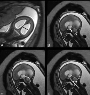
Biometric variables included the transverse cerebellar diameter (TCD), pontine thickness, and pontine height, measured accord-ing to the standards defined by Garel26 and Tilea et al.27The
transverse diameter of the PF (TDPF) was measured according to Woitek et al,17who suggested that this would be a proxy for the
TCD. The midsagittal PF area was measured according to Tsai et al.20The ventricular width (VW) was measured in the coronal
plane according to Garel,26and in case of asymmetry, the largest
value was taken into account. Mamillopontine distance, the level of kinking of the brain stem, medullary length, tentorial length, and width of the cisterna magna were measured as described by Geerdink et al.8The width of the foramen magnum was
de-fined as the distance between the opisthion and the basion. The cerebellar herniation level (CHL) was measured by drawing a perpendicular line from the foramen magnum to the lowest cerebellar portion. In the presence of cerebellar herniation, the deepest portion was measured.20The clivus-supraocciput
an-gle (CSA) was measured according to D’Addario et al.28The
TCD, TDPF, mamillopontine distance, TL, PF area, and CSA are demonstrated inFig 1.
Reproducibility Study
The reproducibility of measurements was determined on a ran-domly chosen subgroup of spinal dysraphism cases (n⫽15/52; referred to as the pilot group). Images were anonymized and up-loaded to a research server29for assessment by M.A. and J.V., a
Posterior Fossa Characteristics
The PF characteristics were determined on the presumed healthy population to obtain normative values. To compare cases of fetal myelomeningocele with the healthy population, we used these normative curves to calculate expected values for the given gesta-tional ages, and the fetal myelomeningocele values were then ex-pressed as observed over the expected ratio.
Short-Term (<7 Days) Postoperative Changes
In this part of the study, we looked at the difference between PF measurements shown to be reproducible in the above part of the study in 23 fetuses who had a fetal operation at our center. These fetuses had preoperative MR imaging and were imaged again within 1 week after the operation. All measurements were per-formed on T2-weighted images in the coronal or sagittal plane of the fetal head. Again, the values were expressed as observed over the expected ratio to determine changes after prenatal treatment that were not attributable to normal growth.
In addition to the posterior fossa, we also evaluated the ventricu-lar width in fetuses with OSD and the difference from the control population. To describe the differential effect of a fetal operation on the parenchyma and ventricles, one can measure changes in the
so-called atriocerebral index (ACi). The ACi is the ratio of the atrial diameter and the cerebral (parenchymal) biparietal diame-ter.24Others called this index the
ventric-ular width index as used in the postnatal literature.18
Statistics
Intraobserver and interobserver vari-ability were analyzed with a 2-way ran-dom ICC using SPSS for Windows, Ver-sion 22.0 (Released 2004; IBM, Armonk, New York). An ICC cutoff value of 0.5 as the lowest acceptable was chosen, taking into account the guidelines for interpre-tation by Cicchetti31and allowing some
variation in view of the limited spatial resolution for assessing such small struc-tures so that borderline parameters can be fully investigated. For the interpreta-tion of ICC values, we followed the guidelines of Koo and Li,30with ICC
val-ues⬍0.5 indicative of poor reliability; values between 0.5 and 0.75, moderate reliability; values between 0.75 and 0.9, good reliability; and values⬎0.90, excel-lent reliability. The normality of GA in the 274 examinations was evaluated using the Shapiro-Wilk test, which indi-cated that GA was not normally distrib-uted. We attempted several transforma-tion models (logarithmic, polynomial, and square root) from which the square root transformation provided the most normally distributed data. Afterward, re-gression analysis was performed on all ex-amined PF characteristics to find normative ranges in correlation with GA. Differences in the reliable parameters between the healthy cohort and the fetuses with OSD were calculated using the Wilcox-on–Mann-Whitney test. The Wilcoxon test was used to analyze dif-ferences in the paired measurements of individual fetuses before and after the operation. All statistics in the PF characteristics section were performed using Analyze-it (Analyze-it for Microsoft Excel 4.81.4; Analyze-it Software, Leeds, UK). APvalue⬍.05 indicated statistical significance.
RESULTS
DemographicsMR imaging examinations in controls were performed at a mean GA of 27.9⫾5.3 weeks (range,18.6 –38.3 weeks). In fetuses with OSD, the mean GA at MR imaging was 23.6⫾0.3 weeks (range, 19.3–27.3 weeks). Descriptive statistics for the study parameters in controls and cases with OSD are shown inTable 1.
Reproducibility Study
The intraobserver ICCs for the PF area, VW, TCD, and CHL were excellent. Conversely, TDPF (0.729), the pontine thickness (0.59),
[image:3.594.58.374.48.382.2]the foramen magnum diameter (0.44), mamillopontine distance (0.66), CSA (0.60), and the width of the cisterna magna (0.48) had a fair-to-good reproducibility. Measurements of the other param-eters showed a low reproducibility (tentorial length) or were un-reliable (level of brain stem kinking, medullar length, and pontine length). Interobserver reproducibility was moderate for TCD, CHL, CSA, and TDPF. PF area and VW had good interrater reliability.
Posterior Fossa Characteristics
All parameters with an intra- and interobserver ICCⱖ0.5 were taken into account for further analysis. Normative curves for VW, PF area, TDPF, TCD, CSA, TCD/TDPF, and CHL were calculated and are shown inTable 2.Figure 2shows the individual observa-tions for cases with OSD, which were all significantly different from what was measured in healthy fetuses (P⬍.0001).
Short-Term (<7 Days) Postoperative Changes
When we considered the observed over expected ratio values, fetal surgery was associated with a significant difference in cerebellar herniation (P ⬍.0001), TCD/TDPF (P⫽ .0002), TCD (P ⫽
.0127), PF area (P⫽.0003), TDPF (P⫽.0127), CSA (P⬍.0001), and VW (P⫽.0002).Figure 3shows the individual observations and boxplots in patients with OSD for all tested parameters, both pre- and postoperatively. In 18/23 (78%) fetuses, the postopera-tive observed over expected ratios of the PF area were improving toward normal compared with the preoperative measurement. The same was true for the observed over expected ratios of the TDPF in 15/23 (65%), for the observed over expected ratios of the TCD in 7/23 (30%), and for the observed over expected TCD/ TDPF ratios in 4/23 (17%) postoperative fetuses. The observed over expected ratio level of cerebellar herniation increased, mean-ing that the Chiari II–associated changes were reduced in 19/23
fetuses at 1 week after the operation. The observed over expected ratios of the CSA increased toward, the normal range in 16/23 (70%) postoperative fetuses. The VW increased in 17/23 (74%) fetuses, but there was no difference in the ACi between the pre-and postoperative examinations (P⫽.46), with a mean of the preoperative measurements of 0.22 and, postoperatively, of 0.23.
DISCUSSION
A large number of structural measurements on MR images of the PF have been suggested to characterize changes attributed to OSD. Some are believed to be clinically relevant due to their rela-tion to the symptoms associated with a small PF, Chiari II mal-formation, and brain stem compression.32These changes are
rep-resented by the degree of cerebellar herniation, mamillopontine distance, brain stem kinking, tentorial hypoplasia, and the con-figuration of the fourth ventricle. These features can be measured reproducibly in the postnatal period.8In the era of prenatal
diag-nosis and fetal surgery, logically, the same measurements are also used. Yet, in case of prenatal surgery, these are measured on midgestational prenatal images at a time when the condition is still progressive and image quality is different. We therefore in-vestigated whether those measurements were reproducible in the window of interest. For instance, we did not evaluate the fourth ventricle because it is hardly visible around 20 –24 weeks in fetuses with OSD. Conversely, we looked at parameters characterizing the brain stem (mamillopontine distance, pontine thickness, pontine length, foramen magnum diameter, level of brain stem kinking, medulla length, tentorial length, and cisterna magna width) and the PF as a whole (TCD, TDPF, PF area, CHL, TCD/TDPF, and CSA). Due to its clinical relevance in fetuses with OSD and the impact on outcome after fetal surgery,33we
also included the VW.
Herein, we conclude that brain stem parameters in fetuses with OSD cannot be measured reliably at midgestation; thus, we were not able to objectively measure brain stem elongation and displacement in utero. For fetuses with OSD, we believe this issue is due to the limited spatial resolution of fetal MR imaging at that point in gestation when the structures involved are in the milli-metric range so that the slightest measurement error has a tre-mendous impact on statistics. Another reason is that in OSD, there is a decrease or even absence of extra-axial CSF, further limiting the contrast resolution of MR imaging.34Contrast
reso-lution is essential for accurate evaluation of small PF structures as well as some additional cerebral lesions due to the presence of different structures (medulla oblongata, pons, vermis, cerebel-lum) in a small area.35There is no difference between balanced
steady-state free-precession sequences and half-Fourier rapid re-laxation with rere-laxation enhancement sequences for measuring the foramen magnum.19Conversely, when we looked at larger
structures (PF area, TCD, and TPFD) and/or with more abundant contrast between fluid and soft tissue (CHL and VW), the ICC values were much better. We confirmed this finding for those parameters typical for the smaller posterior fossa and cerebellar descent in the pathologic subgroup, as others did.17,18,20,22
More-over, the difference with our gestational age–matched healthy pa-tients was highly significant, again as previously described.17
[image:4.594.53.285.65.169.2]Fetal surgery has been shown to reverse those posterior fossa
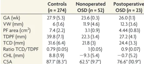
Table 1: Mean and SDs of different parameters for the controls and fetuses with nonoperated and operated spinal dysraphism
Controls (n= 274)
Nonoperated OSD (n= 52)
Postoperative OSD (n= 23) GA (wk) 27.9 (5.3) 23.6 (0.3) 26.0 (1.1)
VW (mm) 6 (1.6) 11.9 (4.6) 12.3 (3.6)
PF area (cm2) 7.4 (2.2) 3.1 (0.9) 4.44 (0.83)
[image:4.594.56.285.207.302.2]TDPF (mm) 39.8 (7.1) 22.3 (3.4) 27.2 (4.1) TCD (mm) 31.6 (6.4) 21.8 (3) 24.4 (3.3) Ratio TCD/TDPF 0.79 (0.05) 1 (0.05) 0.9 (0.07) CHL (mm) 8.8 (1.9) ⫺9.3 (5.4) ⫺0.7 (5.2) CSA 87.1° (8.3°) 62.5° (9.7°) 76.6° (10.9°)
Table 2: Equations of the regression curves with their respective levels of significance and the R2for the TCD, CHL, CSA, TDPF, PF area, VW, and ratio TCD/TDPF
Parameter Regression Curve
P Valuea R2 TCD y⫽64.38–30.58*x⫹4.606*x2 ⬍.0001 0.907
CHL y⫽ ⫺78.26⫹31.6*x–2.849*x2 ⬍.0001 0.117
CSA y⫽21.61⫹12.44*x ⬍.0001 0.265
TDPF y⫽ ⫺65.28⫹19.95*x ⬍.0001 0.894
PF area y⫽14.25–8.537 *x⫹1.369*x2 ⬍.0001 0.875
VW y⫽51.5–17.59*x⫹1.692*x2 .0257 0.027
Ratio TCD/TDPF y⫽3.87–1.215*x⫹0.1192*x2 ⬍.0001 0.243
Note:—x indicates square root (gestational age in weeks); R2
, coefficient of determination.
a
changes.11,14,18Other investigators used the above parameters to
measure those typicallyⱖ4 weeks after the operation.18In the
present study, we acquired images within 2 weeks. In line with ultrasound observations, we quantified significant changes dur-ing that short observation period. Within 1 week, 26% of operated fetuses had a PF area within the normal range, and in 52%, the TCD was normal. Furthermore, the cerebellar herniation level was at or above the foramen magnum in 52% of fetuses, and 70% had a normal CSA. These acute changes in the PF following clo-sure of the defect are in line with the theory of McLone and Knep-per.36In other words, it seems that the effects of fetal surgery on
the PF are already evident and can be quantified very early postoper-atively. They are very likely to persist because others observed the same effects later on and even confirmed them after birth.14,18,37This
outcome might be an interesting proxy for measurement of the effi-cacy of fetal surgery in clinical studies.
In this short-term follow-up study, we observed a postopera-tive increase in ventricular width within 1 week in most patients. Such increase is in line with observations made by others, though several weeks after fetal surgery.18,33They suggest that there is still
a certain degree of obstructive ventricular widening. The dynam-ics of CSF fluid production and resorption in OSD are still poorly understood. It may take some time after fetal surgery for CSF fluid circulation to normalize after stopping its egress.11
In healthy fetuses, the ACi drops dramatically between 24 and 27 weeks, which means that there is, during that time period, a proportional increase in the parenchymal brain component. In our patients undergoing fetal surgery, the ACi remained stable.
[image:5.594.54.531.46.510.2]This might be counterintuitive and contradicts the findings of Rethmann et al.18They measured the ACi and observed a drop in
the ACi; yet, that was 4 weeks after the operation and continued after birth. These contrasting findings can be explained in differ-ent ways. A drop in ACi would suggest a proportional increase in biparietal cerebral diameter, hence a larger parenchymal compo-nent. Conversely, the increase in ACi for a comparable VW in our cohort would suggest that the parenchymal component decreases. Although tempting, both groups cannot be compared because measurements were performed at different gestational ages. In healthy fetuses, the ACi spontaneously declines between 24 and 27 weeks. If fetuses with OSD follow this normal evolution, our find-ings may eventually align with these of Rethmann et al and can be explained by spontaneous and normal evolution. Unfortunately, we have no longitudinal follow-up MR images to further study these observations.
We have looked into prenatal measurements characterizing the brain stem, which, to our knowledge, was not performed in
detail before. We based our evaluation on our own normative values. Furthermore, we documented all parameters in a relatively large pathologic population in the narrow gestational age range that is relevant to prenatal spina bifida repair. Although the num-ber of operated fetuses was not very large, we were able to describe early postoperative changes therein. There are, however, some shortcomings. First, our control fetuses definitely had normal CNS findings on both MR imaging and ultrasound but were not truly fully healthy fetuses. Controls underwent MR imaging be-cause of other congenital abnormalities, presumed not to be as-sociated with CNS abnormalities.
Second, we did not report on advanced MR images, such as DWI and DTI, which may also provide relevant information. We definitely acknowledge the potential of DWI and DTI because they may detect more subtle abnormalities below the anatomic level. Woitek et al38already showed that fetuses with spina bifida
have increased fractional anisotropy compared with normally de-veloping fetuses, however without reporting the functional
[image:6.594.55.532.47.466.2]pact. Although we acquired such sequences in fetuses with spina bifida, we were lacking those in healthy fetuses or the controls used in this study; hence, we could not interpret the findings. Third, we describe in utero findings without correlation to post-natal short- or long-term follow-up or early postpost-natal MR imag-ing confirmation. Although relevant, such a follow-up was be-yond the scope of this study as was a comparison of these posterior fossa measurements with those in a cohort that underwent post-natal repair. Postpost-natal evaluation would most likely have identi-fied additional findings, such as subependymal heterotopias.18,39
These are often missed in utero before as well as after fetal surgery.
CONCLUSIONS
This study showed that the brain stem cannot be reliably charac-terized using the current panel of measurements in fetuses with OSD. Conversely, posterior fossa measurements are demon-strated to be reliable in the evaluation of fetuses with OSD. In addition, these were significantly different from those in the healthy population and changed within 7 days after prenatal sur-gery. This finding advocates for their use in the evaluation of fetuses with OSD on fetal MR imaging on a routine basis before and shortly after a prenatal operation.
Disclosures: Tom Vercauteren—UNRELATED:Employment: University College Lon-don,Comments: main employer for the duration of this work;Grant: Wellcome Trust (WT101957)/Engineering and Physical Sciences Research Council (NS/A000027/ 1*),Comments:Support for Travel to Meetings for the Study or Other Purposes: Wellcome Trust/Engineering and Physical Sciences Research Council,Comments: travel support from the listed grant funding,* Luc De Catte—UNRELATED: Employ-ment: consultant to Feto-Maternal Medicine AZ St-Jan Brugge,Comments: 1 day/2 weeks clinical activity.* Philippe Demaerel—UNRELATED:Board Membership: Edi-torial Board ofNeuroradiology. Jan Deprest—RELATED:Other:Great Ormond Street Hospital Children’s Charity,Comments: This grant pays part of my academic (research) time to my institution*;UNRELATED:Grants/Grants Pending: MEDRI, Wellcome Trust,Comments: We have a grant on the use of advanced imaging techniques in fetal surgery (Wellcome) and a research grant on novel implant mate-rials for Holder of a research chair “POPART” (Pelvic Organ Prolapse - Advanced Research and Technology) - sponsored by MEDRI*; Travel/Accommodations/Meet-ing Expenses Unrelated to Activities Listed: Chiesi Farmaceutici,Comments: paid travel and accommodations to the Sharing Progress in Neonatology 2018 meeting.* *Money paid to the institution.
REFERENCES
1. Garne E, Loane M, Addor M-C, et al.Congenital hydrocephalus: prevalence, prenatal diagnosis and outcome of pregnancy in four European regions.Eur J Paediatr Neurol2010;14:150 –55CrossRef Medline
2. Khoshnood B, Loane M, de Walle H, et al.Long term trends in prev-alence of neural tube defects in Europe: population-based study.
BMJ2015;351:h5949CrossRef Medline
3. Canfield MA, Mai CT, Wang Y, et al; National Birth Defects Preven-tion Network.The association between race/ethnicity and major birth defects in the United States, 1999 –2007.Am J Public Health 2014;104:e14e–23CrossRef Medline
4. Oakeshott P, Hunt GM.Long-term outcome in open spina bifida.
Br J Gen Pract2003;53:632–36Medline
5. Rintoul NE, Sutton LN, Hubbard AM, et al. A new look at myelomeningoceles: functional level, vertebral level, shunting, and the implications for fetal intervention. Pediatrics 2002;109:1–7
CrossRef Medline
6. Mitchell LE, Adzick NS, Melchionne J, et al.Spina bifida.Lancet 2004;364:1885–95CrossRef Medline
7. Barkovich AJ,Congenital malformations of the brain and skull. In: Barkovich AJ, ed, Ovid Technologies, Inc.Pediatric Neuroimaging. 4th ed. Philadelphia: Lippincott Williams & Wilkins; 2005:374 – 84
8. Geerdink N, van der Vliet T, Rotteveel JJ, et al.Essential features of Chiari II malformation in MR imaging: an interobserver reliability study, Part 1.Childs Nerv Syst2012;28:977– 85CrossRef Medline
9. Geerdink N, van der Vliet T, Rotteveel JJ, et al.Interobserver reliabil-ity and diagnostic performance of Chiari II malformation measures in MR imaging, Part 2.Childs Nerv Syst2012;28:987–95CrossRef Medline
10. Biggio JR Jr, Wenstrom KD, Owen J.Fetal open spina bifida: a nat-ural history of disease progression in utero.Prenat Diagn2004;24: 287– 89CrossRef Medline
11. Bruner JP, Tulipan N, Reed G, et al.Intrauterine repair of spina bifida: preoperative predictors of shunt-dependent hydrocephalus.
Am J Obstet Gynecol2004;190:1305–12CrossRef Medline
12. Adzick NS, Thom EA, Spong CY, et al; MOMS Investigators.A ran-domized trial of prenatal versus postnatal repair of myelomeningo-cele.N Eng J Med2011;364:993–1004CrossRef Medline
13. Saleem SN, Said AH, Abdel-Raouf M, et al.Fetal MRI in the evalua-tion of fetuses referred for sonographically suspected neural tube defects (NTDs): impact on diagnosis and management decision.
Neuroradiology2009;51:761–72CrossRef Medline
14. Sutton LN, Adzick NS, Bilaniuk LT, et al.Improvement in hindbrain herniation demonstrated by serial fetal magnetic resonance imag-ing followimag-ing fetal surgery for myelomenimag-ingocele.JAMA1999;282: 1826 –31CrossRef Medline
15. Heuer GG, Moldenhauer JS, Scott Adzick NS.Prenatal surgery for myelomeningocele: review of the literature and future directions.
Childs Nerv Syst2017;33:1149 –55CrossRef Medline
16. Danzer E, Adzick NS.Fetal surgery for myelomeningocele: patient selection, perioperative management and outcomes.Fetal Diagn Ther2011;30:163–73CrossRef Medline
17. Woitek R, Dvorak A, Weber M, et al.MR-based morphometry of the posterior fossa in fetuses with neural tube defects of the spine.PLoS One2014;9:e112585CrossRef Medline
18. Rethmann C, Scheer I, Meuli M, et al.Evolution of posterior fossa and brain morphology after in utero repair of open neural tube defects assessed by MRI. Eur Radiol 2017;27:4571– 80 CrossRef Medline
19. Abele TA, Lee SL, Twickler DM.MR imaging quantitative analysis of fetal Chiari II malformations and associated open neural tube defects: balanced SSFP versus half-Fourier RARE and interobserver reliability.J Magn Reson Imaging2013;38:786 –93CrossRef Medline
20. Tsai T, Bookstein FL, Levey E, et al.Chiari-II malformation: a bio-metric analysis.Eur J Pediatr Surg2002;12(Suppl 1):S12–18Medline
21. Osuagwu FC, Lazareff JA, Rahman S, et al.Chiari I anatomy after ventriculoperitoneal shunting: posterior fossa volumetric eval-uation with MRI. Childs Nerv Syst 2006;22:1451–56 CrossRef Medline
22. Grant RA, Heuer GG, Carrio´n GM, et al.Morphometric analysis of posterior fossa after in utero myelomeningocele repair.J Neurosurg Pediatr2011;7:362– 68CrossRef Medline
23. Chen SC, Simon EM, Haselgrove JC, et al.Fetal posterior fossa volume: assessment with MR imaging. Radiology 2006;238:997– 1003CrossRef Medline
24. Garel C.Methodology.In: Carel C, Delezoide AL, Delezoide V, eds. MRI of the Fetal Brain. Berlin: Springer-Verlag; 2004
25. Saleem SN.Fetal MRI: an approach to practice—a review.J Adv Res 2014;5:507–23CrossRef Medline
26. Garel C.Fetal cerebral biometry: normal parenchymal findings and ventricular size.Eur Radiol2005;15:809 –13CrossRef Medline
27. Tilea B, Alberti C, Adamsbaum C, et al.Cerebral biometry in fetal magnetic resonance imaging: new reference data.Ultrasound Obstet Gynecol2009;33:173– 81CrossRef Medline
28. D’Addario V, Pinto V, Del Bianco A, et al.The clivus-supraocciput angle: a useful measurement to evaluate the shape and size of the fetal posterior fossa and to diagnose Chiari II malformation. Ultra-sound Obstet Gynecol2002;18:146 – 49Medline
collaboration platform for medical imaging research. Comput Methods Programs Biomed2017;139:181–90CrossRef Medline
30. Koo TK, Li MY.A guideline of selecting and reporting intraclass correlation coefficients for reliability research.J Chirop Med2016; 15:155– 63CrossRef Medline
31. Cicchetti DV.Guidelines, criteria, and rules of thumb for evaluat-ing normed and standardized assessment instruments in psychol-ogy.Psychological Assessment1994;6:284 –90CrossRef
32. Stevenson KL.Chiari type II malformation: past, present, and fu-ture.Neurosurg Focus2004;16:E5Medline
33. Tulipan N, Wellons JC 3rd, Thom EA, et al; MOMS Investigators.
Prenatal surgery for myelomeningocele and the need for cerebro-spinal fluid shunt placement.J Neurosurg Pediatr2015;16:613–20
CrossRef Medline
34. Danzer E, Johnson MP, Bebbington M, et al.Fetal head biometry as-sessed by fetal magnetic resonance imaging following in utero myelo-meningocele repair.Fetal Diagn Ther2007;22:1– 6CrossRef Medline
35. Limperopoulos C, Tworetzky W, McElhinney DB, et al.Brain vol-ume and metabolism in fetuses with congenital heart disease: eval-uation with quantitative magnetic resonance imaging and spec-troscopy.Circulation2010;121:26 –33CrossRef Medline
36. McLone DG, Knepper PA.The cause of Chiari II malformation: a unified theory. Pediatr Neurosci 1989;15:1–12 CrossRef Medline
37. Nagaraj UD, Bierbrauer KS, Zhang B, et al.Hindbrain herniation in Chiari II malformation on fetal and postnatal MRI.AJNR Am J Neuroradiol2017;38:1031–36CrossRef Medline
38. Woitek R, Prayer D, Weber M, et al.Fetal diffusion tensor quantifi-cation of brain stem pathology in Chiari II malformation.Eur Ra-diol2016;26:1274 – 83CrossRef Medline