ORIGINAL RESEARCH
Intravascular Frequency-Domain Optical
Coherence Tomography Assessment of
Atherosclerosis and Stent-Vessel Interactions in
Human Carotid Arteries
M.R. Jones G.F. Attizzani C.A. Given II W.H. Brooks M.A. Costa H.G. Bezerra
BACKGROUND AND PURPOSE: Carotid artery–related stroke is largely an embolic disease that has been correlated with inflammation, plaque rupture, and thrombus formation in “vulnerable” atherosclerotic plaque. Nevertheless, current guidelines for carotid revascularization in asymptomatic patients rely on the calculation of stenosis for risk assessment, a parameter that has been viewed with increasing skepticism. Intravascular OCT is an imaging technique that offers high axial resolution (10 m), allowing an unprecedented micron-level assessment of human carotid plaque morphology. This observational article reports the first successful use of the newest iteration of this technology, FDOCT without balloon occlusion to assess human carotid artery disease and carotid stent-vessel interaction in vivo.
MATERIALS AND METHODS:Four patients with asymptomatic carotid artery disease and ambiguous noninvasive and/or angiographic data underwent carotid FDOCT to assess risk and to formulate a treatment strategy.
RESULTS: Findings include the unexpected demonstration of TCFAs, plaque rupture, thrombus, in-flammation, and marked tissue prolapse through stent struts in patients without high-risk factors by conventional criteria, as well as low-risk features in a patient with a high-risk noninvasive study. The procedures were performed without safety issues or special accommodations for vessel occlusion.
CONCLUSIONS:The present study demonstrates the technical feasibility of FDOCT in cervical carotid arteries. As such, this technology holds the promise of not only clarifying ambiguous data in individual patients but of providing data that might call for a future paradigm shift in the assessment of asymptomatic carotid artery disease.
ABBREVIATIONS:CCA⫽common carotid artery; FDOCT⫽frequency-domain optical coherence tomography; NIH⫽neointimal hyperplasia; OCT⫽optical coherence tomography; TCFA⫽thin-cap fibroatheroma; TDOCT⫽time-domain optical coherence tomography
S
troke is the fourth leading cause of death and a leading cause of long-term disability in the United States. Recent estimates list carotid artery atherosclerosis as a culprit in 15%– 20% of these cases.1Almost two-thirds of these strokes affectpatients who were previously asymptomatic.2The challenge to
identify patients with asymptomatic carotid atherosclerosis who are at risk for stroke is daunting because of both the fre-quent occurrence of carotid atherosclerosis in the elderly3and
an increasing awareness that an estimation of the degree of stenosis in a carotid lesion is a poor predictor of clinical out-come.4Numerous studies have suggested that stroke risk may
be best predicted by plaque morphology in these asymptom-atic lesions.5-7The value of MR imaging8and multidetector
CT imaging,4as well as intravascular sonography9to evaluate
carotid disease has been reported but not validated in multi-center trials or accepted into general practice.
Intravascular OCT is an emerging technique recently ap-proved for use in the United States. This imaging technique, analogous to intravascular sonography with the substitution of light for acoustic energy, delivers resolution to 10 –20m, giving it the highest imaging resolution of any currently avail-able intravascular imaging technique.10 Furthermore, the
variable tissue penetration of light (ranging from 1 to 3.5 mm, depending on tissue composition),10 when combined with
pixel intensity provides accurate identification of lipid-rich11 and calcified plaques12and intraluminal thrombus.13In addi-tion, OCT makes it possible to directly visualize and quantify TCFA14and vascular inflammation.15 These important
fea-tures of vulnerable plaque, heretofore largely invisible by other imaging modalities due to their lower resolution, are readily identified and quantified with the micron-scale resolution of OCT. The latest iteration of OCT, FDOCT compared with TDOCT, moreover obviates the need for vessel occlusion by allowing an acquisition speed of up to 25 mm/s.
Detailed description of both systems is beyond the scope of this article. Briefly, while TDOCT uses a moving mirror as its reference arm, FDOCT uses a fixed mirror with a variable frequency light source, allowing simultaneous detection of re-flections from all TE delays, making the system significantly Received October 5, 2011; accepted after revision December 7.
From the Baptist Heart and Vascular Institute (M.R.J., C.A.G., W.H.B.), Central Baptist Hospital, Lexington, Kentucky; and Harrington McLaughlin Heart and Vascular Institute (G.F.A., M.A.C., H.G.B.), University Hospitals, Case Medical Center, Case Western Reserve University, Cleveland, Ohio.
Please address correspondence to Michael R. Jones, MD, FACC, FSCAI, 1720 Nicholasville Rd, Suite 601, Lexington, KY 40503; e-mail: mjones@lexingtoncardio.com
Indicates open access to non-subscribers at www.ajnr.org
faster. The larger (approximately 10 mm) FOV in FDOCT images also allows examinations of larger vessels, such as the carotid arteries.10
We report, for the first time, the successful use of FDOCT in vivo using a nonocclusive technique in the carotid arteries. OCT was used to clarify ambiguous noninvasive and angio-graphic carotid data in 3 cases with the resultant demonstra-tion of interesting findings that suggest that markers for stroke risk heretofore only previously described by ex vivo histo-pathologic studies may be captured in vivo by means of OCT. In a fourth case, OCT ruled out features of plaque complexity previously suggested by a carotid duplex test.
Materials and Methods
Four patients undergoing diagnostic carotid angiography with stan-dard 5F diagnostic catheters underwent ad hoc OCT assessment of ICA lesions. No patient came to the angiographic suite for a planned interventional procedure because it is our policy to perform diagnos-tic carotid and cerebral angiography on all patients before any recom-mendations for revascularization. Indications included the clarifica-tion of abnormal nonspecific carotid angiograms (patients A, C, and D) and the resolution of discordant angiographic-duplex findings (patient B). All patients were asymptomatic. Patient selection and immediate treatment was under the auspices of an interventional car-diologist with coronary OCT experience (M.R.J.), an interventional neuroradiologist (C.A.G.), and an endovascular-trained neurosur-geon (W.H.B.). All patients gave written informed consent for proce-dures performed.
Following angiography and before OCT interrogation, all patients underwent systemic anticoagulation with unfractionated heparin, achieving a target activated clotting time of⬎250 seconds. Three patients (A, C, and D) were studied with7F Arrow-Flex Sheaths (Ar-row International, Reading, Pennsylvania). Patient B was studied us-ing a 6F Shuttle sheath (Cook, Bloomus-ington, Indiana). Embolic pro-tection (NAV-6 Filterwire; Abbott Vascular, Redwood City, California) was used in patients A and C only.
In each case, the delivered guidewires were used to advance a 2.7F C7 Dragonfly catheter (St Jude Medical, St Paul, Minnesota) across the lesion. Data were then acquired with a C7-XR OCT Intravascular Imaging System (St Jude Medical) and digitally stored. All OCT au-tomated pullbacks covered 54 mm of vessel at 20 mm/s. During image acquisition, carotid blood flow was replaced by hand injections of contrast using a 30-mL syringe. Contrast was chosen in lieu of saline because of the efficiency of these high-viscosity agents in displacing signal-intensity-attenuating red blood cells during the data-acquisi-tion period.10The average contrast use was 20 mL per acquisition run. Contrast delivery was achieved in⬍3 seconds per acquisition.
2D images were reconstructed on-line and used to facilitate clin-ical decision-making in the catheterization laboratory. Additional off-line imaging analysis and 3D reconstructions were conducted by 2 experienced OCT analysts blinded to patient characteristics at the Imaging Core Lab, University Hospitals, Case Medical Center, Cleve-land, Ohio (H.G.B. and G.F.A.) by using proprietary software (St Jude Medical). All cross-sectional images (every 0.2 mm) were analyzed after an initial screening for quality and exclusion of any image from analysis in which a portion of the vessel was out of view or in which residual blood impaired analysis. We defined “thrombus” as any mass protruding into the lumen, with an irregular surface and a sharp in-tensity gap between the mass and surrounding tissue.16We also per-formed semiautomated calcium segmentation17 and fibrous cap
quantification in 1 of the cases with a custom-built software by using Matlab (MathWorks, Natick, Massachusetts). Cap thickness was quantified with respect to the centroid of the lumen and rendered in a color code display, as follows: pink (cap thickness⬍65m), green (65–149m), and blue (150 –220m).18
Results
Patient A
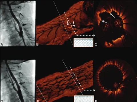
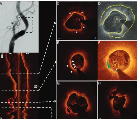
Patient A was a 64-year-old asymptomatic man with 80% angiographic stenosis of the left ICA by NASCET criteria. The patient was considered “high surgical risk” due to se-vere chronic obstructive airways disease and was enrolled in the Abbott-sponsored CHOICE carotid stent registry (http://www.strokecenter.org/trials/clinicalstudies/choice-carotid-stenting-for-high-surgical-risk-patients).
Following placement of a 10⫻40 mm open-cell RX Accu-link carotid stent system (Abbott Vascular), deployed with a 6-mm Viatrac balloon (Abbott Vascular), a postdeployment angiogram demonstrated an unusual eccentric area of non-specific “haziness” in the stented area (Fig 1). OCT was then performed with the demonstration of a large area of severe tissue prolapse without thrombus through the open cells in the Acculink stent (Fig 1B, -C). Prolapse was treated with the placement of a 10⫻ 30 mm closed-cell Xact carotid stent system (Abbott Vascular), with the resolution of prolapse by angiography (Fig 1D) and OCT (Fig 1E, -F).
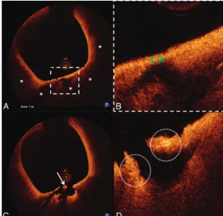
OCT also demonstrated a large TCFA with possible mac-rophage infiltration, plaque rupture, and white thrombus proximal to the stented segment (Fig 2). This segment was not obstructive and could not be detected by angiography. Hence, no additional intervention was performed.
Patient B
Patient B was a 70-year-old asymptomatic man with an abnor-mal right ICA duplex that demonstrated isoechoic and hyper-echoic plaque with marked elevation of flow velocities (peak systolic⫽419 cm/s; peak diastolic⫽165 cm/s) predictive of high-degree ICA stenosis. The contralateral vessel was patent. Cerebral angiography revealed only a 60% right ICA stenosis by NASCET criteria (Fig 3A).
OCT was performed to help determine the severity and complexity of disease, given the discordant findings between noninvasive and invasive studies. OCT revealed a plaque with 55% diameter stenosis at the site of minimal luminal area in the ICA (OCT-derived NASCET criteria). The plaque had ar-eas of calcium (Fig 3) with no signs of plaque instability, such as a large lipid core, thin fibrous cap, erosion, rupture, or thrombus. Intervention was deferred on the basis of such findings.
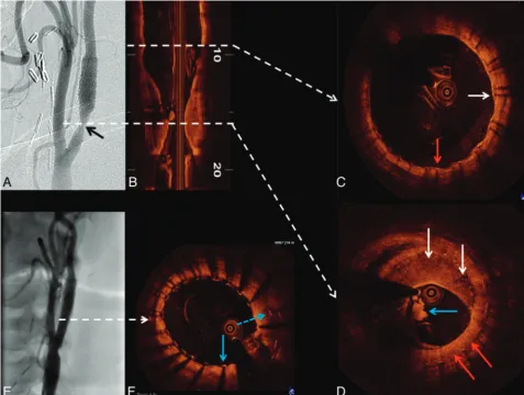
Patient C
Patient C was a 60-year-old man who underwent a left carotid endarterectomy in 1997 and carotid stent placement for ca-rotid restenosis (VIVA Registry: Vivexx stent, C.R. Bard, Mur-ray Hill, New Jersey) in 2006. Carotid angiography performed to evaluate an abnormal ICA duplex study revealed an unusual 60% (NASCET) in-stent restenosis with an area of marked hypolucence in all views (Fig 4A). OCT was undertaken to evaluate these abnormal angiographic findings in a patient
INTERVENTIONAL
ORIGINAL
with a history of both surgical and in-stent restenosis in the target vessel. Findings included a clearly delineated restenotic area with 65% diameter stenosis (OCT-derived NASCET cri-teria) composed of 2 different patterns of neointimal hyper-plasia: a homogeneous signal-intensity-rich area, and a heter-ogeneous region with predominantly poor signal-intensity tissue. Moreover, a large area with irregular contour com-posed of white thrombus (Fig 4D), a marker for clinical risk by pathologic studies, was revealed. Despite the patient’s asymp-tomatic status and with only 60% angiographic stenosis, inter-vention (stent placement with distal embolic protection) was performed (Fig 4E, -F).
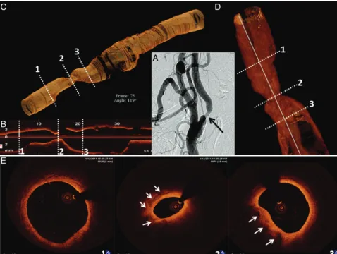
Patient D
Patient D was a 79-year-old woman with asymptomatic right ICA stenosis of moderate severity by duplex examination (ICA peak systolic velocity⫽301 cm/s; diastolic velocity⫽55 cm/s; ICA/CCA ratio ⫽3.8). Diagnostic carotid angiography re-vealed an ulcerated 58% (NASCET) stenosis at the origin of the right ICA (Fig 5A). OCT was performed to assess the le-sion, which had only marginal conventional criteria for revas-cularization. A large complex plaque with extensive
calcifica-tion (Fig 5C, -D) and OCT-derived NASCET measurements of only 40% diameter stenosis was noted. Most important, images also demonstrated large regions of lipid with an exten-sive area of TCFA (Fig 5E, -F) and 2 sites of significant plaque ulceration (Fig 5G, -H). On the basis of these pathologic find-ings and the patient’s age, she was referred for carotid endarterectomy.
Discussion
Ischemic stroke is largely an embolic disease. Nonetheless, current standard-of-practice guidelines suggesting that ca-rotid revascularization with endarterectomy or a stent proce-dure19is beneficial in asymptomatic patients are largely based
on North American20and European21trials. Both trials, lim-iting enrollment to patients with at least 60% angiographic stenosis, reported only modest benefit of an operation over medical management, with a 3-year absolute risk reduction of approximately 3% and no definite correlation between the degree of stenosis and outcomes.
[image:3.594.53.534.45.404.2]plaques. Spangoli et al22examined surgically removed plaques
from patients without symptoms versus those with transient ischemic attack or cerebrovascular accident. They stressed that the presence and severity of clinical events was significantly correlated with thrombus and cap inflammation in ruptured plaques, after assessing endarterectomy specimens in a cohort with similar degrees of angiographic stenosis. Similarly, in an-other histopathologic evaluation, a higher percentage of mac-rophage-rich areas and number of T-cells per square millime-ter in symptomatic-versus-asymptomatic patients with similar degrees of carotid narrowing was described.23
Moreover, using multidetector CT angiography to exam-ine symptomatic atherosclerotic plaques in patients with an-terior circulation disease, Homburg et al24found that culprit
plaque ulcerations were equally distributed between patients with more than and less than 50% carotid stenosis. Further evidence that symptoms from carotid artery disease are corlated with plaque histopathology has been provided by a re-port that established correlation between plaque ulceration, intraluminal thrombus, symptoms, and cerebral microemboli as detected by transcranial Doppler in patients with similar degrees of angiographic stenosis.25Redgrave et al,26in the
his-topathologic study of 526 carotid endarterectomy specimens from recently symptomatic stenosis, found a high prevalence of cap rupture, large lipid core, and attenuated macrophage infiltration, highlighting the fact that these markers for plaque instability were similar to those previously described in unsta-ble coronary artery plaques.
OCT is an emerging technology in the cardiac catheteriza-tion laboratory. Its accuracy in measurements of vascular dimensions,27 in the characterization of atherosclerotic
plaques,11,28 and in the identification of thrombus16 and plaque inflammation15,29has been well demonstrated;
there-fore, it enables imaging of structures that have a potential causative role in cerebrovascular ischemia (eg, thrombus, TCFA with plaque rupture, and inflammation).
In this case series, we report the use of FDOCT to assist in the management of 4 asymptomatic patients in whom a proper course of action was not readily apparent after diag-nostic studies that included conventional angiography. Pa-tient A underwent OCT evaluation to clarify abnormal but nonspecific findings on a carotid angiogram following carotid stent placement, with resultant findings that included marked tissue prolapse through the stent struts and the presence of an otherwise undetectable ruptured TCFA. The 3500-patient CAPTURE (Carotid Acculink/Accunet Post-approval Trial to Uncover unanticipated or Rare Events) carotid stent registry, reporting a 30-day stroke rate of 4.8%, found that only 23% of strokes occurred intraprocedurally,30 raising the possibility
[image:4.594.133.453.44.354.2]full coverage of the segment containing the ruptured TCFA had OCT been performed before stent deployment in this case.
Intervention was, however, deferred in patient B, despite inexplicably high ICA velocities that could otherwise justify revascularization, on the basis of OCT documentation of ⬍60% stenosis and the absence of any features thought to suggest plaque instability by pathologic criteria. Revascular-ization was recommended for patient D, after an OCT evalu-ation of “borderline” carotid duplex and angiographic data demonstrated findings that have been associated with higher risk in the pathology literature.31Patient C, despite the
pres-ence of “low-risk” clinical (restenosis) and angiographic (60% stenosis) markers, was treated with carotid stent placement when an unanticipated large thrombus burden was detected by OCT. This case as well demonstrates the ability of OCT to differentiate normal- and abnormal-appearing tissue on the basis of signal intensity coupled with homogeneity and smoothness of tissue contour. Although these findings have not been previously described in the carotid arteries or vali-dated by OCT, echolucent tissue, described as a “black hole” in intravascular sonography studies after intracoronary brachy-therapy and drug-eluting stent implantation,32is composed of
acellular/hypocellular necrotic tissue, scattered in an extracel-lular matrix rich in proteoglycans but also containing T lym-phocytes and foam cells.33On the basis of known optical prop-erties of different materials, tissue depicting poor signals on OCT images is likely composed of fibrin or organized throm-bus,34though highly organized proteoglycan could also
pro-vide similar images.35
Yoshimura et al36initially described the use of carotid OCT
with balloon occlusion to clarify an ambiguous carotid angio-gram in 2010. More recently, the use of OCT to evaluate ca-rotid artery stent placement was reported in 7 patients for whom the authors emphasized the necessity of proximal bal-loon occlusion (complicated by symptomatic cerebral isch-emia in 1 patient) to obtain acceptable images.37Furthermore,
an early experience with OCT in carotid plaque characteriza-tion was reported in a brief communicacharacteriza-tion that compared OCT with other imaging modalities.38Most recently, a
30-patient cohort undergoing scheduled carotid artery stent placement was studied before and following stent placement with TDOCT.39Again, all patients were imaged after proximal
[image:5.594.54.533.44.404.2]back speed (up to 25 mm/s) compared with TDOCT, enables adequate blood clearance, without the necessity of proximal balloon occlusion. Furthermore, its wider FOV (⬃10 mm) allows imaging of larger arteries.10In our series, only 13% of
all the cross-sections were not suitable for analysis because of blood clearance artifacts or inadequate FOV.
In contrast to these earlier studies, our group also reports the use of data obtained by preintervention FDOCT in clinical decision-making in patients with ambiguous angiographic or noninvasive data. No complications were reported in this case series.
The present cases, although not conclusive, provide impor-tant clinical insights. First, the feasibility of interrogating the carotid vasculature by using high-resolution FDOCT imaging without the need for complex technical adaptations for blood displacement or interruption of carotid flow is shown. Second, these cases are, to the best of our knowledge, the first use of FDOCT to assess plaque morphology and guide, albeit empir-ically, decision-making before planned intervention in human carotid arteries in vivo. Finally, this report also describes the use of “next -generation” analytical imaging software with au-tomated detection and quantification of vascular disease that capitalizes on the 3D nature of FDOCT images.
Several points of caution should be noted in the interpre-tation and use of data presented in this case series. First, ca-rotid artery instrumentation may carry a risk of embolization. While no signals of safety issues in excess of those associated with diagnostic carotid angiography have been reported to date in small series of carotid intravascular sonography and OCT patients,9,36-40the risk versus benefit of this technique will remain uncertain until studies that are appropriately pow-ered to assess safety and efficacy end points are available. Sec-ond, we assumed that OCT histopathologic correlations that have been extensively validated outside of the carotid circula-tion11-16,34,35are also valid in the carotid circulation. Finally,
limited tissue penetration, particularly in lipid-rich plaques, precludes the interpretation and quantification of underlying tissue layers.
In this case series, FDOCT revealed unexpected findings in virtually all patients including marked tissue prolapse through an open-cell carotid stent and the demonstration of ruptured TCFAs and thrombus in lesions defined as “low-risk” accord-ing to current clinical criteria. These unexpected findaccord-ings sug-gest that further studies are needed to evaluate the role of FDOCT in the assessment of asymptomatic carotid artery disease.
[image:6.594.53.531.45.405.2]Conclusions
The present study demonstrates the technical feasibility of FDOCT without balloon occlusion for the assessment of ex-tracranial carotid artery disease. The unique features of this technology, which allow the rapid acquisition of images with unprecedented resolution, make FDOCT a promising tool to help clinicians understand and manage patients with asymp-tomatic carotid artery disease.
Disclosures: Michael R. Jones—RELATED:Consulting Fee or Honorarium: St. Jude Med-ical,UNRELATED:Payment for Lectures (including service on Speakers Bureaus): St. Jude Medical Speakers’ Bureau. Curtis A. Given II—UNRELATED:Payment for Lectures (includ-ing service on Speakers Bureaus):ev3,Comments: I am a proctor and on the Speakers Bureau for Onyx 18, 34, and HD500. Marco A. Costa—UNRELATED:Consultancy: St. Jude Medical,Grants/Grants Pending: St. Jude Medical,*Patents (planned, pending, or issued): a patent of University Hospitals for OCT Software,*OTHER RELATIONSHIPS:prior consul-tant/speaker honorarium fees by Boston Scientific, Cordis, Medtronic, Abbott Vascular. Hiram G. Bezerra—UNRELATED: Consultancy: St. Jude Medical. *Money paid to the institution.
References
1. Roger VL, Go AS, Lloyd-Jones DM, et al, on behalf of the American Heart Association Statistics Committee and Stroke Statistics.Heart disease and stroke statistics: 2011 update—a report from the American Heart Association. Cir-culation2011;123:e2– e220. Epub 2011 Dec 15.
2. Rothwell PM, Warlow CP. Timing of TIAs preceding stroke.Neurology
2005;64:817–20
3. Fine-Edelstein JS, Wolf PA, O’Leary DH, et al.Precursors of extracranial ca-rotid atherosclerosis in the Framingham study.Neurology1994;44:1046 –50 4. Homburg RJ, Rozie S, van Gils M, et al.Association between carotid artery
plaque ulceration and plaque composition evaluated with multidetector CT angiography.Stroke2011;42:367–72
5. Langsfeld M, Gray-Weale AC, Lusby RJ.The role of plaque morphology and diameter reduction in the development of new symptoms in asymptomatic carotid arteries.J Vasc Surg1989;9:548 –57
6. Grønholdt ML, Nordestgaard BG, Schroeder TV, et al.Ultrasonic echolucent carotid plaques predict future strokes.Circulation2001;104:68 –73 7. Topakian R, King A, Kwon SU, et al.Ultrasonic plaque echolucency and
em-boli signals predict stroke in asymptomatic carotid stenosis.Neurology2011; 77:751–58. Epub 2011 Aug 17
[image:7.594.56.535.42.459.2]MR imaging in asymptomatic carotid artery stenosis.AJNRAm J Neuroradiol
2010;31:487–93
9. Irshad K, Millar S, Velu R, et al.Virtual histology intravascular ultrasound in carotid interventions.J Endovasc Ther2007;14:198 –207
10. Bezerra HG, Costa MA, Guagliumi G, et al.Intracoronary optical coherence tomography: a comprehensive review clinical and research applications.
JACC Cardiovasc Interv2009;2:1035– 46
11. Kume T, Akasaka T, Kawamoto T, et al.Assessment of coronary arterial plaque by optical coherence tomography.Am J Cardiol2006;97:1172–75
12. Kume T, Okura H, Kawamoto T, et al.Assessment of the coronary calcification by optical coherence tomography.Eurointervention2011;6:768 –72 13. Kume T, Imanishi T, Takarada S, et al.Assessment of culprit lesion
morphol-ogy in acute myocardial infarction: ability of optical coherence tomography compared with intravascular ultrasound and coronary angioscopy.J Am Coll Cardiol2007;50:933–37. Epub 2007 Aug 20.
14. Kume T, Akasaka T, Kawamoto T, et al.Measurement of the thickness of the fibrous cap by optical coherence tomography.Am Heart J2006;152:755.e1– 4 15. Tearney GJ, Yabushita H, Houser SL, et al.Quantification of macrophage con-tent in atherosclerotic plaques by optical coherence tomography.Circulation
2003;107:113–19
16. Kume T, Akasaka T, Kawamoto T, et al.Assessment of coronary arterial thrombus by optical coherence tomography.Am J Cardiol2006;97:1713–17 17. Wang Z, Kyono H, Bezerra HG, et al.Semiautomatic segmentation and
quan-tification of calcified plaques in intracoronary optical coherence tomography images.J Biomed Opt2010;15:061711
18. Bezerra HG, Attizzani GF, Costa MA.Three-dimensional imaging of fibrous cap by frequency-domain optical coherence tomography.Catheter Cardiovasc Interv2011 Sep 27. [Epub ahead of print]
19. Brott TG, Hobson RW, Howard G, et al.Stenting versus endarterectomy for treatment of carotid-artery stenosis.N Engl J Med2010;363:11–23. Epub 2010 May 26
20.Endarterectomy for asymptomatic carotid artery stenosis: Executive Com-mittee for the Asymptomatic Carotid Atherosclerosis Study. JAMA
1995;273:1421–28
21. Halliday A, Mansfield A, Marro J, et al.Prevention of disabling and fatal strokes by successful carotid endarterectomy in patients with recent neuro-logical symptoms: randomised controlled trial.Lancet2004;363:1491–501 22. Spangoli LG, Mauriello A, Sangiorgi GC, et al.Extracranial thrombotically
active carotid plaque as a risk factor for ischemic stroke.JAMA2004;292: 1845–52
23. Jander S, Sitzer M, Schumann R, et al.Inflammation in high-grade carotid stenosis: a possible role for macrophages and T cells in plaque destabilization.
Stroke1998;29:1625–30
24. Homburg PJ, Rozie S, Van Gils MJ, et al.Atherosclerotic plaque ulceration in the symptomatic internal carotid artery is associated with nonlacunar isch-emic stroke.Stroke2010;41:1151–56
25. Sitzer M, Muller W, Siebler M, et al.Plaque ulceration and lumen thrombus are the main sources of cerebral microemboli in high-grade internal carotid ar-tery stenosis.Stroke1995;26:1231–33
26. Redgrave JN, Lovett JK, Gallager PJ, et al.Histological assessment of 526 symp-tomatic carotid plaques in relation to the nature and timing of ischemic symptoms: the Oxford plaque study.Circulation2006;113:2320 –28 27. Tahara S, Bezerra HG, Baibars M, et al.In vitro validation of new
Fourier-domain optical coherence tomography.Eurointervention2011;6:875– 82 28. Yabushita H, Bouma BE, Houser SL, et al.Characterization of human
athero-sclerosis by optical coherence tomography.Circulation2002;106:1640 – 45 29. MacNeill B, Jang IK, Bouma BE, et al.Focal and multi-focal plaque
macro-phage distributions in patients with acute and stable presentations of coro-nary artery disease.J Am Coll Cardiol2004;44:972–79
30. Fairman R, Gray WH, Scicli AP, et al.The CAPTURE registry: analysis of strokes resulting from carotid artery stenting in the post approval setting— location, severity, and type.Ann Surg2007;246:551–58
31. Finn A, Nakano M, Narula J, et al.Concept of vulnerable/unstable plaque.
Arterioscler Thromb Vasc Biol2010;30:1282–92
32. Costa MA, Sabate M, Angiolillo DJ, et al.Intravascular ultrasound character-ization of the “black hole” phenomenon after drug-eluting stent implanta-tion.Am J Cardiol2006;97:203– 06
33. Kay IP, Ligthart JM, Virmani R, et al.The black hole: echolucent tissue ob-served following intracoronary radiation.Int J Cardiovasc Intervent2003; 5:137– 42
34. Templin C, Meyer M, Muller MF, et al.Coronary optical frequency domain imaging (OFDI) for in vivo evaluation of stent healing: comparison with light and electron microscopy.Eur Heart J2010;31:1792– 801
35. Teramoto T, Fumiaki I, Otake H, et al.Intriguing peri-strut low-intensity area detected by optical coherence tomography after coronary stent deployment.
Circ J2010;74:1257–59
36. Yoshimura S, Kawasaki M, Hatteri A, et al.Demonstration of intraluminal thrombus in the carotid artery by optical coherence tomography: technical case report.Neurosurgery 2010;67(3 suppl operative):onsE305, discussion onsE305.
37. Reimers B, Nikas D, Stabile E, et al.Preliminary experience with optical coher-ence tomography imaging to evaluate carotid artery stents.Eurointervention.
2011;6:98 –105
38. Yoshimura S, Kawasaki M, Yamada K, et al.OCT of human carotid arterial plaques.JACC Cardiovasc Imaging2011;4:432–36
39. Yoshimura S, Kawasaki M, Yamada K, et al.Visualization of internal carotid artery atherosclerotic plaques in symptomatic and asymptomatic patients: a comparison of optical coherence tomography and intravascular ultrasound.
AJNR Am J Neuroradiol2011;33:308 –13