Diagnosis of lung cancer – improving survival rates
ATHEY, V. L., TOD, A. M., SUCKLING, R. and ROGERS, T. K.
Available from Sheffield Hallam University Research Archive (SHURA) at:
http://shura.shu.ac.uk/2529/
This document is the author deposited version. You are advised to consult the publisher's version if you wish to cite from it.
Published version
ATHEY, V. L., TOD, A. M., SUCKLING, R. and ROGERS, T. K. (2010). Diagnosis of lung cancer – improving survival rates. European oncology, 6 (2), 26-30.
Copyright and re-use policy
European Oncology
Diagnosis of Lung Cancer: Improving Survival Rates
Victoria L Athey1,2, Angela M Tod3, Rupert Suckling4, Trevor K Rogers1
1. Doncaster & Bassetlaw Hospitals NHS Foundation Trust 2. University of Sheffield
3. Sheffield Hallam University 4. NHS Doncaster
Author for Correspondence
Victoria Athey
Clinical Research Fellow Chest Clinic
Doncaster Royal Infirmary Armthorpe Road
Doncaster DN2 5LT
v.athey@sheffield.ac.uk 01302 647021
Word Count (excluding references) = 2,250
Key Words: Lung Cancer, Early Diagnosis, Screening, Social Marketing
Abstract
Lung cancer is a major global health burden with high incidence rates but poor long-term survival. Currently the majority of cases are diagnosed at an advanced stage when surgical resection is not feasible.
Screening for lung cancer has been a major focus of research for the last 40 years. Despite this there is still a lack of evidence to promote its use outside of a clinical trial.
Background
Lung cancer is a major worldwide health burden, responsible for 1.3 million deaths in 2004, equating to 2.3% of all deaths. Death rates from lung cancer are predicted to continue to rise, with the disease being responsible for 2.8% of all deaths (1.67 million) by 20151.
Despite advances in treatment, survival rates from lung cancer in the UK have improved by only a few percent in the last 40 years. 5-year survival for patients diagnosed between 1991 and 1993 was 5%2.
Eurocare-43 has highlighted the difference in survival between
England and other European countries. 5-year survival rates in England, for patients diagnosed between 1995 and 1999, were 8.4% compared with the average European rate of 12%. These figures are in even greater contrast to reported 5-year survival rates in the USA of 15.7%, for patients diagnosed between 1995 and 20014. Analysis of
Eurocare-4 also showed that 1-year survival rates in England were lower than the European average, probably reflecting poorer access to care. This would suggest a particular need to promote earlier diagnosis in the UK, in trying to improve survival.
Survival is dependant on disease stage at diagnosis, with marked variation between earlier and later stage disease. 5-year survival for localised disease is around 49% compared with 2% for disease with distant metastases at presentation4. Unfortunately the majority of
lung cancers are already disseminated at the time of presentation4,5.
Screening
Much interest has focussed on diagnosing lung cancer earlier in order to try to improve radical treatment rates and reduce mortality. Initially this interest focussed on screening. The first randomised controlled trial took place in London in the 1960’s6. This looked at a 6-monthly
chest-x-ray for 3 years versus a chest x-ray at the beginning and end of the 3 year period. Diagnosis and resection rates were higher in the group receiving more frequent chest x-rays but lung cancer mortality was similar in both groups. Three American studies7 in the 1970’s and
80’s looked at the use of either chest x-ray alone or in combination with sputum cytology. The Mayo Lung Project8 compared 4-monthly
chest x-ray and sputum cytology to standard care. Participants randomised to the standard care arm were advised to have a chest x-ray and sputum cytology yearly. This showed that resection rates increased by 14% (32% to 46%) in the group undergoing screening when compared to the group receiving standard care alone, but no stage shift was evident. 5-year survival in the screened group reached 33% in comparison to 15% in the non-screened group.
cancer mortality was no different in the 2 groups (3.2/1000 person-years versus 3.0/1000 person-person-years). This lack of improvement in mortality was also evident in the other two studies: the Johns Hopkins8,and Memorial Sloan-Kettering studies9. Both looked at the
addition of 4-monthly sputum cytology to annual chest x-ray. There was also a contemporaneous study10, in Czechoslovakia, comparing
chest x-ray plus sputum cytology every 6 months, for 3 years, with chest x-ray and sputum cytology at the beginning and end of the 3 years. This study essentially replicated the findings of the Mayo Lung Project, with an increased number of cancers detected in the more frequently screened group but with no difference in lung cancer associated mortality11. None of these studies had a ‘no screening’
control group.
Therefore, based on these studies, it would not be possible to recommend either chest x-ray or sputum cytology as a screening test for lung cancer. Indeed, a Cochrane systematic review12 has suggested
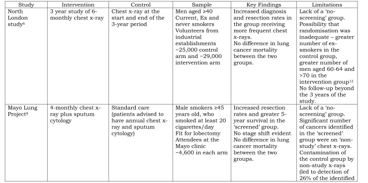
Table 1: Summary of key randomised-controlled trials of the use of chest x-ray with or without sputum cytology for screening for lung cancer
Study Intervention Control Sample Key Findings Limitations North
London study6
3 year study of 6-monthly chest x-ray
Chest x-ray at the start and end of the 3-year period
Men aged ≥40 Current, Ex and never smokers Volunteers from industrial
establishments ~25,000 control arm and ~29,000 intervention arm
Increased diagnosis and resection rates in the group receiving more frequent chest x-rays.
No difference in lung cancer mortality between the two groups.
Lack of a ‘no
-screening’ group.
Possibility that randomisation was inadequate – greater number of
ex-smokers in the control group, greater number of men aged 60-64 and >70 in the
intervention group12
No follow-up beyond the 3 years of the study.
Mayo Lung Project8
4-monthly chest x-ray plus sputum cytology
Standard care (patients advised to have annual chest x-ray and sputum cytology)
Male smokers ≥45
years old, who smoked at least 20 cigarettes/day Fit for lobectomy Attendees at the Mayo clinic
~4,600 in each arm
Increased resection rates and greater 5-year survival in the
‘screened’ group.
No stage shift evident No difference in lung cancer mortality between the two groups.
Lack of a ‘no
-screening’ group.
Significant number of cancers identified
in the ‘screened’ group were on ‘non
cancers in this group). Johns Hopkins Lung Project8,13
Annual chest x-ray plus 4-monthly sputum cytology
Annual chest x-ray Male smokers(≥20 cigarettes/day)
aged ≥45 years
~5,200 in each arm
No difference in the number of lung cancers diagnosed, resection rates or lung cancer mortality between the two groups.
Greater number of squamous cell carcinoma identified in the intervention group.
Underpowered to evaluate the efficacy of sputum cytology as a screening test12
Memorial Sloan-Kettering Cancer Center Lung Cancer Screening Programme8,9
Annual chest x-ray plus 4-monthly sputum cytology
Annual chest x-ray Male smokers ≥45 years old.
Current (or within the last year) smokers of ≥20 cigarettes/day ~5,000 in each arm
No difference in the number of lung cancers diagnosed, resection rates or lung cancer mortality between the two groups
Underpowered to evaluate the efficacy of sputum cytology as a screening test12
Kubík et al 198610
3 year study of 6-monthly chest x-ray and sputum
cytology followed by annual chest x-ray for 3 years
Chest x-ray and sputum cytology at the beginning and end of the 3 year period, followed by annual chest x-ray for 3 years
Men aged 40-64 Attending the chest clinic
Lifetime cigarette consumption of >150,000 cigarettes ~3,170 men in each arm
Increased rate of diagnosis in the more frequently screened group during years 1-3.
No stage shift
No difference in lung cancer mortality between the 2 groups
Lack of a ‘no
Chest x-rays are less sensitive for the detection of lung cancer than CT. Evaluation of the chest x-rays taken as part of the Mayo Lung Project identified that 90% of peripheral lung cancers and 65-70% of central tumours, that were detected by 4-monthly chest x-ray had, in retrospect, been visible on previous x-rays14. Interest has therefore
moved to the use of computed tomography (CT) for screening. This was made possible by the advent of low-dose spiral CT, which reduced both the radiation dose and scan time15. Early reports showed
increased rates of detection over chest x-ray, with the vast majority of detected tumours being stage I 16,17,18.The largest observational report
of CT screening is the International Early Lung Cancer Action Project (I-ELCAP)19. A total of 31,567 participants, aged over 40 and deemed
to be at lung cancer risk due to either cigarette smoking or occupational exposure, had a baseline CT. All patients had to be fit to undergo thoracic surgery, if required. 27,456 repeat scans were performed between 7 and 18 months after the previous screening. 479 cancers were identified, 405 on the initial scan and 74 on annual screening. There were 5 interim diagnoses. Overall 85% of tumours were clinical stage I with 72% confirmed pathological stage I. 10-year lung cancer survival, for all participants, was 80%, increasing to 88% in those with clinical stage I disease.
However, a key criticism of CT is that it identifies nodules that will ultimately turn out to be non-malignant. During the prevalence screen in the I-ELCAP study19, 13% of CTs identified non-calcified nodules
requiring further investigation, including serial CTs, PET-CT and percutaneous biopsy. Only 9.6% of these participants were subsequently proven to have lung cancer. Even higher rates of benign nodule identification have been quoted in other studies20,21,22,23 and
up to 20% of invasive procedures following CT screening are for benign disease 20,21,23.
An additional concern is overdiagnosis, such that patients receive treatment for slowly growing tumours that may never have caused them any problems in their lifetime, a phenomenon that is already recognised in other screening programmes24,25. Several studies have
calculated tumour volume doubling times for screen-detected cancers. These have shown that many of the screen-identified tumours are slow growing, with doubling times well in excess of the 40-70 days calculated from epidemiological data of non-screen identified cancers26.
At present there is insufficient evidence to recommend low dose CT as a screening test for lung cancer, although there are several randomised controlled trials currently underway seeking definitively to answer this question 27,28,29.
autofluorescence bronchoscopy. One observational study looked at the use of bronchoscopy, along with CT, as a primary tool in screening20,30. Volunteer current and former smokers underwent
sputum induction for quantitative cytometry and CT before being offered autofluorescence bronchoscopy. 561 subjects were enrolled in the study, with 378 undergoing bronchoscopy. 14 primary lung cancers were identified of which 4 (29%) were CT occult and only detected by autofluorescence bronchoscopy. All of these CT occult cancers were squamous cell carcinomas. Because of the observational nature of the study, the significance of the use of this approach on mortality is unknown.
Biological tools, such as testing serum for tumour-associated antibodies, detection of gene-promoter hypermethylation in sputum samples, exhaled breath volatile organic compounds and detection of novel proteins in serum or sputum are also in development31.
Unfortunately, none of these is currently ready for use in clinical practice.
At the current time, no form of screening for lung cancer can be recommended.
Symptom recognition and reporting
Interest has now switched to looking at whether lung cancer can be diagnosed earlier in its natural history by focussing on promoting symptom recognition and reporting. 90% of patients are symptomatic at the time of diagnosis32, often experiencing multiple
symptoms33,34,35. Many of those presenting will have been
symptomatic for many months, with reported delays to healthcare of up to 2 years33. Much work has focussed on investigating this, with
reported median delays from onset of symptoms to presenting to health care ranging from 7 to 31 days35,36,37,38,39. Public knowledge of
lung cancer symptoms generally appears to be poor35,37,40,41. Patients
often develop symptoms but are unaware that they could be related to a sinister cause: it appears that between 50 and 75% of lung cancer patients may not be aware of the significance of their symptoms35,37.
Only when further symptoms develop, or their general health deteriorates, will they seek advice33,35. In particular, systemic
symptoms such as lethargy and weight loss seem to be associated with longer delays, whereas haemoptysis tends to prompt a more rapid response33,39.
It has also been noted that even those deemed to be at risk of lung cancer, predominantly current and ex-smokers, do not always perceive themselves to be at risk35,41. Even when patients recognise a
smoker, being unsure as to whether the symptom/change experienced
is ‘normal’, putting the symptom down to being part of the ageing
process, minimising symptoms, stoicism and the difficulty of separating out current changes in health care from co-morbid conditions40,41.
Delays have also been identified once patients present to their primary care team, with many patients having to present on more than one occasion before onward referral/further investigation. This is despite clear advice in the British National Institute for Clinical Excellence (NICE) guidelines regarding chest x-ray referral42. The delay from first
presentation to referral to a respiratory specialist has been reported to range from a mean of 34 days38 to 73 days (range 0->175)37,36,39,43,44.
Bowen & Raynor’s study37 also showed that, of the 76% of patients
who first consulted their own family doctor, only one third of patients were referred following their initial consultation, with a further third referred by another doctor in the practice, suggesting a 2nd
consultation. A Danish study looked at potential reasons for the delay in onward referral of symptomatic patients and identified several contributing factors. In patients with co-morbid diseases, symptoms were often ascribed to this rather than potential lung cancer45. Chest
x-rays reported as normal were associated with a longer delay, with primary care teams being falsely reassured. 25% of lung cancer diagnoses in the UK are made during an acute admission, despite the patient having presented previously to their primary healthcare team with a symptom that could be indicative of lung cancer46.
Improving the early diagnosis of lung cancer in Britain has become a government priority, with the formation of the National Awareness and Early Detection Initiative (NAEDI), a key component of the 2007 cancer reform strategy47. This hypothesizes that delays lead to more
advanced disease at diagnosis with associated poor 1- and 5-year survival rates and potentially avoidable deaths. Abdel-Rehman et al calculated that if UK survival rates were similar to those in Europe, then nearly 1000 deaths/year, within 5 years of the diagnosis of lung cancer, could be avoided48.
The NAEDI pathway highlights many areas, which could be targeted in order to try to promote earlier diagnosis49.
Figure 1 The NAEDI pathway49 (From Richards 2009)
(JPEG file)
commercial marketing techniques to change individual and organisational behaviours and policies50.
Similar approaches have already been used in other cancers, an example of which is the West of Scotland Cancer Awareness Project (WoSCAP)51. This project used a mass media campaign combined with
general practice education to raise awareness of the symptoms of oral and colorectal cancer. Awareness of symptoms was improved and, in those presenting who were aware of the campaign, presentation was timelier in 60%.
An initial social marketing pilot has been carried out in lung cancer, in Doncaster, the largest metropolitan borough in the UK, which has a high rate of lung cancer52,53. And a high rate of social deprivation. The
social marketing campaign and primary care education programme was initially designed as a way of addressing a recognised health inequality. Six areas, covered by eleven general practice surgeries, with the highest lung cancer risk, were identified. In these areas, brief intervention training was undertaken with the general practitioners, practice nurses and local pharmacists. Following this there was a public awareness campaign launched comprising leaflets, advertising on bus banners and billboards (Figure 2), local media events and coughing bus stops.
Figure 2 Poster used on billboards
(JPEG file)
This project increased awareness of the importance of seeking medical advice for a prolonged cough and resulted in a statistically significant increase in chest x-ray referrals. Lung cancer diagnosis rates were also increased although this was not at a statistically significant level. No stage shift was evident but the numbers at different lung cancer stages were too small for subgroup analysis53,54.
Conflict of Interest
The authors declare no conflict of interest
References
1 World Health Organisation. The global burden of disease: 2004 update. 2008. Geneva: WHO press.
http://www.who.int/healthinfo/global_burden_disease/2004_report_update/en /index.html
2 Quinn M, Babb P, Brock A et al. Cancer Trends in England and Wales 1950-1999. SMPS No 66. Office of National Statistics. The Stationary Office. London. 2001.
3 Sant M, Allemani C, Santaquilani M et al, EUROCARE working group.
EUROCARE-4 Survival of cancer patients diagnosed in 1995-1999. Results and Commentary. Eur J of Cancer 2009;45:931-991.
4 SEER Cancer Statistics Review 1975-2006 Section 15: lung and bronchus. National Cancer Institute.
http://seer.cancer.gov/csr/1975_2006/results_merged/sect_15_lung_bronchus. pdf
5 Cancer Research UK. Lung cancer and smoking statistics: Key facts. http://info.cancerresearchuk.org/cancerstats/types/lung/index.htm
6 Brett GZ. The value of lung cancer detection by 6-monthly chest radiographs. Thorax 1968;23:414-420.
7 Berlin NI, Buncher CR, Fontana RS et al. The National Cancer Institute Cooperative Early Cancer Detection program. Results of the initial screen (prevalence). Early lung cancer detection: Introduction. Am Rev Respir Dis 1984;130:545-49.
8 Fontana RS, Sanderson DR, Woolner LB et al. Screening for lung cancer: a critique of the Mayo Lung Project. Cancer 1991;67(4 Suppl):1155-1164. 9 Melamed MR, Flehinger BJ, Zaman MB et al. Screening for early lung cancer.
Results of the Memorial Sloan-Kettering study in New York. Chest 1984;86:44-53.
10 Kubík A, PoláK J. Lung Cancer Detection: Results of a Randomized Prospective Study in Czechoslovakia. Cancer 1986;57:2427-2437.
11 Kubík AK, Parkin DM, Zatloukal P. Czech Study on Lung Cancer Screening: Post-Trial Follow Up of Lung Cancer Deaths Up to Year 15 Since Enrollment. Cancer 2000;89(Suppl 11):2363-2368.
12 Manser R, Irving LB, Stone C et al. Screening for lung cancer. Cochrane Database of Systematic Reviews 2004, Issue 1. Art. No.: CD001991. DOI: 10.1002/14651858.CD001991.pub2
13 Frost JK, Ball WC Jr, Levin ML et al. Early lung cancer detection: results of the initial (prevalence) radiologic and cytologic screening in the Johns Hopkins study. Am Rev Respir Dis 1984;130:549-54
14 Muhm JR, Miller WE, Fontana RS et al. Lung cancer detected during a screening programme using four-month chest radiographs. Radiology 1983;148:609-615. 15 Midthun DE, Jett JR. Chapter 4: Screening for lung cancer. In Thoracic
Malignancies. Spiro SG, Huber RM and Janes SM (eds) Eur Respir Mon 2009;44:57-70
16 Kaneko M, Eguchi K, Ohmatsu et al. Peripheral lung cancer: screening and detection with low-dose spiral CT versus radiography. Radiology 1996;201:798-802
18 Henschke CI, McCauley DI, Yankelevitz DF et al. Early Lung Cancer Action Project: overall design and findings from baseline screening. Lancet
1999;354:99-105.
19 The International Early Lung Cancer Action Programme Investigators. Survival of Patients with Stage I Lung Cancer Detected on CT Screening. N Engl J Med 2006;355:1763-1771.
20 McWilliams A, Mayo J, MacDonald S et al. Lung cancer screening: a different paradigm. Am J Respir Crit Care Med 2003;168:1167-1173.
21 Diederich S, Wormanns D, Semik M et al. Screening for early lung cancer with low dose spiral CT: prevalence in 817 asymptomatic smokers. Radiology 2002;222:773-781
22 Swensen SJ, Jett JR, Hartman TE et al. CT screening for lung cancer: five-year prospective experience. Radiology 2005;235:259-265.
23 Crestanello JA, Allen MS, Jett JR et al. Thoracic surgical operations in patients enrolled in a computed tomography screening trial. J Thorac Cardiovasc Surg 2004;128:254-259
24 Zackrisson S, Andersson I, Janzon L et al. Rate of over-diagnosis of breast cancer 15 years after end of Mälmo mammographic screening trial: follow-up study. BMJ, doi:10.1136/bmj.38764.572569.7C (published 3 March 2006)
25 Draisma G, Boer R, Otto SJ et al. Lead times and overdetection due to prostate-specific antigen screening: estimates from the European randomized study of screening for prostate cancer. J Natl Cancer Inst 2003;95:868-78.
26 Bach PB, Silvestri GA, Hanger M et al. Screening for Lung Cancer: ACCP evidence-based clinical practice guidelines (2nd Edition). Chest
2007;132:69S-77S
27 Aberle DR, Black WC, Golding JG et al. Experimental design and outcomes of the National Lung Screening Trial (NLST): a multicenter randomized controlled trial of the helical CT vs chest x-ray for lung cancer screening (abstract). Am J Respir Crit Care Med 2003;167:A736
28 UK lung cancer screening trial (UKLS) – Feasibility study and protocol development. http://www.hta.ac.uk/1752
29 van Iersel CA, de Koning HJ, Draisma G et al. Risk-based selection from the general population in a screening trial: Selection criteria, recruitment and power for the Dutch-Belgian randomised lung cancer multi-slice CT screening trial (NELSON). Int J Cancer 2006;120:868-874.
30 McWilliams AM, Mayo JR, Ahn MI et al. Lung cancer screening using multi-slice thin-section computed tomography and autofluorescence bronchoscopy. J Thorac Oncol 2006;1:61-68.
31 Ghosal R, Kloer P, Lewis KE. A review of novel biological tools used in screening for the early detection of lung cancer. Postgrad Med J 2009;85:358-363.
32 NHS Executive. Referral guidelines for suspected cancer. Department of Health. London. 2002
33 Corner J, Hopkinson J, Fitzsimmons D et al. Is late diagnosis of lung cancer inevitable? Interview study of patients’ recollections of symptoms before diagnosis. Thorax 2005;60:314-319.
34 Buccheri G, Ferrigno D. Lung cancer: Clinical presentation and specialist referral time. Eur Respir J 2004;24:98-904.
35 Smith SM, Campbell NC, MacLeod U et al. Factors contributing to the time taken to consult with symptoms of lung cancer: a cross sectional study. Thorax
2009;64:523-531
36 Koyi H, Hillerdal G, Brandén E. Patient’s and doctors’ delays in the diagnosis of chest tumours. Lung Cancer 2002;35:53-57.
37 Bowen EF, Raynor CFJ. Patient and GP led delays in the recognition of symptoms suggestive of lung cancer. Lung Cancer 2002;37:227-228. 38 Salomaa E-R, Sällinen S, Hiekkanen H et al. Delays in the diagnosis and
39 Lövgren M, Leveälahti H, Tishelman C et al. Time spans from first symptom to treatment in patients with lung cancer – The influence of symptoms and demographic characteristics. Acta Oncologica 2008;47:397-405.
40 Corner J, Hopkinson J, Roffe J. Experience of health changes and reasons for delay in seeking care: A UK study of the months prior to the diagnosis of lung cancer. Social Science and Medicine 2006;62:1381-1391.
41 Tod A, Craven J, Allmark P. Diagnostic delay in lung cancer: a qualitative study. Journal of Advanced Nursing 2008;61(3):336-343.
42 National Institute for Clinical Excellence. Lung Cancer: NICE guidelines. 2005. http://www.nice.org.uk/CG024NICEguidelines
43 Jones RVH, Dudgeon TA. Time between presentation and treatment of six common cancers: a study in Devon. Br J Gen Pract 1992;42:419-422.
44 Annakkaya AN, Arbak P, Balbay O et al. Effect of symptom-to-treatment interval on prognosis in lung cancer. Tumori 2007;93:61-7.
45 Bjerager M, Palshof T, Dahl R et al. Delay in the diagnosis of lung cancer in general practice. Br J Gen Pract 2006;56:863-868.
46 Barrett J, Hamilton W. Pathways to the diagnosis of lung cancer in the UK: a cohort study. BMC Family Practice 2008;9:31
47 Department of Health: Cancer Reform Strategy, Crown, 2007.
http://www.dh.gov.uk/prod_consum_dh/groups/dh_digitalassets/documents/d igitalasset/dh_081007.pdf
48 Abdel-Rehman M, Stockton D, Rachet B et al. What if cancer survival in Britain was the same as in Europe: how many deaths are avoidable? Br J Cancer 2009;101:S115-124
49 Richards MA. The National Awareness and Early Detection Initiative in England: assembling the evidence. Br J Cancer 2009;101:S1-4.
50 Evans WD, McCormack L. Applying Social Marketing in Health Care:
Communicating Evidence to Change Consumer Behaviour. Med Decis Making 2008;28:781.
51 Hastings G, McDermott L. Putting social marketing into practice. BMJ 2006;332:1210-1212.
52 Tod AM, Suckling R. Early Lung Cancer Identification in Doncaster (ELCID). Lung Cancer 2009;63(1):S19
53Rogers TK, Athey V, Tod A, Walters SJ, Suckling R. Early detection of lung cancer: A social marketing evaluation. Thorax 2009;64(suppl IV):A21