1,5-Dimethyl-2-phenyl-4-[phenyl(pyri-din-2-ylamino)methyl]-1
H
-pyrazol-3(2
H
)-one
K. Krishnakumar,aS. Franklin,b* G. Venkatesa Prabhua and T. Balasubramanianc
aDepartment of Chemistry, National Institute of Technology, Tiruchirappalli 620 015, Tamilnadu, India,bDepartment of Physics, Bishop Heber College (Autonomous), Tiruchirappalli 620 017, Tamilnadu, India, andcDepartment of Physics, National Institute of Technology, Tiruchirappalli 620 015, Tamilnadu, India
Correspondence e-mail: xrdfrank@yahoo.co.in
Received 22 August 2012; accepted 15 October 2012
Key indicators: single-crystal X-ray study;T= 273 K; mean(C–C) = 0.003 A˚; Rfactor = 0.040;wRfactor = 0.095; data-to-parameter ratio = 11.4.
In the title compound, C23H22N4O, the pyrazole ring makes dihedral angles of 45.57 (11) with the attached phenyl ring,
and 83.98 (10) and 67.85 (10) , respectively, with the other
phenyl ring and the pyridyl ring. The pyridyl ring makes a dihedral angle of 80.15 (10)with the adjacent phenyl ring. In
the crystal, N—H O hydrogen bonds supplemented by weak C—H O hydrogen bonds link the molecules into chains which run parallel to thea-axis direction.
Related literature
For the origin of the material studied, see: Vijayan (1971); Singh & Vijayan (1973). For related structures, see: Singh & Vijayan (1974, 1976); Tordjman et al. (1991); Yadav et al.
(2003); Li & Zhang (2004); Wen (2005); Sunet al.(2007).
Experimental
Crystal data
C23H22N4O Mr= 370.45
a= 5.701 (5) A˚
b= 12.485 (5) A˚
c= 26.736 (5) A˚
V= 1903.0 (19) A˚3
MoKradiation
= 0.08 mm1
T= 273 K 0.30.20.2 mm
Data collection
Bruker Kappa APEXII CCD diffractometer
Absorption correction: multi-scan (SADABS; Bruker, 1999)
Tmin= 0.976,Tmax= 0.984
14432 measured reflections 2959 independent reflections 2337 reflections withI> 2(I)
Rint= 0.036
Refinement
R[F2> 2(F2)] = 0.040 wR(F2) = 0.095 S= 1.01 2959 reflections 259 parameters
H atoms treated by a mixture of independent and constrained refinement
max= 0.18 e A˚
3
min=0.19 e A˚
[image:1.610.50.259.542.715.2]3
Table 1
Hydrogen-bond geometry (A˚ ,).
D—H A D—H H A D A D—H A
N3—H3A O1i 0.85 (3) 2.16 (3) 2.982 (3) 161 (2) C18—H18 O1i
0.93 2.58 3.335 (3) 138 C23—H23C O1ii
0.96 2.59 3.512 (3) 162
Symmetry codes: (i)x1;y;z; (ii)x;yþ1 2;zþ
3 2.
Data collection:APEX2(Bruker, 2004); cell refinement:APEX2 and SAINT-Plus (Bruker, 2004); data reduction:SAINT-Plus and XPREP (Bruker, 2004); program(s) used to solve structure: SHELXS97(Sheldrick, 2008); program(s) used to refine structure: SHELXL97 (Sheldrick, 2008); molecular graphics: ORTEP-3 for Windows(Farrugia, 1997) andMercury(Macraeet al., 2006); software used to prepare material for publication:PLATON(Spek, 2009).
The authors thank the SAIF at IITM Chennai, sponsored by DST India, for providing analytical facilities.
Supplementary data and figures for this paper are available from the IUCr electronic archives (Reference: GO2065).
References
Bruker (1999).SADABS. Bruker AXS Inc., Madison, Wisconsin, USA. Bruker (2004).APEX2,SAINT-PlusandXPREP. Bruker AXS Inc., Madison,
Wisconsin, USA.
Farrugia, L. J. (1997).J. Appl. Cryst.30, 565.
Li, Z.-X. & Zhang, X.-L. (2004).Acta Cryst.E60, o2199–o2200.
Macrae, C. F., Edgington, P. R., McCabe, P., Pidcock, E., Shields, G. P., Taylor, R., Towler, M. & van de Streek, J. (2006).J. Appl. Cryst.39, 453–457. Sheldrick, G. M. (2008).Acta Cryst.A64, 112–122.
Singh, T. P. & Vijayan, M. (1973).Acta Cryst.B29, 714–720. Singh, T. P. & Vijayan, M. (1974).Acta Cryst.B30, 557–562. Singh, T. P. & Vijayan, M. (1976).Acta Cryst.B32, 2432–2437. Spek, A. L. (2009).Acta Cryst. D65, 148–155.
Sun, Y.-F., Li, J.-K., Zheng, Z.-B. & Wu, R.-T. (2007).Acta Cryst.E63, o2522– o2523.
Tordjman, I., Durif, A. & Masse, R. (1991).Acta Cryst.C47, 351–353. Vijayan, M. (1971).Curr. Sci.40, 262–264.
Wen, P. (2005).Acta Cryst.E61, o2918–o2920.
Yadav, P. N., Demertzis, M. A., Kovala-Demertzi, D., Skoulika, S. & West, D. X. (2003).Inorg. Chim. Acta,349, 30–36.
Structure Reports
Online
supporting information
Acta Cryst. (2012). E68, o3193 [doi:10.1107/S1600536812042936]
1,5-Dimethyl-2-phenyl-4-[phenyl(pyridin-2-ylamino)methyl]-1
H
-pyrazol-3(2
H
)-one
K. Krishnakumar, S. Franklin, G. Venkatesa Prabhu and T. Balasubramanian
S1. Comment
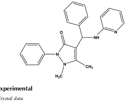
Antipyrine (2,3-dimethyl-1-phenylpyrazol-5-one) is one of the best known pyrazole derivatives used as pain-relieving medicine. It is the first pyrazole derivative to be introduced as an analgesic and antipyretic drug. Antipyrine and its derivatives have been long known for their wide spectrum of biological activities. Their roles in biological processes have become a topic of study in recent years. Though rarely used alone, nowadays, on account of its toxicity, antipyrine forms part of some combination products used as pain-relieving medicines. Many of its derivatives like amidopyrine and metamizol are well known and widely used analgesics. Therefore a detailed knowledge of the structure and the possible modes of interaction of antipyrine are important in elucidating the molecular mechanism of the action of pain-relieving medicines. The crystal structure of antipyrine, its derivatives and some of its metallic complexes have already been reported (see related literature). Presently, the crystal structure of 2-amino pyridino benzyl antipyrine {1,2-dihydro-2,3-dimethyl-1-phenyl-4-(phenyl (pyridin-2-yl amino) methyl)pyrazol-5-one} has been elucidated. The conformational features and the hydrogen bonding analysis have been analysed in order to have a further insight in to their chemical and pharmaceutical aspects. The chemical scheme of 2-amino pyridino benzyl antipyrine is shown.
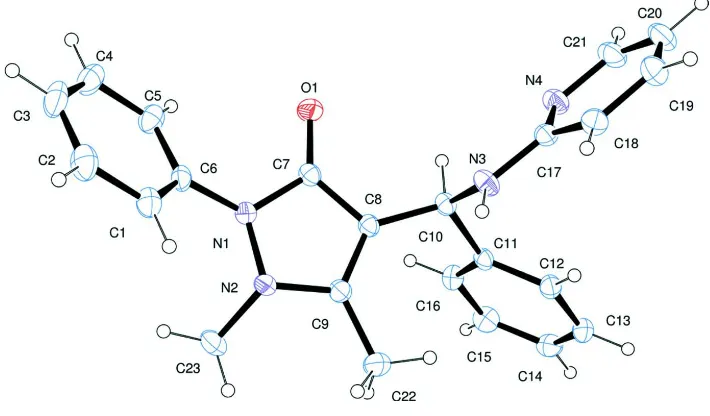
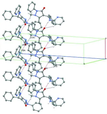
An ORTEP-3 (Farrugia, 1997) diagram of the asymmetric unit of 2-amino pyridino benzyl antipyrine (APBA) is shown in Figure 1. In APBA, the antipyrine, benzene and pyridyl amine molecules are bridged by C10 which is the asymmetric sp3 carbon in the structure. Bond lengths and angles are comparable with those of the related structures reported. Four flat fragments are present in the structure elucidated. The dihedral angles between these flat rings A (C1—C6), B (N1, N2, C7—C9), C (C11—C16) and D (C17—C21, N4) are A/B= 45.57 (11)°, A/C= 54.96 (10)°, A/D= 82.10 (11)°, B/C= 83.98 (10)°, B/D= 67.85 (10)° and C/D= 80.15 (10)°. These values show that the pyrazole (B), benzene (C) and pyridyl (D) rings are almost perpendicular to each other. The crystal packing of the compound APBA is stabilized by N3– H3A···O1 and C18—H18···O1 hydrogen bonds which link the molecules into one-dimensional chains which run parallel to the a-axis, Table 1, Figure 2. In addition there is a short contact C23–H23C···O1.
S2. Experimental
For APBA, which crystallizes in the space group P212121, the Friedel equivalents were merged in the absence of
significant anomalous scattering effects prior to the final refinement cycles and the absolute structure was assigned arbitrarily. The 002 reflection was omitted because it was obscured by the beamstop. The H atom attached to N3 was refined isotropically. H atoms attached to C atoms were included in calculated positions and treated as riding atoms, with C—H=0.93 to 0.96Å, and with Uiso(H)=1.5Ueq(C) for those attached to methyl H atoms and Uiso(H)=1.2Ueq(C) for the
[image:3.610.127.482.202.410.2]remaining H atoms. The positions of the methyl hydrogens and that attached to N3 were checked on a final difference map.
Figure 1
Figure 2
Part of the crystal structure of APBA, showing the hydrogen bonding patterns. For the sake of clarity, only H atoms involved in hydrogen bonding are shown. Dashed lines represent hydrogen bonds.
1,5-Dimethyl-2-phenyl-4-[phenyl(pyridin-2-ylamino)methyl]-1H- pyrazol-3(2H)-one
Crystal data
C23H22N4O
Mr = 370.45
Orthorhombic, P212121
Hall symbol: P 2ac 2ab
a = 5.701 (5) Å
b = 12.485 (5) Å
c = 26.736 (5) Å
V = 1903.0 (19) Å3
Z = 4
F(000) = 784
Dx = 1.293 Mg m−3
Mo Kα radiation, λ = 0.71073 Å Cell parameters from 3758 reflections
θ = 2.2–25.4°
µ = 0.08 mm−1
T = 273 K Block, orange 0.3 × 0.2 × 0.2 mm
Data collection
Bruker Kappa APEXII CCD diffractometer
Graphite monochromator
Absorption correction: multi-scan (SADABS; Bruker, 1999)
2337 reflections with I > 2σ(I)
Rint = 0.036
θmax = 29.5°, θmin = 2.2°
k = −17→17
l = −36→34
Refinement
Refinement on F2
Least-squares matrix: full
R[F2 > 2σ(F2)] = 0.040
wR(F2) = 0.095
S = 1.01 2959 reflections 259 parameters 0 restraints
Primary atom site location: structure-invariant direct methods
Secondary atom site location: difference Fourier map
Hydrogen site location: inferred from neighbouring sites
H atoms treated by a mixture of independent and constrained refinement
w = 1/[σ2(F
o2) + (0.0435P)2 + 0.2362P]
where P = (Fo2 + 2Fc2)/3
(Δ/σ)max < 0.001
Δρmax = 0.18 e Å−3
Δρmin = −0.19 e Å−3
Special details
Geometry. All e.s.d.'s (except the e.s.d. in the dihedral angle between two l.s. planes) are estimated using the full covariance matrix. The cell e.s.d.'s are taken into account individually in the estimation of e.s.d.'s in distances, angles and torsion angles; correlations between e.s.d.'s in cell parameters are only used when they are defined by crystal symmetry. An approximate (isotropic) treatment of cell e.s.d.'s is used for estimating e.s.d.'s involving l.s. planes.
Refinement. Refinement of F2 against ALL reflections. The weighted R-factor wR and goodness of fit S are based on F2,
conventional R-factors R are based on F, with F set to zero for negative F2. The threshold expression of F2 > σ(F2) is used
only for calculating R-factors(gt) etc. and is not relevant to the choice of reflections for refinement. R-factors based on F2
are statistically about twice as large as those based on F, and R- factors based on ALL data will be even larger.
Fractional atomic coordinates and isotropic or equivalent isotropic displacement parameters (Å2)
x y z Uiso*/Ueq
H10 0.1045 0.1232 0.6192 0.036* C11 −0.0829 (3) 0.25022 (15) 0.59317 (7) 0.0282 (4) C12 −0.2876 (4) 0.25263 (16) 0.56545 (7) 0.0331 (4) H12 −0.4044 0.2023 0.5715 0.04* C13 −0.3200 (4) 0.32958 (17) 0.52860 (8) 0.0390 (5) H13 −0.458 0.3303 0.51 0.047* C14 −0.1491 (4) 0.40456 (17) 0.51947 (8) 0.0410 (5) H14 −0.1703 0.4559 0.4947 0.049* C15 0.0535 (4) 0.40313 (17) 0.54730 (8) 0.0420 (5) H15 0.1691 0.4541 0.5414 0.05* C16 0.0869 (4) 0.32708 (16) 0.58385 (7) 0.0355 (4) H16 0.2247 0.3273 0.6025 0.043* C17 −0.2525 (4) 0.00363 (14) 0.60187 (7) 0.0319 (4) C18 −0.4543 (4) −0.06045 (16) 0.60472 (8) 0.0400 (5) H18 −0.5674 −0.0482 0.6291 0.048* C19 −0.4802 (5) −0.14120 (17) 0.57079 (9) 0.0497 (6) H19 −0.6112 −0.1855 0.572 0.06* C20 −0.3093 (5) −0.15665 (18) 0.53444 (9) 0.0527 (7) H20 −0.3239 −0.2107 0.5107 0.063* C21 −0.1212 (5) −0.09074 (17) 0.53461 (8) 0.0473 (6) H21 −0.007 −0.1017 0.5103 0.057* C22 −0.2613 (4) 0.35812 (17) 0.69792 (9) 0.0460 (6) H22A −0.1975 0.4263 0.6884 0.069* H22B −0.36 0.367 0.7267 0.069* H22C −0.3518 0.3294 0.6708 0.069* C23 0.0838 (5) 0.39199 (16) 0.78084 (9) 0.0508 (6) H23A 0.1981 0.4314 0.762 0.076* H23B 0.1424 0.3794 0.814 0.076* H23C −0.0591 0.4325 0.7827 0.076* H3A −0.332 (5) 0.0995 (19) 0.6557 (9) 0.049 (7)*
Atomic displacement parameters (Å2)
U11 U22 U33 U12 U13 U23
C11 0.0338 (9) 0.0309 (9) 0.0200 (9) 0.0000 (8) 0.0012 (8) −0.0035 (7) C12 0.0328 (10) 0.0361 (9) 0.0305 (11) −0.0005 (9) −0.0017 (8) −0.0032 (8) C13 0.0420 (12) 0.0446 (11) 0.0304 (11) 0.0070 (10) −0.0078 (9) −0.0045 (9) C14 0.0567 (14) 0.0375 (11) 0.0288 (11) 0.0090 (11) 0.0039 (10) 0.0052 (8) C15 0.0476 (13) 0.0390 (11) 0.0396 (12) −0.0055 (11) 0.0071 (10) 0.0057 (9) C16 0.0335 (10) 0.0400 (11) 0.0330 (11) −0.0036 (9) −0.0023 (8) 0.0006 (8) C17 0.0415 (10) 0.0263 (9) 0.0278 (10) 0.0024 (8) −0.0037 (8) 0.0026 (7) C18 0.0429 (12) 0.0348 (10) 0.0423 (13) −0.0010 (9) −0.0038 (10) 0.0007 (8) C19 0.0548 (14) 0.0384 (12) 0.0558 (15) −0.0077 (11) −0.0115 (13) −0.0040 (10) C20 0.0776 (18) 0.0353 (11) 0.0452 (14) 0.0009 (13) −0.0092 (13) −0.0140 (10) C21 0.0679 (16) 0.0385 (11) 0.0354 (12) 0.0068 (12) 0.0022 (11) −0.0090 (9) C22 0.0517 (13) 0.0461 (12) 0.0403 (13) 0.0179 (11) −0.0038 (11) −0.0047 (9) C23 0.0748 (17) 0.0324 (11) 0.0452 (13) 0.0050 (12) −0.0110 (13) −0.0129 (9)
Geometric parameters (Å, º)
O1—C7 1.231 (2) C10—H10 0.98 N1—C7 1.399 (2) C11—C12 1.383 (3) N1—N2 1.405 (2) C11—C16 1.386 (3) N1—C6 1.422 (2) C12—C13 1.389 (3) N2—C9 1.369 (2) C12—H12 0.93 N2—C23 1.467 (2) C13—C14 1.373 (3) N3—C17 1.369 (2) C13—H13 0.93 N3—C10 1.450 (3) C14—C15 1.374 (3) N3—H3A 0.85 (3) C14—H14 0.93 N4—C17 1.332 (3) C15—C16 1.376 (3) N4—C21 1.346 (3) C15—H15 0.93 C1—C6 1.377 (3) C16—H16 0.93 C1—C2 1.379 (3) C17—C18 1.403 (3) C1—H1 0.93 C18—C19 1.364 (3) C2—C3 1.368 (4) C18—H18 0.93 C2—H2 0.93 C19—C20 1.390 (4) C3—C4 1.367 (4) C19—H19 0.93 C3—H3 0.93 C20—C21 1.352 (4) C4—C5 1.385 (3) C20—H20 0.93
C4—H4 0.93 C21—H21 0.93
C5—C6 1.382 (3) C22—H22A 0.96
C5—H5 0.93 C22—H22B 0.96
C7—C8 1.434 (3) C22—H22C 0.96 C8—C9 1.358 (3) C23—H23A 0.96 C8—C10 1.502 (3) C23—H23B 0.96 C9—C22 1.486 (3) C23—H23C 0.96 C10—C11 1.530 (3)
C9—N2—N1 106.73 (15) C11—C12—H12 119.7 C9—N2—C23 121.87 (16) C13—C12—H12 119.7 N1—N2—C23 114.84 (18) C14—C13—C12 120.2 (2) C17—N3—C10 122.43 (17) C14—C13—H13 119.9 C17—N3—H3A 118.4 (17) C12—C13—H13 119.9 C10—N3—H3A 116.5 (17) C13—C14—C15 119.4 (2) C17—N4—C21 116.5 (2) C13—C14—H14 120.3 C6—C1—C2 119.8 (2) C15—C14—H14 120.3 C6—C1—H1 120.1 C14—C15—C16 120.7 (2) C2—C1—H1 120.1 C14—C15—H15 119.7 C3—C2—C1 120.4 (2) C16—C15—H15 119.7 C3—C2—H2 119.8 C15—C16—C11 120.6 (2) C1—C2—H2 119.8 C15—C16—H16 119.7 C4—C3—C2 119.8 (2) C11—C16—H16 119.7 C4—C3—H3 120.1 N4—C17—N3 117.50 (19) C2—C3—H3 120.1 N4—C17—C18 122.95 (18) C3—C4—C5 120.8 (2) N3—C17—C18 119.54 (19) C3—C4—H4 119.6 C19—C18—C17 118.3 (2) C5—C4—H4 119.6 C19—C18—H18 120.9 C6—C5—C4 119.0 (2) C17—C18—H18 120.9 C6—C5—H5 120.5 C18—C19—C20 119.4 (2) C4—C5—H5 120.5 C18—C19—H19 120.3 C1—C6—C5 120.20 (19) C20—C19—H19 120.3 C1—C6—N1 120.80 (19) C21—C20—C19 118.0 (2) C5—C6—N1 118.96 (19) C21—C20—H20 121 O1—C7—N1 123.31 (17) C19—C20—H20 121 O1—C7—C8 130.83 (18) N4—C21—C20 124.9 (2) N1—C7—C8 105.83 (16) N4—C21—H21 117.6 C9—C8—C7 107.59 (17) C20—C21—H21 117.6 C9—C8—C10 130.63 (19) C9—C22—H22A 109.5 C7—C8—C10 121.76 (17) C9—C22—H22B 109.5 C8—C9—N2 110.70 (17) H22A—C22—H22B 109.5 C8—C9—C22 129.95 (19) C9—C22—H22C 109.5 N2—C9—C22 119.35 (17) H22A—C22—H22C 109.5 N3—C10—C8 109.99 (15) H22B—C22—H22C 109.5 N3—C10—C11 114.21 (16) N2—C23—H23A 109.5 C8—C10—C11 113.27 (15) N2—C23—H23B 109.5 N3—C10—H10 106.2 H23A—C23—H23B 109.5 C8—C10—H10 106.2 N2—C23—H23C 109.5 C11—C10—H10 106.2 H23A—C23—H23C 109.5 C12—C11—C16 118.57 (18) H23B—C23—H23C 109.5
C3—C4—C5—C6 −1.4 (3) C7—C8—C10—C11 −136.16 (18) C2—C1—C6—C5 −1.8 (3) N3—C10—C11—C12 −0.6 (2) C2—C1—C6—N1 175.87 (19) C8—C10—C11—C12 −127.61 (19) C4—C5—C6—C1 2.7 (3) N3—C10—C11—C16 −177.14 (17) C4—C5—C6—N1 −175.04 (19) C8—C10—C11—C16 55.9 (2) C7—N1—C6—C1 −121.8 (2) C16—C11—C12—C13 0.9 (3) N2—N1—C6—C1 18.7 (3) C10—C11—C12—C13 −175.58 (18) C7—N1—C6—C5 55.9 (3) C11—C12—C13—C14 −0.3 (3) N2—N1—C6—C5 −163.60 (16) C12—C13—C14—C15 −0.4 (3) N2—N1—C7—O1 −173.02 (17) C13—C14—C15—C16 0.4 (3) C6—N1—C7—O1 −29.3 (3) C14—C15—C16—C11 0.2 (3) N2—N1—C7—C8 5.19 (19) C12—C11—C16—C15 −0.9 (3) C6—N1—C7—C8 148.89 (17) C10—C11—C16—C15 175.72 (18) O1—C7—C8—C9 176.9 (2) C21—N4—C17—N3 178.70 (18) N1—C7—C8—C9 −1.2 (2) C21—N4—C17—C18 −0.2 (3) O1—C7—C8—C10 −1.4 (3) C10—N3—C17—N4 10.0 (3) N1—C7—C8—C10 −179.44 (16) C10—N3—C17—C18 −171.04 (18) C7—C8—C9—N2 −3.4 (2) N4—C17—C18—C19 0.6 (3) C10—C8—C9—N2 174.63 (18) N3—C17—C18—C19 −178.4 (2) C7—C8—C9—C22 176.7 (2) C17—C18—C19—C20 −0.8 (3) C10—C8—C9—C22 −5.2 (4) C18—C19—C20—C21 0.7 (4) N1—N2—C9—C8 6.6 (2) C17—N4—C21—C20 0.1 (3) C23—N2—C9—C8 141.4 (2) C19—C20—C21—N4 −0.3 (4)
Hydrogen-bond geometry (Å, º)
D—H···A D—H H···A D···A D—H···A
N3—H3A···O1i 0.85 (3) 2.16 (3) 2.982 (3) 161 (2)
C18—H18···O1i 0.93 2.58 3.335 (3) 138
C23—H23C···O1ii 0.96 2.59 3.512 (3) 162