2-(1
H
-Indol-3-yl)acetohydrazide
Lala Rukh Sidra,aIslam Ullah Khan,a* Muhammad Yarb and Jim Simpsonc
a
Materials Chemistry Laboratory, Department of Chemistry, GC University, Lahore 54000, Pakistan,bInterdisciplinary Research Center in Biomedical Materials, COMSATS Institute of Information Technology, Lahore 54000, Pakistan, and cDepartment of Chemistry, University of Otago, PO Box 56, Dunedin, New Zealand Correspondence e-mail: [email protected]
Received 24 September 2012; accepted 5 October 2012
Key indicators: single-crystal X-ray study;T= 296 K; mean(C–C) = 0.003 A˚; Rfactor = 0.046;wRfactor = 0.122; data-to-parameter ratio = 16.8.
In the title compound C10H11N3O, the mean plane of the
indole ring system (r.m.s. deviation 0.0131 A˚ ) subtends a dihedral angle of 87.27 (5) to the almost planar
acetohy-drazide substituent (r.m.s. deviation 0.0291 A˚ ). In the crystal, bifurcated N—H (O,N) and N—H N hydrogen bonds involving the pyrrole N–H grouping combine to form zigzag chains along a. Additional N—H O contacts from the hydrazide N–H group augmented by C—H interactions link the molecules into chains along the a axis. The overall effect of these contacts is a three-dimensional network structure with molecules stacked along theb-axis direction.
Related literature
For the use of hydrazides in the synthesis of heterocyclic compounds, see: Narayana et al. (2005a,b) and in the production of pharmaceuticals, see: Liu et al. (2006). For related structures, see: Butcheret al.(2007); Hou (2009); Li & Ban (2009); Sarojiniet al.(2007a,b,c,d).
Experimental
Crystal data
C10H11N3O
Mr= 189.22
Orthorhombic,Pbca a= 12.1599 (7) A˚ b= 9.6153 (4) A˚ c= 16.2345 (8) A˚
V= 1898.16 (16) A˚3
Z= 8
MoKradiation
= 0.09 mm1
T= 296 K
0.170.140.11 mm
Data collection
Bruker APEXII CCD area detector diffractometer
8600 measured reflections
2329 independent reflections 1294 reflections withI> 2(I) Rint= 0.039
Refinement
R[F2> 2(F2)] = 0.046
wR(F2) = 0.122 S= 1.00 2329 reflections 139 parameters
H atoms treated by a mixture of independent and constrained refinement
max= 0.16 e A˚3
[image:1.610.311.565.257.313.2]min=0.16 e A˚ 3
Table 1
Hydrogen-bond geometry (A˚ ,).
Cg2 is the centroid of the C1–C6 benzene ring.
D—H A D—H H A D A D—H A
N1—H1N O10i
0.80 (2) 2.21 (2) 2.927 (2) 149.4 (19) N1—H1N N3i
0.80 (2) 2.50 (2) 3.126 (2) 136.6 (19) N2—H2N O10ii
0.89 (2) 2.20 (2) 3.0799 (19) 166.3 (17) C9—H9A Cg2iii 0.97 2.73 3.644 (2) 157
Symmetry codes: (i)xþ1 2;yþ
1
2;zþ1; (ii)xþ 1 2;y
1
2;z; (iii)x;y 3 2;z
1 2.
Data collection:APEX2(Bruker 2005); cell refinement:APEX2
andSAINT(Bruker 2005); data reduction:SAINT; program(s) used
to solve structure:SHELXS97(Sheldrick, 2008); program(s) used to
refine structure:SHELXL97(Sheldrick, 2008); molecular graphics:
ORTEP-3(Farrugia, 1997) andMercury(Macraeet al., 2008);
soft-ware used to prepare material for publication:SHELXL97,enCIFer
(Allenet al., 2004),PLATON(Spek, 2009),publCIF(Westrip 2010).
The authors acknowledge the Higher Education Commis-sion of Pakistan for the purchase of the diffractometer.
Supplementary data and figures for this paper are available from the IUCr electronic archives (Reference: HG5253).
References
Allen, F. H., Johnson, O., Shields, G. P., Smith, B. R. & Towler, M. (2004).J. Appl. Cryst.37, 335–338.
Bruker (2005).APEX2,SAINTandSADABS. Bruker AXS Inc., Madison, Wisconsin, USA.
Butcher, R. J., Jasinski, J. P., Narayana, B., Sunil, K. & Yathirajan, H. S. (2007). Acta Cryst.E63, o3652.
Farrugia, L. J. (1997).J. Appl. Cryst.30, 565. Hou, J.-L. (2009).Acta Cryst.E65, o851.
Li, C.-M. & Ban, H.-Y. (2009).Acta Cryst.E65, o883.
Liu, F., Stephen, A. G., Adainson, C. S., Gousset, K., Aman, M. J., Freed, E. O., Fisher, R. J. & Burke, T. R. Jr (2006).Org. Lett.8, 5165–5168.
Macrae, C. F., Bruno, I. J., Chisholm, J. A., Edgington, P. R., McCabe, P., Pidcock, E., Rodriguez-Monge, L., Taylor, R., van de Streek, J. & Wood, P. A. (2008).J. Appl. Cryst.41, 466–470.
Narayana, B., Ashalatha, B. V., Vijayaraj, K. K., Fernandes, J. & Sarojini, B. K. (2005a).Bioorg. Med. Chem.13, 4638–4644.
Narayana, B., Vijayaraj, K. K., Ashalatha, B. V. & Suchetha Kumari, N. (2005b).Pharmazie,338, 373–377.
Sarojini, B. K., Mustafa, K., Narayana, B., Yathirajan, H. S. & Bolte, M. (2007d).Acta Cryst.E63, o4419.
Sarojini, B. K., Narayana, B., Sunil, K., Yathirajan, H. S. & Bolte, M. (2007b). Acta Cryst.E63, o3551.
organic compounds
o3140
Sidraet al. doi:10.1107/S1600536812041694 Acta Cryst.(2012). E68, o3140–o3141Acta Crystallographica Section E Structure Reports
Online
Acta Cryst.E63, o3862–o3863.
Sarojini, B. K., Yathirajan, H. S., Sunil, K., Narayana, B. & Bolte, M. (2007a). Acta Cryst.E63, o3487.
supporting information
sup-1
Acta Cryst. (2012). E68, o3140–o3141supporting information
Acta Cryst. (2012). E68, o3140–o3141 [doi:10.1107/S1600536812041694]
2-(1
H
-Indol-3-yl)acetohydrazide
Lala Rukh Sidra, Islam Ullah Khan, Muhammad Yar and Jim Simpson
S1. Comment
Hydrazides are useful precursors in the synthesis of several heterocyclic compounds. (Narayana et al., 2005a,b). They are also intermediates in the production of many pharmaceutically important compounds (Liu et al., 2006). The structures of a number of hydrazides and their derivatives have also been reported (Butcher et al., 2007; Hou, 2009; Li & Ban, 2009; Sarojini et al., 2007a,b,c,d).
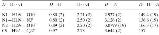
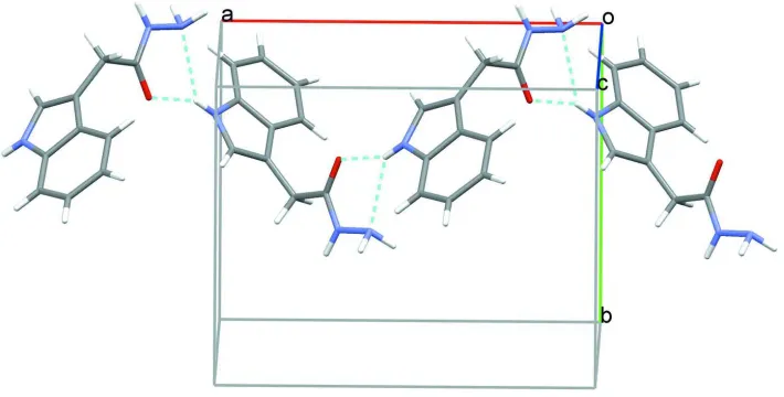
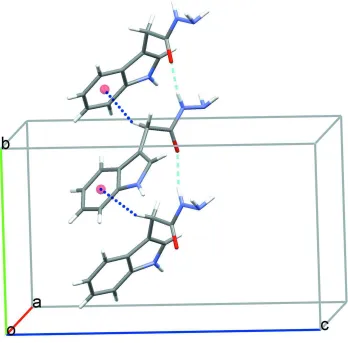
In the title hydrazide compound, the indole ring system is planar (r.m.s. deviation 0.0131 Å) and subtends an angle of 87.27 (5)° to the C9, C10, O10, N2, N3 acetohydrazide substituent which is also planar (r.m.s. deviation 0.0291 Å). In the crystal structure, bifurcated N1–H1N···O10 and N1–H1N···N6 hydrogen bonds together form zigzag chains along a, Table 1, Fig 2. Additional N2–H2N···O10 contacts augmented by C9–H9A···π interactions link the molecules into rows along b, Fig 3. The overall effect of these contacts is a three dimensional network structure with molecules stacked along the b axis, Fig 4.
S2. Experimental
Indole 3-methyl ester (500 mg, 2.6 mmole, 1eq) was added to hydrazine hydrate (80%, 4eq) in ethanol. The reaction mixture was refluxed for 2–3 h, allowed to cool and poured into 100 ml of chilled water. The resulting solid was filtered, dried and re-crystallized from ethanol to obtain the product (300 mg, 60%), mp: 143°C. The purity of the compound was confirmed using thin layer chromatography Rf: 0.18, (n-hexane: ethyl acetate). Crystals of the title compound suitable for X-ray analysis were grown from a solution in ethanol at room temperature.
S3. Refinement
N bound H atoms were located in difference Fourier maps and their coordinates were refined with Uiso=1.2Ueq (N). All
H-atoms bound to carbon were refined using a riding model with d(C—H) = 0.93 Å, for aromatic and 0.97 Å for CH2 H
Figure 1
The structure of the title compound showing the atom numbering scheme with displacement ellipsoids drawn at the 50% probability level
Figure 2
[image:4.610.129.484.310.490.2]supporting information
[image:5.610.133.481.72.415.2]sup-3
Acta Cryst. (2012). E68, o3140–o3141Figure 3
Molecules linked into rows along the b by N–H···O hydrogen bonds (dashed lines) and C–H···π contacts (dotted lines).
Figure 4
[image:5.610.130.482.457.629.2]Crystal data
C10H11N3O
Mr = 189.22
Orthorhombic, Pbca
Hall symbol: -P 2ac 2ab
a = 12.1599 (7) Å
b = 9.6153 (4) Å
c = 16.2345 (8) Å
V = 1898.16 (16) Å3
Z = 8
F(000) = 800
Dx = 1.324 Mg m−3
Mo Kα radiation, λ = 0.71073 Å Cell parameters from 1251 reflections
θ = 3.0–22.1°
µ = 0.09 mm−1
T = 296 K Prism, colorless 0.17 × 0.14 × 0.11 mm
Data collection
Bruker APEXII CCD area detector diffractometer
Radiation source: fine-focus sealed tube Graphite monochromator
φ and ω scans
8600 measured reflections 2329 independent reflections
1294 reflections with I > 2σ(I)
Rint = 0.039
θmax = 28.3°, θmin = 3.0°
h = −16→14
k = −12→12
l = −20→21
Refinement
Refinement on F2
Least-squares matrix: full
R[F2 > 2σ(F2)] = 0.046
wR(F2) = 0.122
S = 1.00 2329 reflections 139 parameters 0 restraints
Primary atom site location: structure-invariant direct methods
Secondary atom site location: difference Fourier map
Hydrogen site location: inferred from neighbouring sites
H atoms treated by a mixture of independent and constrained refinement
w = 1/[σ2(F
o2) + (0.0512P)2 + 0.1536P]
where P = (Fo2 + 2Fc2)/3
(Δ/σ)max < 0.001
Δρmax = 0.16 e Å−3
Δρmin = −0.16 e Å−3
Special details
Geometry. All e.s.d.'s (except the e.s.d. in the dihedral angle between two l.s. planes) are estimated using the full covariance matrix. The cell e.s.d.'s are taken into account individually in the estimation of e.s.d.'s in distances, angles and torsion angles; correlations between e.s.d.'s in cell parameters are only used when they are defined by crystal symmetry. An approximate (isotropic) treatment of cell e.s.d.'s is used for estimating e.s.d.'s involving l.s. planes.
Refinement. Refinement of F2 against ALL reflections. The weighted R-factor wR and goodness of fit S are based on F2,
conventional R-factors R are based on F, with F set to zero for negative F2. The threshold expression of F2 > σ(F2) is used
only for calculating R-factors(gt) etc. and is not relevant to the choice of reflections for refinement. R-factors based on F2
are statistically about twice as large as those based on F, and R- factors based on ALL data will be even larger.
Fractional atomic coordinates and isotropic or equivalent isotropic displacement parameters (Å2)
x y z Uiso*/Ueq
supporting information
sup-5
Acta Cryst. (2012). E68, o3140–o3141C3 0.3892 (2) 0.4202 (2) 0.79725 (14) 0.0683 (6) H3 0.3980 0.4878 0.8377 0.082* C4 0.29916 (19) 0.3318 (2) 0.80094 (13) 0.0664 (6) H4 0.2479 0.3428 0.8430 0.080* C5 0.28434 (16) 0.22822 (19) 0.74347 (12) 0.0542 (5) H5 0.2242 0.1687 0.7466 0.065* C6 0.36154 (14) 0.21440 (16) 0.68030 (11) 0.0418 (4) C7 0.37296 (14) 0.12255 (16) 0.61199 (11) 0.0437 (4) C8 0.46342 (15) 0.16439 (18) 0.57039 (12) 0.0512 (5) H8 0.4894 0.1235 0.5222 0.061* C9 0.29943 (16) 0.00197 (17) 0.59225 (12) 0.0526 (5) H9A 0.2600 −0.0243 0.6418 0.063* H9B 0.3448 −0.0764 0.5762 0.063* C10 0.21718 (14) 0.02925 (16) 0.52500 (11) 0.0404 (4) O10 0.18296 (11) 0.14649 (11) 0.50855 (8) 0.0575 (4) N2 0.18364 (13) −0.08386 (15) 0.48613 (10) 0.0505 (4) H2N 0.2143 (16) −0.165 (2) 0.4996 (11) 0.061* N3 0.09921 (17) −0.07433 (16) 0.42677 (12) 0.0631 (5) H3N1 0.1226 (17) −0.128 (2) 0.3824 (13) 0.076* H3N2 0.0381 (19) −0.125 (2) 0.4406 (14) 0.076*
Atomic displacement parameters (Å2)
U11 U22 U33 U12 U13 U23
N1 0.0422 (9) 0.0513 (10) 0.0711 (12) −0.0088 (7) 0.0042 (9) 0.0060 (9) C1 0.0431 (10) 0.0411 (9) 0.0548 (11) −0.0031 (7) −0.0091 (9) 0.0057 (9) C2 0.0620 (14) 0.0526 (11) 0.0699 (15) −0.0112 (10) −0.0161 (12) −0.0005 (10) C3 0.0867 (17) 0.0570 (13) 0.0612 (14) 0.0031 (12) −0.0161 (13) −0.0101 (11) C4 0.0741 (15) 0.0708 (14) 0.0542 (13) 0.0138 (12) 0.0036 (11) 0.0026 (11) C5 0.0498 (11) 0.0527 (11) 0.0600 (12) −0.0022 (9) 0.0011 (10) 0.0136 (10) C6 0.0415 (9) 0.0341 (8) 0.0497 (10) 0.0006 (7) −0.0065 (8) 0.0085 (8) C7 0.0442 (10) 0.0335 (8) 0.0534 (11) 0.0010 (7) −0.0056 (9) 0.0087 (8) C8 0.0537 (11) 0.0422 (10) 0.0577 (12) 0.0064 (8) 0.0021 (10) 0.0009 (9) C9 0.0625 (12) 0.0328 (9) 0.0624 (12) −0.0039 (8) −0.0095 (10) 0.0070 (8) C10 0.0444 (10) 0.0282 (8) 0.0485 (10) −0.0012 (7) 0.0029 (8) 0.0015 (7) O10 0.0671 (9) 0.0300 (6) 0.0752 (9) 0.0049 (6) −0.0198 (7) −0.0020 (6) N2 0.0627 (10) 0.0292 (8) 0.0594 (10) 0.0020 (7) −0.0114 (9) −0.0020 (7) N3 0.0782 (13) 0.0455 (10) 0.0656 (12) −0.0004 (8) −0.0177 (11) −0.0082 (8)
Geometric parameters (Å, º)
C3—H3 0.9300 C10—N2 1.322 (2) C4—C5 1.377 (3) N2—N3 1.411 (2) C4—H4 0.9300 N2—H2N 0.89 (2) C5—C6 1.397 (2) N3—H3N1 0.93 (2) C5—H5 0.9300 N3—H3N2 0.92 (2)
C8—N1—C1 108.72 (15) C8—C7—C6 106.48 (15) C8—N1—H1N 124.2 (15) C8—C7—C9 127.50 (18) C1—N1—H1N 125.9 (15) C6—C7—C9 126.01 (16) N1—C1—C2 130.74 (18) C7—C8—N1 110.48 (17) N1—C1—C6 107.46 (16) C7—C8—H8 124.8 C2—C1—C6 121.79 (18) N1—C8—H8 124.8 C3—C2—C1 117.75 (19) C7—C9—C10 114.65 (14) C3—C2—H2 121.1 C7—C9—H9A 108.6 C1—C2—H2 121.1 C10—C9—H9A 108.6 C2—C3—C4 121.66 (19) C7—C9—H9B 108.6 C2—C3—H3 119.2 C10—C9—H9B 108.6 C4—C3—H3 119.2 H9A—C9—H9B 107.6 C5—C4—C3 121.2 (2) O10—C10—N2 123.07 (16) C5—C4—H4 119.4 O10—C10—C9 122.82 (15) C3—C4—H4 119.4 N2—C10—C9 114.10 (14) C4—C5—C6 118.58 (18) C10—N2—N3 119.81 (15) C4—C5—H5 120.7 C10—N2—H2N 118.4 (12) C6—C5—H5 120.7 N3—N2—H2N 121.8 (12) C5—C6—C1 119.03 (17) N2—N3—H3N1 105.6 (13) C5—C6—C7 134.12 (16) N2—N3—H3N2 112.8 (15) C1—C6—C7 106.84 (16) H3N1—N3—H3N2 97.9 (18)
C8—N1—C1—C2 179.29 (19) C5—C6—C7—C8 177.68 (18) C8—N1—C1—C6 0.1 (2) C1—C6—C7—C8 −1.15 (18) N1—C1—C2—C3 179.47 (19) C5—C6—C7—C9 −3.4 (3) C6—C1—C2—C3 −1.5 (3) C1—C6—C7—C9 177.72 (16) C1—C2—C3—C4 −0.5 (3) C6—C7—C8—N1 1.3 (2) C2—C3—C4—C5 1.6 (3) C9—C7—C8—N1 −177.58 (16) C3—C4—C5—C6 −0.7 (3) C1—N1—C8—C7 −0.9 (2) C4—C5—C6—C1 −1.2 (2) C8—C7—C9—C10 −79.8 (2) C4—C5—C6—C7 −179.92 (18) C6—C7—C9—C10 101.5 (2) N1—C1—C6—C5 −178.42 (15) C7—C9—C10—O10 −26.0 (3) C2—C1—C6—C5 2.3 (3) C7—C9—C10—N2 155.06 (17) N1—C1—C6—C7 0.62 (18) O10—C10—N2—N3 −4.6 (3) C2—C1—C6—C7 −178.61 (16) C9—C10—N2—N3 174.30 (17)
supporting information
sup-7
Acta Cryst. (2012). E68, o3140–o3141N1—H1N···N3i 0.80 (2) 2.50 (2) 3.126 (2) 136.6 (19)
N2—H2N···O10ii 0.89 (2) 2.20 (2) 3.0799 (19) 166.3 (17)
C9—H9A···Cg2iii 0.97 2.73 3.644 (2) 157