Heavy metals belong to undesirable substances in water environment. They are known for their ability of persistent accumulation in animal tis-sues. Although these substances are also present in unpolluted waters, their natural concentrations are incomparably lower than those in heavily pol-luted areas. They can influence fish organisms and disturb the process of reproduction (Pundir and Saxena, 1992; Thomas, 1993; Mukherjee et al., 1994). Cd is one of the most dangerous among heavy metals (Łukjanienko, 1974). It is introduced into waters with sewage and dusts from industri-al production, mine waters and zinc metindustri-allurgy. A significant quantity of Cd in water comes also from agricultural production (phosphate fertilis-ers). The concentration of Cd in inland waters can dramatically increase during a run-off episode. For example Olsvik et al. (2000) reported that in the Naustebekken River (Norway) Cd concentration increased from 90 ng/l at the low water level to
170 ng/l during the run-off episode. Cd has a wide spectrum of negative effects on the fish organ-ism, including the modulation of the immunore-sponse (O’Neill, 1981; Saxena et al., 1992; Macia and Hernández, 1995), pituitary hormone secre-tion (Mikołajczyk et al., 1990; Thomas, 1990) and ovarian functions (Victor et al., 1986; Singh, 1989; Thomas, 1990, 1993; Mukherjee et al., 1994). Cd was found to influence the activity of many en-zymes, including those which participate in the process of ovarian steroidogenesis (Mukherjee et al., 1992, 1994). Waterborne Cd was found to delay the hatching of fish larvae, to decrease the number of hatched larvae and to increase the number of deformed larvae (Witeska et al., 1995; Sarnowski, 2000; Hallare et al., 2005).
The goal of our investigation was to evaluate the influ-ence of Cd on the last stage of Prussian carp Carassius auratus gibelio (subspecies of goldfish) oocyte matu-ration in vitro, both spontaneous and stimulated with
The influence of cadmium on Prussian carp oocyte
maturation, development of eggs and hatching
P. Szczerbik, T. Mikołajczyk, M. Sokołowska-Mikołajczyk, M. Socha,
J. Chyb, P. Epler
Department of Ichthyobiology and Fisheries, University of Agriculture, Kraków, Poland
ABSTRACT: The influence of cadmium (Cd) on the last stage of Prussian carp (Carassius auratus gibelio B.) oocyte maturation and embryonic development was investigated. The (postvitellogenic) oocytes were incubated with carp pituitary homogenate (Chh), 17α,20β-dihydroxy-4-pregnen-3-one (17α,20βOH-P) and Cd at the concentrations of 20, 100 and 200μM. Cd was found to stimulate spontaneous oocyte maturation, however it inhibited Chh-stimulated maturation. Cd had no influence on the effects of 17α,20βOH-P (MIS – maturation inducing steroid). It seems that the action of Cd on the last stage of oocyte maturation takes place before the MIS production. In an in vivo study Prussian carp eggs were incubated in the water con-taining Cd at the concentrations of 0, 0.01, 0.1, 1 or 10 mg/l. There were no differences between the final numbers of hatched larvae in the respective groups, however Cd at the highest concentration accelerated hatching and increased the number of deformed larvae. These results suggest that Prussian carp eggs are very resistant to waterborne Cd.
carp pituitary homogenate (Chh) or 17α,20β -dihy-droxy-4-pregnen-3-one (17α,20βOH-P), known as MIH or MIS (maturation inducing hormone/ster-oid). Steroid hormone secretion was also determined: 17α,20β-dihydroxy-4-pregnen-3-one (17α,20βOH-P), 17β-estradiol (E2) and testosterone (T).
The influence of Cd on embryonic development (development of activated eggs) was investigated in an in vivo experiment. Egg mortality, number of hatching larvae and percentage of deformed larvae were measured.
MATERIAL AND METHODS
Incubation of oocytes
Oocytes (postvitellogenic ones) obtained from 6 se-xually mature goldfish females (subspecies: Prussian carp Carassius auratus gibelio B.) were used in the experiment.Fish were sacrificed by decapitation and then samples of the ovary were taken and placed in chilled Cortland medium (BSS) (Wolf and Quimby, 1969) Small groups of oocytes were separated me-chanically, washed twice with ice cold medium and placed in glass culture dishes. There were 12 experi-mental groups (Table 1). Each group consisted of six
incubation dishes. In each dish the oocytes from one female were placed. The incubation was performed in an incubator at 18°C for 24 hours. The oocytes were incubated in BSS in a combination (except the con-trol group) with Cd at the concentrations of 20, 100 and 200µM, carp pituitary homogenate (Chh) at the concentration of 100 µg/ml or 17α,20β -dihydroxy-4-pregnen-3-one (17α,20βOH-P) (SIGMA Chemical Co., USA) at the concentration of 100 µg/ml (Ta- ble 1). Chh was prepared from lyophilised pituitary glands obtained from sexually mature common carp (Cyprinus carpio) females.
After incubation, the oocytes were fixed in Serr’s fluid (600 ml of alcohol 96% + 300 ml of formalde-hyde + 100 ml of acetic acid) and immersed in tur-pentine oil for transparency. Then the oocytes were examined for the germinal vesicle (GV) position: in the centre, shifted, located peripherally or those after the germinal vesicle break down (GVBD) (Table 2). Steroid hormone levels in the media were deter-mined by the ELISA method as described below.
Determination of steroid hormone levels
[image:2.595.64.538.99.273.2]The levels of ovarian steroids were determined by an immunoenzymatic method (ELISA). Before
Table 1. Incubation of the oocytes; design of the experiment
Control group BSS
Spontaneous maturation
BSS + Cd (20µM) BSS + Cd (100µM) BSS + Cd (200µM)
Chh stimulated oocyte maturation
BSS + Chh (100 µg/ml)
BSS + Chh (100 µg/ml) + Cd (20µM) BSS + Chh (100 µg/ml) + Cd (100µM) BSS + Chh (100 µg/ml) + Cd (200µM)
17α,20βOH-P stimulated oocyte maturation
BSS + 17α,20βP (100 ng/ml)
BSS + 17α,20βP (100 ng/ml) + Cd (20µM) BSS + 17α,20βP (100 ng/ml) + Cd (100µM) BSS + 17α,20βP (100 ng/ml) + Cd (200µM)
Table 2. Evaluation of the oocyte maturation
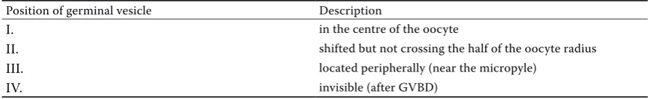
Position of germinal vesicle Description
I. in the centre of the oocyte
II. shifted but not crossing the half of the oocyte radius III. located peripherally (near the micropyle)
[image:2.595.62.531.688.760.2]this determination the following reagents were pre-pared:
1. Phosphate buffer (pH = 7.2; 0.1M) 2. Carbonate buffer (pH = 9.6; 0.05M)
3. PBST buffer (phosphate buffer + NaCl 1% + Tween 20; 0.1%)
4. PBSTA buffer (PBST + gelatine 0.1% + chlorhexi-dine 0.05%)
5. TMB – tetramethylbenzidine solution (10 ml of acetate buffer 0.2M, pH = 4.0 + citric acid 0.07 ml 0.1M + tetramethylbenzidine 0.1 ml + H2O2 30% 0.005 ml)
The determination consisted of:
1. Coating: 200 µl of carbonate buffer (contain-ing a monoclonal antibody) was added into each well of the microplates (NUNC A/S, Denmark). For the determination of 17β-estradiol (E2) and testosterone (T) monoclonal anti rabbit IgG γ chain specific antibodies (SIGMA Chemical Co., USA) at the concentration of 1:2 000 were used. For the determination of 17α,20β -dihydroxy-4-pregnen-3-one (17α,20βOH-P) monoclonal anti rabbit/sheep IgG antibodies (SIGMA Chemical Co., USA) at the concentration of 1:5 000 were used. Microplates were incubated at 4°C for 14 to 16 hours.
2. Saturation (of the free binding sites on the mi-croplates): 200 µl of the casein solution was added into each well in the microplates. Microplates were incubated at 37°C for 2 hours and then they were washed with PBST and dried.
3. Incubation – incubation of samples and stand-ards with antibodies: coated microplates were washed with PBST buffer and dried. 20 µl of sam-ples (duplicated) and/or standards at the concen-trations 0.1, 0.3, 1, 3, 10, 30, 100, 300, 1 000 ng/ml (17β-estradiol) and 0.1, 0.3, 1, 3, 10, 30, 100, 300 ng/ml (17α,20βOH-P), respectively, were added into the wells in the microplates. Samples and standards were incubated with the specific antibodies (50 µl per well): anti-E2 (a gift from Dr. Yaron, Israel) at the concentration of 1:400 000 (for 17β-estradiol), anti-T (Steraloids, USA) at the concentration of 1:6 000 (for testosterone) or anti-17α,20βOH-P (a gift from Dr. Scott, England) at the concentration of 1:20 000 (for 17α,20βOH-P) and 50 µl of tracer (complex of hormone and peroxidase) at the concentration of 1:24 000 (for 17β-estradiol and testosterone) and 1:16 000 for 17α,20βOH-P. Maximal binding was determined in the wells where no samples were added and non-specific binding was determined
in the wells with no specific antibodies. Micro-plates were incubated at 4°C for 16 hours. 4. Enzymatic substrate decomposition: microplates
were washed with PBST and dried. TMB solu-tion (150 µl) was added into each well and the microplates were incubated (in darkness) at 25°C for 30 minutes.
5. Arresting of the enzymatic substrate decomposi-tion: 50 µl of 2M H2SO4 was added into each well and the microplates stayed at room temperature for 5 minutes.
6. Measurement of optic density: an EL 311 read-er (BIO-TEK Instruments) at a wave length of 450 nm was used.
Incubation of activated eggs
The eggs obtained from 4 Prussian carp (Carassius auratus gibelio B.) females were placed in Petri dishes, containing water with no Cd addition and activated (not fertilized) with mixed common carp (Cyprinus carpio) sperm (obtained from 3 males). 10 µl of sperm was added into each dish. After 1 minute water was replaced with water (45 ml) containing Cd at the concentrations of 0 (control group), 0.01, 0.1, 1 and 10 mg Cd/l. About 150 eggs were incu-bated in each dish. The samples of eggs obtained from each female were divided into two dishes and incubated separately (they were duplicated). There were 5 experimental groups. Each group consisted of 8 dishes. Fungi Stop Konzentrat (TETRA, Germany) at the concentration of 75 µl/l of water was used to prevent fungal diseases of eggs. Mortality, hatch-ing rate, number of hatched larvae and number of deformed larvae were determined.
Statistical analysis
The results were analysed by means of one-way ANOVA followed by Duncan’s multiple range test at P ≤ 0.05 significance level.
RESULTS
The influence of cadmium on oocyte maturation
by the GV position. The percentage of oocytes with GV in position I in the group incubated with BSS alone was significantly higher in comparison with the groups incubated with 100 and 200µM of cadmium (Figure 1A). On the other hand, the
percentage of oocytes with GV in position “IV” (after GVBD) was the highest in the group incu-bated with 100µM of cadmium and it was signifi-cantly higher in comparison with the remaining groups.
90 80 70 60 50 40 30 20 10 0
O
oc
yt
es
(%
)
BSS BSS + Chh + BSS + Chh + BSS + Chh + BSS + Ch + 17α, 20βOH-P 17α, 20βOH-P 17α, 20βOH-P 17α, 20βOH-P 20µM Cd 100µM Cd 200µM Cd
1C 90 80 70 60 50 40 30 20 10 0
O
oc
yt
es
(%
)
BSS BSS + Chh BSS + Chh + BSS + Chh + BSS + Ch + 20µM Cd 100µM Cd 200µM Cd
1B 90 80 70 60 50 40 30 20 10 0
O
oc
yt
es
(%
)
BSS BSS + 20µM Cd BSS + 100µM Cd BSS + 200µM Cd
1A Figure 1. The influence of
cadmium on oocyte matura-tion. (1A): spontaneous oocyte maturation. (1B): Chh stimulated oocyte mat-uration. (1C): 17α,20β
-OH- P stimulated oocyte maturation. Data are ex-pressed as means ± SEM (n = 6).
Significant differences (P ≤ 0.05) are marked with:
– significantly more from the remaining groups; –
significantly more than in the groups incubated with 100 and 200µM Cd; ▲ – sig-nificantly more than in the group incubated with BSS +
Chh
[image:4.595.68.414.82.677.2]Significant acceleration of oocyte maturation was found in the group incubated with Chh or 17α,20βOH-P in comparison with the group in-cubated in BSS alone (Figures 1B and 1C). It was visible as a decrease in the number of oocytes with GV in position I and II and an increase in the number of oocytes with GV located peripher-ally or those after GVBD. In the groups incubated with Chh and cadmium the inhibition of Chh-stimulated oocyte maturation was observed: the percentage of oocytes with GV in position II was significantly higher in all cadmium treated groups in comparison with the group incubated with Chh alone (Figure 1C). There was no effect of cadmium on 17α,20βOH-P action.
The influence of cadmium on steroid hormone secretion
No influence of cadmium on spontaneous Chh or 17α,20βOH-P-stimulated steroid hormone secretion was found. The level of 17β-estradiol ranged from 1.29 ng/ml in the groups incubated with BSS alone to 4.5 ng/ml in the groups incu-bated with Chh. The level of 17α,20βOH-P in all groups was very low (under the detectable level). Cadmium did not modify the spontaneous tes-tosterone secretion either. After 24 hours of in-cubation testosterone levels were about 0 ng/ml in all groups. Higher levels were detected in the
groups incubated with Chh (0.3–0.5 ng/ml). In the group incubated with Chh alone and with Chh and cadmium, testosterone levels were significantly higher in comparison with the group incubated with BSS alone.
The influence of cadmium on the development of activated eggs
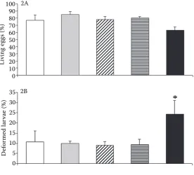
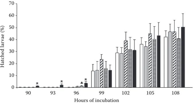
After the first day of incubation the percentage of living eggs ranged from 63.17 in the group incu-bated at the concentration of 10 mg Cd/l to 84.77 in the group incubated at the concentration of 0.01 mg Cd/l. There were no significant differences between the cadmium treated groups and the con-trol group (Figure 2A). The first larvae hatched in the group incubated at the concentration of 10 mg Cd/l after 90 hours of incubation (Figure 3). At this time as well as after 93 and 96 hours the number of hatched larvae in this group was significantly higher than in the remaining groups (except the group incubated at the concentration of 1 mg Cd/1 after 96 hours). There were no significant dif-ferences between the respective groups after 99, 102, and 105 hours. There were no differences in the final numbers of hatched larvae either (after 108 hours). The highest percentage of deformed larvae (significantly higher in comparison with the other groups) was found in the group incubated at 10 mg Cd/l (Figure 2B).
100 90 80 70 60 50 40 30 20 10 0
Li
vi
ng
e
gg
s (
%
)
35 30 25 20 15 10 5 0
D
ef
or
m
ed
la
rv
ae
(%
)
2A
[image:5.595.66.343.517.767.2]2B
Figure 2. The influence of cadmium on the development of Prussian carp eggs and hatching. (2A): The percent-age of living eggs after the first day of incubation. (2B) The percentage of deformed larvae. Data are expressed as means ± SEM (n = 4). Asterisk (∗)
DISCUSSION
It is known that Cd can act as a reproductive disrupter in animals (Singhal et al., 1985). It was found to interfere with ovarian steroidogenesis in mammals (Paksy et al., 1992) and fish (Mukherjee et al., 1994). Cd was found to stimulate the spon-taneous maturation of Prussian carp oocytes in our study. We suppose that such an effect could come from the chemical similarity of Cd to calcium. Calcium is a very important regulator of ovarian steroidogenesis (Veldhuis and Klase, 1982; Tsang and Carnegie, 1983, 1984; Kleiss-San Francisco and Schuetz, 1987). The calcium channel block-ers were shown to inhibit the forskolin (a direct adenyl cyclase activator) stimulated testosterone production in goldfish (Van Der Kraak, 1991). It was found that Cd affected calcium homeostasis by activating or inhibiting several calcium-related enzymes (Beyersmann and Hechtenberg, 1997). It can participate in many calcium-dependent proc-esses (by competition or replacement of calcium ions) (Viarengo, 1985; Lohmann and Beyersmann, 1994). Thus Cd could intensify the action of cal-cium ions in conditions of our experiment and it could stimulate the spontaneous migration of ger-minal vesicle in this way. On the other hand, we observed the inhibition of Chh-stimulated oocyte maturation. Luteinizing hormone (LH) is the ac-tive fraction of Chh. One of the effects of the LH action on oocyte is an increase in the activity of 20β-hydroxysteroid dehydrogenase (20βHSD)
– this enzyme is responsible for the conversion of 17α-hydroxyprogesterone (17αOHP) to 17α,20β -dihydroxy-4-pregnen-3-one (17α,20βOH-P), which is known as MIS (maturation inducing steroid). Calcium is one of the activators of LH-stimulated 20βHSD activity (Nagahama, 1990; Nagahama et al., 1994). LH also caused de novo 20βHSD sis by the mechanisms depending on RNA synthe-sis (Nagahama et al., 1994). It is possible that the inhibition of the Chh-stimulated oocyte matura-tion by Cd, observed in our experiment, is due to a disturbance of RNA synthesis (action at the level of 20βHSD gene, resulting in the lower production of 20βHSD) or to a decrease in this enzyme activ-ity. This second hypothesis is strongly supported by the results of the investigations indicating that Cd decreases the activity of many enzymes, includ-ing 5-3β and 17β-hydroxysteroid dehydrogenase in the common carp ovary (Mukherjee et al., 1992, 1994). Other investigators also reported that Cd inhibited LH-stimulated steroidogenesis and ovar-ian maturation in fish, for example in common carp (Cyprinus carpio) females (Mukherjee et al., 1994). No influence of Cd on 17α,20βOH-P action was detected. These results suggest that Cd acts by the cells of the follicular envelope before this steroid is produced.
Cd accelerated the hatching of goldfish larvae in our in vivo experiment. Many other investigators observed that Cd delayed hatching, for example in common carp Cyprinus carpio (Witeska et al., 1995) and rainbow trout Oncorhynchus mykiss (Sarnowski, 70
60
50
40
30
20
10
0
H
at
ch
ed
la
rv
ae
(%
)
[image:6.595.66.408.86.268.2]90 93 96 99 102 105 108 Hours of incubation
2000). In our experiment relatively high concentra-tions of Cd (from 0.01 to 10 mg/l) were used. These concentrations were significantly higher than, for example, those used by Witeska et al. (1995) (from 0.001 to 0.05 mg Cd/l). We observed the accelera-tion of hatching at the concentraaccelera-tion of 10 mg/l. Sikorska and Ługowska (2005) also observed the acceleration of common carp larvae hatching after exposure to a high concentration of Cd (0.2 mg/l or higher). Woodworth and Pascoe (1982) observed earlier (premature) hatching after the exposition of rainbow trout (Oncorhynchus mykiss) fertilized eggs to Cd. Thus it is possible that such an effect occurs at rather relatively high concentrations of Cd or in more sensitive species. We have several hypotheses on the effect of Cd accelerated hatch-ing. Some enzymes of the perivitelline fluid are responsible for the enclosing of calcium into pro-tein structures which build the egg envelope (Zotin, 1958; Yamamoto, 1960; Fisher, 1963). Cd may bind to the functional groups of these enzymes or be enclosed into protein structures, replacing calcium. Thus the acceleration of hatching may be a result of the disturbance of the egg envelope hardening.
Chorionase (the hatching enzyme) plays a key role in the process of hatching. Many data suggest that an increase in intracellular calcium is necessary for the induction of this enzyme secretion (Schoots et al., 1983; Yamagami, 1997). It is possible that Cd can act as an additive activator of chorionase (for the chemical similarity of Cd and calcium) and that it intensifies the action of calcium ions in this way. Moreover, heavy metals were found to damage the hatching glands (Miś et al., 1995, 1996; Miś and Bigaj, 1997), causing probably an earlier release of chorionase from damaged secretory granules. Cd can also change the respiratory conditions of embryo, by the neglection of gas exchange across the chorion (oxygen transport). Such an effect may come from the sedimentation of metal salt on the egg surface. It is known that oxygen deficit in water can cause the acceleration of hatching (Yamagami, 1970; Hagenmaier, 1972), partly as a result of the earlier release of chorionase (Hagenmaier, 1972; DiMichelle et al., 1981). It is well documented that the most of waterborne Cd (and also other heavy metals) is absorbed by the chorion (Westernhagen and Dethlefsen, 1975; Beattle and Pascoe, 1978; Michibata, 1981; Nakagawa and Ishio, 1989), how-ever, when chorionase starts to dissolve the chorion, Cd is released and could be a source of toxic stress for embryos (Sikorska and Ługowska, 2005).
It was found that Cd and other heavy metals caused a dose-dependent inhibition of fish egg swelling during their water hardening (Sikorska and Ługowska, 2005). It led to a reduction in the space (inside the egg) available to an embryo. It may be the reason for earlier hatching, decrease in the size of larvae and increase in the number of de-formed larvae. We found a statistically significant increase in the number of deformed larvae only at the highest Cd concentration (10 mg/l), however Cd at any other concentration did not decrease the final number of hatched larvae. We suppose that such a result may come from the high resist-ance of Prussian carp against Cd as well as from the toxic effect of Cd on water fungi and bacteria which are dangerous for egg development in nor-mal conditions. Gauthier et al. (2006) observed that Cd prolonged the hatching time and increased the mortality of fathead minnows (Pimephales promelas) larvae under field conditions (in con-taminated lakes) but not in laboratory exposures. These authors concluded that this was a result of higher stress in natural environments, which could sensitize fish to Cd and other contaminants (Gauthier et al., 2006). It is worth noticing that in our experiment Cd was added into water after the activation of eggs with common carp sperm. The design of our experiment allowed us to eliminate the effect of Cd on sperm ability to the activation of eggs in this way.
The results of our investigations indicate that Cd can influence the last stage of Prussian carp oocyte maturation. The results suggest that Cd acts in the period before the production of maturation inducing steroid. Activated Prussian carp eggs were shown to be surprisingly highly resistant to water-borne Cd. It may be one of the reasons (together with a gynogenetic model of reproduction) for the spread of this species in the waters of Poland.
REfERENCES
Beattle J.H., Pascoe D. (1978): Cd uptake by rainbow trout, Salmo gairdneri eggs and alevin. J. Fish Biol., 13, 631–637.
Beyersmann D., Hechtenberg S. (1997): Cd, gene regula-tion and cellular signalling in mammalian cells. Toxicol. Appl. Pharmacol., 144, 247–261.
Fisher K.C. (1963): The formation and properties of the external membrane of the trout egg. Roy. Soc. Canada, 1, 323–332.
Gauthier C., Couture P., Pyle G.G. (2006): Metal effects on fathead minnows (Pimephales promelas) under field and laboratory conditions. Ecotoxicol. Environ. Saf., 63, 353–364.
Hagenmaier H.E. (1972): Zum Schlupfprozess bei Fishen. II. Gewinnung und Charakterisierung des Schlupf-sekretes bei der regenbogenforelle (Salmo gairdneri
Rich.). Experimentia, 28, 1214–1215.
Hallare A.V., Schirling M., Luckenbach T., Köhler H.R., Triebskorn R. (2005): Combined effects of temperature and Cd on developmental parameters and biomarker responses in zebrafish (Danio rerio) embryos. J. Therm. Biol., 30, 7–17.
Kleiss-San Francisco S., Schuetz A. (1987): Sources of calcium and the involvement of calmodulin during steroidogenesis of the amago salmon (Onchorhynchus rhodurus). Gen. Comp. Endocrinol., 72, 39–53. Lohmann R.D., Beyersmann D. (1994): Effect of zinc and
cadmium on apoptic DNA fragmentation in isolated bovine liver nuclei. Environ. Health Perspect., 102, 269–271.
Łukjanienko W.J. (1974): Toksykologia ryb. PWRiL, Warszawa.
Macia M., Hernández M. (1995): Modulation of the ad-herence of human polymorphonuclear leukocytes by Cd and nickel: sexual differences. Arch. Environ. Con-tam. Toxicol., 29, 15–19.
Michibata H. (1981): Effect of water hardness on the tox-icity of Cd to the eggs of the teleost Oryzias latipes. Bull. Environ. Contam. Toxicol., 27, 187–192.
Mikołajczyk T., Weil C., Epler P., Breton B. (1990): In-volvement of voltage-dependent calcium channels (VDCC) in the action of GnRH on GtH release in com-mon carp (Cyprinus carpio L.): comparison with K+ action. Reprod. Nutr. Dev., 30, 619–628.
Miś J., Bigaj J. (1997): Hatching glands of carp (Cyprinus carpio L.) embryos from the eggs incubated at various concentrations of zinc or copper. Pol. Arch. Hydrobiol., 44, 153–155.
Miś J., Bieniarz K., Epler P., Sokołowska-Mikołajczyk M., Chyb J. (1995): Incubation of fertilized common carp (Cyprinus carpio L.) eggs in different concentrations of copper. Pol. Arch. Hydrobiol., 42, 269–276.
Miś J., Bieniarz K., Epler P., Sokołowska-Mikołajczyk M. (1996): Incubation of fertilized common carp (Cyprinus carpio L.) eggs in different concentrations of zinc. Pol. Arch. Hydrobiol., 43, 79–86.
Mukherjee D., Guha D., Kumar V. (1992): Effect of certain toxicants on gonadotropin-induced ovarian
non-es-terified cholesterol depletion and steroidogenic enzyme stimulation of the common carp Cyprinus carpio in vitro. Biomed. Environ. Sci., 5, 92–98.
Mukherjee D., Kumar V., Chakraborti P. (1994): Effect of mercuric chloride and Cd chloride on gonadal function and its regulation in sexually mature common carp
Cyprinus carpio. Biomed. Environ. Sci., 7, 13–24. Nagahama Y. (1990): Endocrine control of oocyte
matu-ration in teleosts. In: Epple A., Scanes C.G., Stetson M.H. (eds.): Progress in Comparative Endocrinology. New York, Wiley-Liss, 385–392.
Nagahama Y., Yoshikuni M., Yamashita M., Nagahama Y. (1994): Regulation of oocyte maturation in fish. In: Sherwood N.M, Hew C.L., Parrell A.P., Randall D.J. (eds.): Fish Physiology. Mol. Endocrinol. Fish New York, 13, 393–439.
Nakagawa H., Ishio S. (1989): Effect of water hardness on the toxicity and accumulation of Cd in eggs of medaka Oryzias latipes. Nippon Suisan Gakkai Shi, 55, 321–326.
Olsvik P.A., Gundersen P., Andersen R.A., Zachariasen K.E. (2000): Metal accumulation and metallothionein in two populations of brown trout, Salmo trutta, ex-posed to different natural water environments during a run-off episode. Aquat. Toxicol., 50, 301–316. O’Neill G.J. (1981): Effects of intraperitoneal lead and Cd
on the humoral response of Salmo trutta. Bull. Environ. Comtam. Toxicol., 27, 42–48.
Paksy K., Varga B., Lazar P. (1992): Cadmium interferes with steroid biosynthesis in rat granulosa and luteal cells in vitro. Biometals, 5, 245–250.
Pundir R., Saxena A.B. (1992): Chronic toxic exposure of Cd on the pituitary gland of fish Puntius ticto and pattern of recoupment. J. Environ. Biol., 13, 69–74. Sarnowski P. (2000): Wpływ miedzi i kadmu na rozwój
embrionalny pstrąga tęczowego. Komunikaty Rybackie, 6, 30–31.
Saxena M.P., Gopal K., Jones W., Ray P.K. (1992): Immune responses to Aeromonas hydrophila in catfish ( Heterop-neustis fossilis) exposed to Cd and hexchlorocyclohexane. Bull. Environ. Contam. Toxicol., 48, 194–201.
Schoots A.F.M., Evertse P.A.C., Denuce J.M. (1983): Ul-trastructural changes in hatching gland cells of pike embryos (Esox lucius L.) and evidence for their degen-eration by apopresis. Cell Tissue Res., 229, 573–589. Singh H. (1989): Interaction of xenobiotics with
repro-ductive endocrine functions in a protogynous teleost,
Monopterus albus. Mar. Environ. Res., 28, 285–289. Singhal R.L., Vijayvargiya R., Shukla G.S. (1985): Toxic
Sikorska J., Ługowska K. (2005): Wpływ kadmu na rozwój embrionalny karpia (Cyprinus carpio L.). Komunikaty Rybackie, 3, 6–8.
Thomas P. (1990): Teleost model for studying the effects of chemicals on female reproductive endocrine func-tion. J. Exp. Zool., 4, 126–128.
Thomas P. (1993): Effect of cadmium on gonadotropin secretion from Atlantic croaker pituitaries incubated
in vitro. Mar. Environ. Res., 35, 141–145.
Tsang B.K., Carnegie J.A. (1983): Calcium requirement in the gonadotrophic regulation of rat granulosa cell pro-gesterone production. Endocrinology, 113, 763–769. Tsang B.K., Carnegie J.A. (1984): Calcium dependent
reg-ulation of progesterone production: effects of the calcium ionophore A23187, prostaglandin E2, dl-isoproterenol and cholera toxin. Biol. Reprod., 30, 787–794.
Van Der Kraak G. (1991): Role of calcium in the control of steroidogenesis in preowulatory ovarian follicles of the goldfish. Gen. Comp. Endocrinol., 81, 268–275. Veldhuis J.D., Klase P.A. (1982): Mechanisms by which
calcium ions regulate the steroidogenic actions of lutei-nizing hormone in isolated ovarian cells in vitro. En-docrinology, 111, 1–6.
Viarengo A. (1985): Biochemical effects of trace metals. Mar. Pollut. Bull., 16, 153–158.
Victor B., Mahalingam S., Sarojini R. (1986): Toxicity of mercury and Cd on oocyte differentiation and vitello-genesis of the teleost, Lepidocephalichthys thermalis
(Bleeker). J. Environ. Biol., 7, 209–214.
Westernhagen H.V., Dethlefsen V. (1975): Combined ef-fects of Cd and salinity on development and survival of flounder eggs. J. Marine Biol. Ass. UK, 55, 945– 957.
Witeska M., Jezierska B., Chaber J. (1995): The influence of Cd on common carp embryos and larvae. Aquacul-ture, 129, 129–132.
Wolf K., Quimby M.C. (1969): Fish cell and tissue culture. In: Hoar W.S., Randall D.J. (eds.): Fish Physiology. Academic Press, New York, 253–312.
Woodworth J., Pascoe D. (1982): Cd toxicity to rainbow trout, Salmo gairdneri Richardson: a study of eggs and alevins. J. Fish Biol., 21, 47–57.
Yamagami K. (1970): A method for rapid and quantitative determination of hatching enzyme (chorionase) activ-ity of the medaka, Oryzias latipes. Annot. Zool. Jnp., 43, 1–9.
Yamagami K. (1997): A short history of the hatching en-zyme studies in medaka. Fish Biol. J. Medaka, 9, 5–15. Yamamoto T. (1960): Physiology of fertilization in fish
eggs. Int. Rev. Cytol., 12, 361–405.
Zotin A.I. (1958): The mechanism of hardening of the sal-monid egg membrane after fertilization or spontaneous activation. J. Embryol. Exp. Morphol., 6, 546–568.
Received: 2007–02–19 Accepted after corrections: 2007–07–24
Corresponding Author
Dr. Paweł Szczerbik, Department of Ichthyobiology and Fisheries, University of Agriculture, ul. Spiczakowa 6, 30 199 Kraków-Mydlniki, Poland