3-Oxo-3-(piperidin-1-yl)propanenitrile
Hoong-Kun Fun,a*‡Ching Kheng Quah,a§Hatem A. Abdel-Azizb and Hazem A. Ghabbourb
a
X-ray Crystallography Unit, School of Physics, Universiti Sains Malaysia, 11800 USM, Penang, Malaysia, andbDepartment of Pharmaceutical Chemistry, College of Pharmacy, King Saud University, PO Box 2457, Riyadh 11451, Saudi Arabia Correspondence e-mail: [email protected]
Received 24 July 2012; accepted 8 August 2012
Key indicators: single-crystal X-ray study;T= 296 K; mean(C–C) = 0.002 A˚; Rfactor = 0.053;wRfactor = 0.128; data-to-parameter ratio = 12.9.
In the title compound, C8H12N2O, the piperidine ring exhibits
a chair conformation and its least-squares plane (all atoms) makes a dihedral angle of 32.88 (12)with the propanenitrile unit (r.m.s. deviation = 0.001 A˚ ). In the crystal, molecules are linked by C—H O hydrogen bonds, forming chains along [001].
Related literature
For ring conformations, see: Cremer & Pople (1975). For background to piperidine derivatives, see: Andrews et al. (2008); Abdel-Aziz & Mekawey (2009); Abdel-Aziz et al. (2009, 2011). For the synthesis, see: Whitehead & Traverso (1955).
Experimental
Crystal data
C8H12N2O Mr= 152.20 Monoclinic,P21=c a= 9.7106 (2) A˚ b= 8.9468 (2) A˚ c= 9.8487 (2) A˚
= 101.425 (1)
V= 838.69 (3) A˚3 Z= 4
CuKradiation
= 0.66 mm1 T= 296 K
0.700.620.39 mm
Data collection
Bruker SMART APEXII CCD diffractometer
Absorption correction: multi-scan (SADABS; Bruker, 2009) Tmin= 0.656,Tmax= 0.783
5110 measured reflections 1300 independent reflections 1222 reflections withI> 2(I) Rint= 0.030
Refinement
R[F2> 2(F2)] = 0.053 wR(F2) = 0.128 S= 1.12 1300 reflections
101 parameters
H-atom parameters constrained
max= 0.20 e A˚
3
min=0.30 e A˚
3
Table 1
Hydrogen-bond geometry (A˚ ,).
D—H A D—H H A D A D—H A
C7—H7A O1i 0.97 2.23 3.1922 (17) 170
Symmetry code: (i)x;yþ1 2;z
1 2.
Data collection:APEX2(Bruker, 2009); cell refinement:SAINT (Bruker, 2009); data reduction:SAINT; program(s) used to solve structure: SHELXTL (Sheldrick, 2008); program(s) used to refine structure:SHELXTL; molecular graphics:SHELXTL; software used to prepare material for publication:SHELXTLandPLATON(Spek, 2009).
The authors thank the Deanship of Scientific Research and the Research Center, College of Pharmacy, King Saud University. HKF and CKQ thank Universiti Sains Malaysia (USM) for the Research University Grant (No. 1001/PFIZIK/ 811160).
Supplementary data and figures for this paper are available from the IUCr electronic archives (Reference: HB6912).
References
Abdel-Aziz, H. A., Abdel-Wahab, B. F., El-Sharief, M. A. M. Sh. & Abdulla, M. M. (2009).Monatsh. Chem.140, 431–437.
Abdel-Aziz, H. A. & Mekawey, A. A. I. (2009).Eur. J. Med. Chem.44, 3985– 4997.
Abdel-Aziz, H. A., Ng, S. W. & Tiekink, E. R. T. (2011).Acta Cryst.E67, o2172.
Andrews, D. M., Stokes, E. S. E., Carr, G. R., Matusiak, Z. S., Roberts, C. A., Waring, M. J., Brady, M. C., Chresta, C. M. & East, S. J. (2008).Bioorg. Med. Chem. Lett.18, 2580–2854.
Bruker (2009).APEX2,SAINTandSADABS. Bruker AXS Inc., Madison, Wisconsin, USA.
Cremer, D. & Pople, J. A. (1975).J. Am. Chem. Soc.97, 1354–1358. Sheldrick, G. M. (2008).Acta Cryst.A64, 112–122.
Spek, A. L. (2009).Acta Cryst.D65, 148–155.
Whitehead, C. W. & Traverso, J. J. (1955).J. Am. Chem. Soc.20, 5867–5872. Acta Crystallographica Section E
Structure Reports Online
supporting information
Acta Cryst. (2012). E68, o2726 [doi:10.1107/S1600536812035015]
3-Oxo-3-(piperidin-1-yl)propanenitrile
Hoong-Kun Fun, Ching Kheng Quah, Hatem A. Abdel-Aziz and Hazem A. Ghabbour
S1. Comment
Piperidines are an important class of heterocycles found in numerous natural products and medicinal structures (Andrews
et al., 2008). In continuation of our interest in the chemistry of piperidines (Abdel-Aziz & Mekawey, 2009; Abdel-Aziz et
al., 2009, 2011), we report here the crystal structure of the title compound.
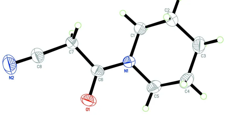
In the title molecule, Fig. 1, the piperidin-1-yl ring (N1/C1-C5) exhibits a chair conformation, puckering parameters
(Cremer & Pople, 1975) Q = 0.5455 (18) Å; Θ = 1.84 (19)° and φ = 113 (6)Å, and its least square plane makes a dihedral
angle of 32.88 (12)° with the propanenitrile unit (N2/C6-C8, r.m.s. deviation = 0.001 Å).
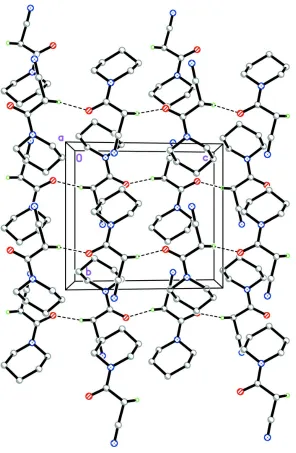
In the crystal (Fig.2), molecules are linked via C7–H7A···O1 hydrogen bonds (Table 1), forming chains along [001].
S2. Experimental
The title compound was prepared by the reaction of ethyl cyanoacetate with piperidine according to the reported method
(Whitehead et al., 1955). Colourless blocks were obtained by slowly evaporating an ethanol solution at room
temperature.
S3. Refinement
[image:2.610.125.484.458.645.2]All H atoms were positioned geometrically and refined using a riding model with C–H = 0.97 Å and Uiso(H) = 1.2 Ueq(C).
Figure 1
Figure 2
The crystal structure of the title compound, viewed along the a axis. H atoms not involved in hydrogen bonds (dashed
lines) have been omitted for clarity.
3-Oxo-3-(piperidin-1-yl)propanenitrile
Crystal data
C8H12N2O Mr = 152.20
Monoclinic, P21/c Hall symbol: -P 2ybc
a = 9.7106 (2) Å
b = 8.9468 (2) Å
c = 9.8487 (2) Å
β = 101.425 (1)°
V = 838.69 (3) Å3 Z = 4
F(000) = 328
Dx = 1.205 Mg m−3
Cu Kα radiation, λ = 1.54178 Å Cell parameters from 2826 reflections
θ = 4.6–70.9°
T = 296 K Block, colourless
0.70 × 0.62 × 0.39 mm
Data collection
Bruker SMART APEXII CCD diffractometer
Radiation source: fine-focus sealed tube Graphite monochromator
φ and ω scans
Absorption correction: multi-scan (SADABS; Bruker, 2009)
Tmin = 0.656, Tmax = 0.783
5110 measured reflections 1300 independent reflections 1222 reflections with I > 2σ(I)
Rint = 0.030
θmax = 63.0°, θmin = 4.7°
h = −11→11
k = −7→10
l = −11→11
Refinement
Refinement on F2 Least-squares matrix: full
R[F2 > 2σ(F2)] = 0.053 wR(F2) = 0.128 S = 1.12 1300 reflections 101 parameters 0 restraints
Primary atom site location: structure-invariant direct methods
Secondary atom site location: difference Fourier map
Hydrogen site location: inferred from neighbouring sites
H-atom parameters constrained
w = 1/[σ2(F
o2) + (0.0759P)2 + 0.0948P] where P = (Fo2 + 2Fc2)/3
(Δ/σ)max = 0.001 Δρmax = 0.20 e Å−3 Δρmin = −0.30 e Å−3
Extinction correction: SHELXTL (Sheldrick, 2008), Fc*=kFc[1+0.001xFc2λ3/sin(2θ)]-1/4 Extinction coefficient: 0.82 (4)
Special details
Geometry. All esds (except the esd in the dihedral angle between two l.s. planes) are estimated using the full covariance matrix. The cell esds are taken into account individually in the estimation of esds in distances, angles and torsion angles; correlations between esds in cell parameters are only used when they are defined by crystal symmetry. An approximate (isotropic) treatment of cell esds is used for estimating esds involving l.s. planes.
Refinement. Refinement of F2 against ALL reflections. The weighted R-factor wR and goodness of fit S are based on F2, conventional R-factors R are based on F, with F set to zero for negative F2. The threshold expression of F2 > 2sigma(F2) is used only for calculating R-factors(gt) etc. and is not relevant to the choice of reflections for refinement. R-factors based on F2 are statistically about twice as large as those based on F, and R- factors based on ALL data will be even larger.
Fractional atomic coordinates and isotropic or equivalent isotropic displacement parameters (Å2)
x y z Uiso*/Ueq
N1 0.24331 (12) 0.06319 (12) 0.22467 (11) 0.0473 (4)
N2 0.08305 (19) 0.56540 (17) 0.19855 (19) 0.0850 (6)
O1 0.24717 (12) 0.26280 (12) 0.36568 (9) 0.0599 (5)
C1 0.19317 (15) −0.01701 (16) 0.09524 (15) 0.0514 (5)
H1A 0.1452 0.0519 0.0257 0.062*
H1B 0.1266 −0.0934 0.1097 0.062*
C2 0.31440 (19) −0.08861 (19) 0.04488 (17) 0.0637 (5)
H2A 0.3752 −0.0113 0.0205 0.076*
H2B 0.2787 −0.1468 −0.0377 0.076*
C3 0.39850 (19) −0.1892 (2) 0.15449 (19) 0.0683 (6)
H3A 0.3414 −0.2738 0.1705 0.082*
C4 0.44535 (18) −0.10440 (19) 0.28805 (19) 0.0673 (6)
H4A 0.4908 −0.1729 0.3594 0.081*
H4B 0.5136 −0.0293 0.2751 0.081*
C5 0.32379 (19) −0.0296 (2) 0.33513 (16) 0.0635 (5)
H5A 0.2627 −0.1051 0.3621 0.076*
H5B 0.3590 0.0322 0.4155 0.076*
C6 0.20841 (13) 0.20271 (15) 0.25256 (12) 0.0425 (5)
C7 0.11643 (16) 0.28844 (15) 0.13497 (14) 0.0491 (5)
H7A 0.1588 0.2852 0.0537 0.059*
H7B 0.0250 0.2410 0.1116 0.059*
C8 0.09924 (16) 0.44374 (17) 0.17336 (16) 0.0558 (5)
Atomic displacement parameters (Å2)
U11 U22 U33 U12 U13 U23
N1 0.0568 (7) 0.0441 (7) 0.0387 (7) 0.0043 (5) 0.0038 (5) 0.0000 (4)
N2 0.0915 (12) 0.0557 (10) 0.1066 (14) 0.0150 (8) 0.0165 (9) −0.0146 (8)
O1 0.0771 (8) 0.0609 (8) 0.0407 (7) −0.0035 (5) 0.0090 (5) −0.0114 (4)
C1 0.0560 (8) 0.0436 (8) 0.0495 (8) 0.0026 (6) −0.0017 (6) −0.0064 (6)
C2 0.0770 (11) 0.0552 (10) 0.0575 (9) 0.0141 (7) 0.0097 (8) −0.0108 (7)
C3 0.0657 (10) 0.0527 (10) 0.0837 (13) 0.0142 (7) 0.0077 (8) −0.0032 (8)
C4 0.0628 (10) 0.0564 (10) 0.0736 (11) 0.0085 (7) −0.0083 (8) 0.0097 (7)
C5 0.0808 (11) 0.0605 (10) 0.0448 (9) 0.0074 (7) 0.0018 (7) 0.0103 (6)
C6 0.0476 (7) 0.0455 (8) 0.0364 (7) −0.0053 (5) 0.0133 (5) −0.0027 (5)
C7 0.0615 (9) 0.0436 (9) 0.0428 (8) 0.0051 (6) 0.0115 (6) −0.0030 (5)
C8 0.0597 (9) 0.0496 (10) 0.0600 (9) 0.0057 (6) 0.0167 (7) −0.0037 (6)
Geometric parameters (Å, º)
N1—C6 1.3361 (18) C3—H3A 0.9700
N1—C1 1.4597 (16) C3—H3B 0.9700
N1—C5 1.4650 (17) C4—C5 1.508 (3)
N2—C8 1.134 (2) C4—H4A 0.9700
O1—C6 1.2271 (16) C4—H4B 0.9700
C1—C2 1.508 (2) C5—H5A 0.9700
C1—H1A 0.9700 C5—H5B 0.9700
C1—H1B 0.9700 C6—C7 1.5224 (18)
C2—C3 1.514 (2) C7—C8 1.458 (2)
C2—H2A 0.9700 C7—H7A 0.9700
C2—H2B 0.9700 C7—H7B 0.9700
C3—C4 1.508 (2)
C6—N1—C1 125.76 (11) C5—C4—H4A 109.2
C6—N1—C5 119.75 (11) C3—C4—H4A 109.2
C1—N1—C5 113.99 (12) C5—C4—H4B 109.2
N1—C1—C2 110.43 (11) C3—C4—H4B 109.2
N1—C1—H1A 109.6 H4A—C4—H4B 107.9
N1—C1—H1B 109.6 N1—C5—H5A 109.4
C2—C1—H1B 109.6 C4—C5—H5A 109.4
H1A—C1—H1B 108.1 N1—C5—H5B 109.4
C1—C2—C3 111.34 (14) C4—C5—H5B 109.4
C1—C2—H2A 109.4 H5A—C5—H5B 108.0
C3—C2—H2A 109.4 O1—C6—N1 123.45 (12)
C1—C2—H2B 109.4 O1—C6—C7 119.88 (12)
C3—C2—H2B 109.4 N1—C6—C7 116.67 (11)
H2A—C2—H2B 108.0 C8—C7—C6 111.28 (11)
C4—C3—C2 110.49 (13) C8—C7—H7A 109.4
C4—C3—H3A 109.6 C6—C7—H7A 109.4
C2—C3—H3A 109.6 C8—C7—H7B 109.4
C4—C3—H3B 109.6 C6—C7—H7B 109.4
C2—C3—H3B 109.6 H7A—C7—H7B 108.0
H3A—C3—H3B 108.1 N2—C8—C7 177.51 (18)
C5—C4—C3 111.85 (14)
C6—N1—C1—C2 131.80 (14) C3—C4—C5—N1 −53.1 (2)
C5—N1—C1—C2 −56.33 (17) C1—N1—C6—O1 175.87 (13)
N1—C1—C2—C3 55.29 (18) C5—N1—C6—O1 4.4 (2)
C1—C2—C3—C4 −54.4 (2) C1—N1—C6—C7 −4.54 (19)
C2—C3—C4—C5 53.3 (2) C5—N1—C6—C7 −175.99 (12)
C6—N1—C5—C4 −132.36 (14) O1—C6—C7—C8 6.13 (18)
C1—N1—C5—C4 55.23 (18) N1—C6—C7—C8 −173.48 (11)
Hydrogen-bond geometry (Å, º)
D—H···A D—H H···A D···A D—H···A
C7—H7A···O1i 0.97 2.23 3.1922 (17) 170