Original Article
ELANE
is highly expressed in leukemia
patients and predicts poor survival
Yanli Zhao
1,3*, Lining Si
2,3*, Weihua Zhang
3, Wei Huang
4, Rong Wang
11Department of Medicine, Qinghai University, Xining, Qinghai Province, China; 2Department of Critical-Care Medi-cine, Affiliated Hospital of Qinghai University, Xining, Qinghai Province, China; 3Department of Pathophysiology, Harbin Medical University, Harbin, Heilongjiang Province, China; 4Division of Cellular Therapy, Duke University, Durham, USA. *Equal contributors.
Received March 18, 2018; Accepted October 11, 2018; Epub April 15, 2019; Published April 30, 2019
Abstract:ELANE (Elastase, Neutrophil Expressed), which has been identified as an oncogene in various tumors, is associated with neutropenia, both cyclic and autosomal dominant forms. However, little is known about ELANE on leukemia progression. To better understand the role of ELANE in leukemia progression, the expression level of ELANE, as well as correlation and survival analyses, was performed in patients with leukemia. Gene expression of ELANE on the normal monocytes and myeloid leukemia samples was obtained and compared via Oncomine. Corre-lation of ELANE and proto-oncogenes includes MYC and TP53 was assessed. The clinical prognostic value of ELANE was found to be elevated using Metzeler Acute myeloid leukemia microarray. As a result, ELANE was found to be highly expressed in the leukemia samples compared to normal monocytes. ELANE was found to be positively corre-lated with MYC expression, whereas it was negatively correcorre-lated with TP53 expression. Additionally, high expression of ELANE was associated with a relatively shorter survival time in leukemia patients, suggesting that ELANE is an oncogene in leukemia development. In conclusion, ELANE acts as a pro-oncogene in leukemia, which provides a potential therapeutic target.
Keywords: ELANE, leukemia, expression, correlation, survival rate
Introduction
Leukemia occurs in 2.3 million people and has
caused 353,500 deaths by 2018 [1]. Four main
types of leukemia - acute lymphoblastic
leuke-mia (ALL), acute myeloid leukeleuke-mia (AML),
chron-ic lymphocytchron-ic leukemia (CLL) and chronchron-ic my-
eloid leukemia (CML) have been identified in
clinic. Epidemic studies revealed that many risk
factors, such as smoking, ionizing radiation,
chemicals, prior chemotherapy, and even Down
syndrome can be ascribed to the
carcinogene-sis and progression of leukemia [2]. However,
the exact cause of leukemia is still unknown.
ELANE
is a serine protein-coding gene that
pro-vides instructions for making a protein called
neutrophil elastase [3]. Previously, numerous
studies have reported roles for the
ELANE
mu-
tation in pathological processes such as in-
flammatory diseases [4] and cancers [5].
Ph-armacological inhibitors such as alpha
2-anti-plasmin and some natural products (curcumin
and EGCG) have been well documented to
counteract and decrease damage caused by
neutrophil elastase [6, 7]. However, whether
ELANE
can be used as a marker for leukemia
diagnostics is still unclear. In this present stu-
dy, to clarify gene expression changes that
are common in cancer tissues or differ among
cancer tissues, we analyzed leukemia patient
samples downloaded from GEO database. Ex-
pression, correlation, and survival analyses of
ELANE
associated with leukemia
were
per-formed and
ELANE
was identified as a
poten-tial therapeutic target against leukemia.
Material and methods
ELANE expression analysis
Figure 1. ELANE mRNA levels in the Stegmaier leukemia microarray. Expres-sion analysis for ELANE was performed using student t-test method via com-paring acute myeloid leukemia (n = 9, group 3) and monocyte samples (n = 3, group 1). P value and fold change were extracted directly without any manual modification. In this figure, p value was 2.34E-5 and the fold change of ELANE in acute myeloid leukemia was 28.425 compared with monocyte samples in Stegmaier leukemia microarray.
using the online open access
Oncomine database (https://
www.oncomine.org/). The
p
value and fold change were
extracted directly from the
Oncomine analysis without
any manual modification.
Microarray correlation
analy-sis
To address the role of
ELANE
in leukemia,
MYC
and
TP53
were used as internal
refer-ences for the Pearson
correla-tion analysis of
ELANE.
Clinical significance of ELANE
in leukemia patients
Overall survival (OS) of
ELA-
NE
in leukemia patients was
accessed using a data set
of patients originally derived
from Metzeler acute myeloid
leukemia microarray as
previ-ously described [8, 9].
Statistical analysis
[image:2.612.91.376.469.706.2]Gene expression was con-
sidered to be dramatically
changed if the
p
-value was
less than 5%. For survival
analysis, the overall survival
rate was performed using a
log-rank test and considered
to be different if the
p-
value
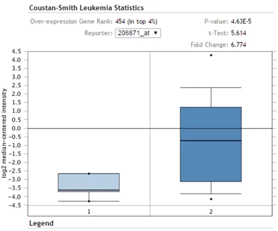
Figure 2.ELANE mRNA levels in the Coustan-Smith leukemia mi-croarray. Expression analysis for
ELANE was performed using stu-dent t-test method via comparing T-cell childhood acute lympho-blastic leukemia (n = 46, group 2) and CD10 positive CD19-pos-itive hematogone (n = 4, group 1) samples. P value and fold change were extracted directly without any manual modification. In this figure, p value was 4.63E-5 and the fold change of ELANE
was less than 5% based on the one-way ANOVA
[image:3.612.92.376.102.354.2]with Tukey’s post-test.
mia cell growth and increase the overall surviv-
al time in leukemia patient. As shown in Figures
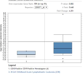
Figure 3. ELANE mRNA levels in the Coustan-Smith leukemia microarray. Ex-pression analysis for ELANE was performed using student t-test method via comparing B-cell childhood acute lymphoblastic leukemia (n = 238, group 2) and CD10 positive CD19 positive hematogone (n = 4, group 1) samples. P value and fold change were extracted directly without any manual modifica -tion. In this figure, p value was 0.002 and the fold change of ELANE in acute myeloid leukemia was 4.251 compared with monocyte samples in the Steg-maier leukemia microarray.
Figure 4. Correlation analysis of ELANE and TP53 expression in Herold chron-ic lymphocytchron-ic leukemia (n = 44). Correlation analysis was performed using a linear regression method according to the expression of ELANE and TP53 in the Herold chronic lymphocytic leukemia microarray. In this figure, the best-fit vales of the slopes were -0.02377 ± 0.03255 (A. ELANE ID 206871_at vs. TP53 ID 201746_at) and -0.0009251 ± 0.004012 (B. ELANE ID 206871_at
vs.TP53 ID 211300_s_at), which indicated that ELANE was negatively cor-related with TP53 in Herold chronic lymphocytic leukemia microarray.
Results
ELANE mRNA is highly
ex-pressed in patients with
leu-kemia
To compare
ELANE
expres-sion in leukemia patient
sam-ples, the original Series Ma-
trix. txt File(s) of Stegmaier
leukemia [10] and
Coustan-Smith leukemia [11] were
downloaded from Gene Ex-
pression Omnibus (GEO). Ori-
ginal intensity files were per
-formed MAS 5.0
normaliza-tion before further analysis.
As shown in Figure 1, mRNA
level of
ELANE
was highly
expressed in acute myeloid
leukemia samples compar-
ed to the normal monocyte
cells, with a total of 28.428
fold change in Stegmaier leu-
kemia.
Furthermore, mRNA level of
ELANE
was also found to be
significantly increased in T-
and B-cell childhood acute
myeloid leukemia samples
compared to the CD10
+and
CD19
+hematogone (Figures
2 and 3).
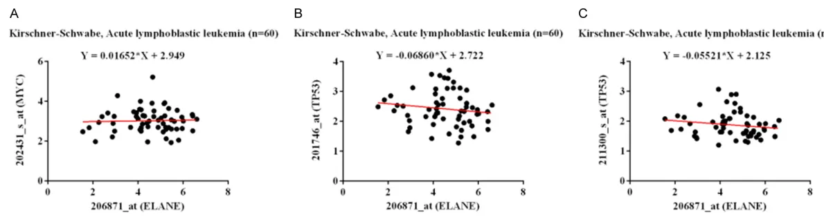
ELANE is positively correlated
with pro-oncogene MYC
ex-pression and negatively
corre-lated with tumor suppressor
TP53 expression in leukemia
samples
To address the role of
ELANE
in leukemia progression,
cor-relation analysis of
ELANE
[image:3.612.92.376.464.587.2]4-6, expression of
ELANE
was found to be
consistently negatively correlated with
TP53
expression. Additionally,
ELANE
was positively
correlated with the expression of
MYC
(Figure
5A). In conclusion, our data strongly suggested
that
ELANE
served as an oncogene in leukemia
patients.
suppressor
TP53
, suggesting that
ELANE
may
act as an oncogene in leukemia. Finally, this
prediction was validated by the survival
analy-sis in Metzeler acute myeloid leukemia patients.
TP53
[16] (tumor protein with MW ~53 kDa) is
a tumor suppressor gene found on
chromo-Figure 6. Correlation analysis of ELANE and TP53 expression in Metzeler acute myeloid leukemia (n = 79). In this figure, the best-fit vales of the slopes were -0.005027 ± 0.005507 (A. ELANE ID 206871_at vs.TP53 ID 201746_ at) and -0.0001598 ± 0.001214 (B. ELANE ID 206871_at vs. TP53 ID 201746_at), which indicates that ELANE is negatively correlated with TP53
[image:5.612.91.376.72.179.2]in Metzeler acute myeloid leukemia microarray.
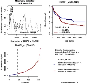
Figure 7. Overall survival analysis of ELANE in the Metzeler acute myeloid leukemia 2 dataset. Survival analysis was performed using a log-rank test. In this figure, high and low groups of ELANE were defined using the expression of ELANE (ID: 206871_at) in Metzeler acute myeloid leukemia 2 microarray with a cut-off of 2336.28. For high expression of ELANE group (expression >2336.28, red line), the overall survival time was shorter compared with the low expression group (expression ≤2336.28, blue line) with p value of 0.17.
Low expression of ELANE
sug-gested a good prognosis in
leukemia patients
The clinical significance of
ELANE
was further validated
by Metzeler acute myeloid
leukemia patients [8]. Both
of
Figures 7 and 8 indicated
that mRNA level of
ELANE
with a relatively lower expre-
ssion predicted a good
sur-vival in leukemia patients
compared to the high
expres-sion group.
Discussion
Microarrays have enabled re-
searchers to conduct
large-scale quantitative
assess-ments of gene expression,
defining the transcriptome of
a multitude of cellular types
and states [15]. In addition,
gene expression microarrays
provide a snapshot of all the
transcriptional activity in a
biological sample. Collective-
ly, the Gene Expression Om-
nibus (GEO) at the National
Center for Biotechnology In-
formation (NCBI) has emerg-
ed as the leading fully public
repository for gene
expres-sion data. To better address
the role of
ELANE
in leuke-
mia progression, in this
pres-ent study, we delineated the
expression, correlation and
survival of
ELANE
using the
datasets downloaded from
GEO. As a result,
ELANE
was
found to be highly expressed
in leukemia sample. In
addi-tion, mRNA level of
ELANE
[image:5.612.90.376.274.545.2]some 17 that is involved in DNA repair [17, 18],
cell cycle arrest [19], and apoptosis [20], and
its overexpression is a hallmark of many human
cancers [21].
TP53
drew initial attention from
cancer community because its expression was
frequently detected in acute myeloid leukemia
(AML) patients and closely connected with the
prognosis in leukemia patients [22, 23]. It has
been validated that a higher expression of the
wild-type
TP53
in leukemia may accelerate the
apoptotic process. In our study,
ELANE
was
found to be negatively correlated with
TP53
expression, which indicated that
ELANE was
an
oncogenic gene in leukemia progression.
Unlike
TP53
,
MYC
(c-
MYC
) is a tumor
suppres-sor that codes for a transcription factor. A
mutated version of
MYC
is found in cancers
[24], which finally causes
MYC
to be
constitu-tively expressed. Malfunctions in
MYC
have
also been found in carcinoma of the cervix,
colon, breast, lung and stomach [25].
MYC
is
thus viewed as a promising target for anti-
We would like to thank Gene Expression Om-
nibus (GEO) and Oncomine for making their
data readily available to the scientific commu
-nity. This work was supported by National Na-
tural Science Foundation of China (No. 8106-
0162, No. 81460284, No. 81460051, No. 31-
560039), Natural Science Foundation of Qing-
hai Provincial (No. 2014-ZJ-944Q, No. 2015-
ZJ-744, 2015-ZJ-929Q, 2015-ZJ-731), Constru-
ction of Laboratory of Qinghai Province (No.
2015-ZJ-Y23). The funders had no role in study
design, data collection and analysis, decision
to publish, or preparation of the manuscript.
Disclosure of conflict of interest
None.
[image:6.612.91.376.73.338.2]Address correspondence to: Rong Wang, Depart- ment of Biochemistry, Medical College of Qinghai University, No. 16 Kunlun Road, Chengxi District, Xining 810001, Qinghai Province, China. Tel: +86-971-6108393; Fax: +86-+86-971-6108393; E-mail: qh- [email protected]
Figure 8. Overall survival analysis of ELANE in the Metzeler acute myeloid leukemia 1 dataset. Survival analysis was performed using a log-rank test. In this figure, high and low groups of ELANE were defined using the expression of ELANE (ID: 206871_at) in Metzeler acute myeloid leukemia 1 microarray with a cut-off of 9607.86. For the high expression of ELANE group (expres-sion >9607.86, red line), the overall survival time was shorter compared with the low expression group (expression ≤9607.86, blue line) with p value of 0.019.
cancer drugs. In our study,
ELANE
was found to be
posi-tively correlated with
MYC
ex-
pression, which further
vali-dated our hypothesis of the
oncogenic role of
ELANE
in
leukemia progression.
Microarray technology is es-
sentially a measurement tool
for measuring expressions of
genes and gene expressions
could be employed as
predic-tors of patient survival. In this
study, the clinical significance
of
ELANE
was further
validat-ed by Metzeler acute myeloid
leukemia patients. Finally, we
discovered that low express-
ion of
ELANE
predicted a good
survival in leukemia patients.
In conclusion, in this study,
the role of
ELANE
in leuke-
mia patients was determin-
ed using the public available
microarray datasets and de-
monstrate that
ELANE
might
be a potential therapeutic
tar-get against leukemia.
References
[1] GBD 2015 Disease and Injury Incidence and Prevalence Collaborators. Global, regional, and national incidence, prevalence, and years lived with disability for 310 diseases and inju-ries, 1990-2015: a systematic analysis for the Global Burden of Disease Study 2015. Lancet 2016; 388: 1545-1602.
[2] Vardiman JW, Thiele J, Arber DA, Brunning RD, Borowitz MJ, Porwit A, Harris NL, Le Beau MM, Hellstrom-Lindberg E, Tefferi A and Bloomfield CD. The 2008 revision of the World Health Or-ganization (WHO) classification of myeloid neo -plasms and acute leukemia: rationale and im-portant changes. Blood 2009; 114: 937-51. [3] Takahashi H, Nukiwa T, Yoshimura K, Quick
CD, States DJ, Holmes MD, Whang-Peng J, Knutsen T and Crystal RG. Structure of the hu-man neutrophil elastase gene. J Biol Chem 1988; 263: 14739-47.
[4] Pham CT. Neutrophil serine proteases: specific regulators of inflammation. Nat Rev Immunol 2006; 6: 541-50.
[5] Sato T, Takahashi S, Mizumoto T, Harao M, Aki-zuki M, Takasugi M, Fukutomi T and Yamashita J. Neutrophil elastase and cancer. Surg Oncol 2006; 15: 217-22.
[6] Fan S, Xu Y, Li X, Tie L, Pan Y and Li X. Opposite angiogenic outcome of curcumin against isch-emia and Lewis lung cancer models: in silico, in vitro and in vivo studies. Biochim Biophys Acta 2014; 1842: 1742-54.
[7] Xiaokaiti Y, Wu H, Chen Y, Yang H, Duan J, Li X, Pan Y, Tie L, Zhang L and Li X. EGCG reverses human neutrophil elastase-induced migration in A549 cells by directly binding to HNE and by regulating alpha1-AT. Sci Rep 2015; 5: 11494. [8] Metzeler KH, Hummel M, Bloomfield CD,
Spiekermann K, Braess J, Sauerland MC, Hei-necke A, Radmacher M, Marcucci G, Whitman SP, Maharry K, Paschka P, Larson RA, Berdel WE, Büchner T, Wörmann B, Mansmann U, Hiddemann W, Bohlander SK, Buske C; Cancer and Leukemia Group B; German AML Coopera-tive Group. An 86-probe-set gene-expression signature predicts survival in cytogenetically normal acute myeloid leukemia. Blood 2008; 112: 4193-201.
[9] Zheng Z, Fan S, Zheng J, Huang W, Gasparetto C, Chao NJ, Hu J and Kang Y. Inhibition of thio-redoxin activates mitophagy and overcomes adaptive bortezomib resistance in multiple my-eloma. J Hematol Oncol 2018; 11: 29. [10] Stegmaier K, Ross KN, Colavito SA, O’Malley S,
Stockwell BR and Golub TR. Gene expression-based high-throughput screening (GE-HTS) and application to leukemia differentiation. Nat Genet 2004; 36: 257-63.
[11] Coustan-Smith E, Song G, Clark C, Key L, Liu P, Mehrpooya M, Stow P, Su X, Shurtleff S, Pui CH, Downing JR and Campana D. New markers for minimal residual disease detection in acute lymphoblastic leukemia. Blood 2011; 117: 6267-76.
[12] Herold T, Jurinovic V, Metzeler KH, Boulesteix AL, Bergmann M, Seiler T, Mulaw M, Thoene S, Dufour A, Pasalic Z, Schmidberger M, Schmidt M, Schneider S, Kakadia PM, Feuring-Buske M, Braess J, Spiekermann K, Mansmann U, Hiddemann W, Buske C and Bohlander SK. An eight-gene expression signature for the predic-tion of survival and time to treatment in chron-ic lymphocytchron-ic leukemia. Leukemia 2011; 25: 1639-45.
[13] Herold T, Mulaw MA, Jurinovic V, Seiler T, Met-zeler KH, Dufour A, Schneider S, Kakadia PM, Spiekermann K, Mansmann U, Hiddemann W, Buske C, Dreyling M and Bohlander SK. High expression of MZB1 predicts adverse progno-sis in chronic lymphocytic leukemia, follicular lymphoma and diffuse large B-cell lymphoma and is associated with a unique gene expres-sion signature. Leuk Lymphoma 2013; 54: 1652-7.
[14] Kirschner-Schwabe R, Lottaz C, Todling J, Rhe-in P, Karawajew L, Eckert C, von Stackelberg A, Ungethum U, Kostka D, Kulozik AE, Ludwig WD, Henze G, Spang R, Hagemeier C and Seeger K. Expression of late cell cycle genes and an in-creased proliferative capacity characterize ve- ry early relapse of childhood acute lympho-blastic leukemia. Clin Cancer Res 2006; 12: 4553-61.
[15] Govindarajan R, Duraiyan J, Kaliyappan K and Palanisamy M. Microarray and its applications. J Pharm Bioallied Sci 2012; 4 Suppl 2: S310-2. [16] Matlashewski G, Lamb P, Pim D, Peacock J,
Crawford L and Benchimol S. Isolation and characterization of a human p53 cDNA clone: expression of the human p53 gene. EMBO J 1984; 3: 3257-62.
[17] Lindemann A, Takahashi H, Patel AA, Osman AA and Myers JN. Targeting the DNA damage response in OSCC with TP53 mutations. J Dent Res 2018; 97: 635-644.
[18] Sen T, Gay CM and Byers LA. Targeting DNA damage repair in small cell lung cancer and the biomarker landscape. Transl Lung Cancer Res 2018; 7: 50-68.
[19] Lin D, Meng L, Xu F, Lian J, Xu Y, Xie X, Wang X, He H, Wang C and Zhu Y. Enhanced wild-type p53 expression by small activating RNA dsP53-285 induces cell cycle arrest and apoptosis in pheochromocytoma cell line PC12. Oncol Rep 2017; 38: 3160-6.
G and Hilal G. Different TP53 mutants in p53 overexpressed epithelial ovarian carcinoma can be associated both with altered and unal-tered glycolytic and apoptotic profiles. Cancer Cell Int 2018; 18: 14.
[21] Bieging KT, Mello SS and Attardi LD. Unravel-ling mechanisms of p53-mediated tumour suppression. Nat Rev Cancer 2014; 14: 359-70.
[22] Hou HA, Chou WC, Kuo YY, Liu CY, Lin LI, Tseng MH, Chiang YC, Liu MC, Liu CW, Tang JL, Yao M, Li CC, Huang SY, Ko BS, Hsu SC, Chen CY, Lin CT, Wu SJ, Tsay W, Chen YC and Tien HF. TP53 mutations in de novo acute myeloid leukemia patients: longitudinal follow-ups show the mu-tation is stable during disease evolution. Blood Cancer J 2015; 5: e331.
[23] Kadia TM, Jain P, Ravandi F, Garcia-Manero G, Andreef M, Takahashi K, Borthakur G, Jabbour E, Konopleva M, Daver NG, Dinardo C, Pierce S, Kanagal-Shamanna R, Patel K, Estrov Z, Cortes J and Kantarjian HM. TP53 mutations in newly diagnosed acute myeloid leukemia: clinicomolecular characteristics, response to therapy, and outcomes. Cancer 2016; 122: 3484-91.
[24] Dang CV. MYC on the path to cancer. Cell 2012; 149: 22-35.