R E S E A R C H
Open Access
Dynamic changes in DNA modification states
during late gestation male germ line
development in the rat
Catherine M Rose
1, Sander van den Driesche
2, Richard M Sharpe
2, Richard R Meehan
3and Amanda J Drake
1*Abstract
Background:Epigenetic reprogramming of fetal germ cells involves the genome-wide erasure and subsequent re-establishment of DNA methylation. Mouse studies indicate that DNA demethylation may be initiated at embryonic day (e) 8 and completed between e11.5 and e12.5. In the male germline, DNA remethylation begins around e15 and continues for the remainder of gestation whilst this process occurs postnatally in female germ cells. Although 5-methylcytosine (5mC) dynamics have been extensively characterised, a role for the more recently described DNA modifications (5-hydroxymethylcytosine (5hmC), 5-formylcytosine (5fC) and 5-carboxylcytosine (5caC)) remains unclear. Moreover, the extent to which the developmental dynamics of 5mC reprogramming is conserved across species remains largely undetermined. Here, we sought to describe this process during late gestation in the male rat.
Results:Using immunofluorescence, we demonstrate that 5mC is re-established between e18.5 and e21.5 in the rat, subsequent to loss of 5hmC, 5fC and 5caC, which are present in germ cells between e14.5 and e16.5. All of the evaluated DNA methyl forms were expressed in testicular somatic cells throughout late gestation. 5fC and 5caC can potentially be excised through Thymine DNA Glycosylase (TDG) and repaired by the base excision repair (BER) pathway, implicating 5mC oxidation in active DNA demethylation. In support of this potential mechanism, we show that TDG expression is coincident with the presence of 5hmC, 5fC and 5caC in male germ cell development. Conclusion:The developmental dependent changes in germ cell DNA methylation patterns suggest that they are linked with key stages of male rat germline progression.
Keywords:Germ cells, Rat, DNA modification, 5-methylcytosine, 5-hydroxymethylcytosine, 5-formylcytosine, 5-carboxylcytosine, Thymine DNA Glycosylase
Background
Methylation of the cytosine base in DNA (DNA methy-lation) is an essential epigenetic mark in mammals that contributes to the regulation of transcription, chromatin organisation and histone modification deposition [1,2]. DNA methylation at regulatory regions, including pro-moters, is associated with stable transcriptional silencing of genes and transposons, genomic imprinting and X in-activation [3,4]. In order to give rise to functional gametes, primordial germ cells (PGC) undergo extensive epigenetic
reprogramming including erasure of DNA methylation and extensive chromatin remodelling, a process which is thought to be necessary to remove potential epimutations and to erase parental imprints in this cell lineage [5-8]. In the mouse, PGCs specified from epiblast cells at around
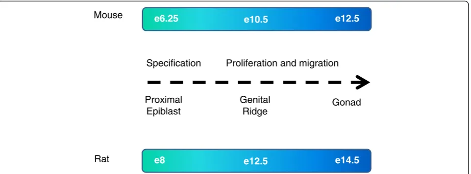
embryonic day (e) 6.25–7 migrate along the developing
hind-gut endoderm (from e8.5) and colonise the genital ridges from e10.5 [8] (Figure 1). The precise timings in the rat are less well-documented but may occur slightly later than in the mouse (Figure 1) [9]. During mouse em-bryogenesis, DNA methylation is established by e6 and is thought to contribute to stable lineage commitment [10-12]. The erasure of DNA methylation in PGCs ap-pears to be initiated from approximately e8.5 and is com-pleted by approximately e13.5 [6,13,14] and occurs at the
* Correspondence:[email protected]
1Endocrinology Unit, University/BHF Centre for Cardiovascular Science,
University of Edinburgh, The Queen's Medical Research Institute, 47 Little France Crescent, Edinburgh EH16 4TJ, UK
Full list of author information is available at the end of the article
majority of the genome, with the exception of certain loci, some of which correspond to retrotransposons [15,16]. Erasure of DNA methylation is followed by a period of remethylation, the timing of which differs between the male and female germlines. In the mouse, DNA remethy-lation is initiated at e15.5 and continues until e18.5 and beyond (at least at imprinted genes and repeats) in male germ cells, whereas in females, the process occurs after birth [14,16-21]. Although epigenetic reprogramming has been extensively investigated in mouse germ cells, the ex-tent to which this process is conserved across species re-mains largely undetermined. Notably, the degree of active demethylation of cytosine methylation in the sperm gen-ome prior to forming a functional zygotic nucleus varies in different mammals; for example, demethylation is ex-tensive in the mouse, but in contrast, there is no observ-able demethylation of the sheep male pronucleus at any point in the first cell cycle [22-24].
The potential pathways governing active removal of 5-methylcytosine (5mC) are now being elucidated with the recent discovery of further modified forms of cytosine nucleotides including hydroxymethylcytosine (5hmC), 5-formylcytosine (5fC) and 5-carboxylcytosine (5caC), which are sequentially produced from 5mC by the action of the ten-eleven translocase (TET) family of iron, abscorbic acid andα-ketoglutarate-dependent dioxygenases [25-29]. 5hmC-modified CpGs are significantly enriched at the bodies of actively transcribing genes in many tissues (but not all) and are present to some degree at enhancer ele-ments and a small cohort of regions spanning an anno-tated transcriptional start site [30-34] whilst the levels of 5fC and 5caC are low in somatic cells, perhaps in part due to their potential inhibitory effect on transcription [35]. Although the dynamics of 5mC, 5hmC, 5fC and 5caC
have been investigated in fertilised zygotes from a variety of organisms [36-40], there are few reported studies for developing fetal germ cells [13]. A recent study has shown that DNA demethylation in mouse PGCs entails conver-sion of 5mC to 5hmC by TET1 and TET2 with the loss of 5mC largely complete by e11.5 [13]. The progressive de-cline in 5hmC in the absence of enrichment of 5fC and 5caC suggests that subsequent demethylation may occur by a replication-dependent mechanism in the mouse. Any role for 5hmC, 5fC and/or 5caC in the later stages of germline epigenetic reprogramming is not known.
Here, we have investigated the spatiotemporal relation-ship between 5mC, 5hmC, 5fC and 5caC specifically during late (e14.5–e21.5) male fetal rat germ cell development by immunofluorescence using previously validated antibodies [13,14,41-44]. Although the timing of germ cell specifica-tion is not as well described in the rat as in the mouse, germ cells are present in the rat embryonic testis by e14.5 (Figure 1). We firstly wished to address if male germ cell development in the rat is associated with global demethyl-ation and subsequent remethyldemethyl-ation as in the mouse and, secondly, if 5hmC, 5fC and 5caC are present in a pattern that is indicative of a developmental-associated function in the rat germline. Since 5fC and 5caC can
both be ‘repaired’ by Thymine DNA Glycosylase (TDG)
to produce unmodified cytosine, we also sought to assess whether TDG was present in the rat germline during late fetal development to potentially mediate active DNA de-methylation [26,45].
Results
Immunohistochemistry for 5mC and DAZL
Several commonly used germ cell markers are not com-patible with the hydrochloric acid (HCl) antigen retrieval
e6.25 e10.5 e12.5
Gonad Genital Ridge Proximal Epiblast Mouse Rat
Proliferation and migration Specification
e8 e12.5 e14.5
of modified cytosines for use in immunofluorescence (IF). In order to clearly show the localisation of 5mC to germ cells at all stages of development, we used the spe-cific germ cell cytoplasmic marker DAZL, which survives HCl antigen retrieval, in combination with 5mC for im-munohistochemistry (IHC) (Figure 2). The testis contains
different cellular compartments; germ cells (shown with cytoplasm stained for DAZL in blue) are rounded in shape and contained within the seminiferous cords, which also contain Sertoli cells (Figure 2). The hormone-producing Leydig cells, peritubular myoid cells and other interstitial cell types are located outside of the seminiferous cords in
the interstitium. There was no/little detectable 5mC in germ cells between e14.5 and e18.5 (Figure 2A,B,C,D,E), suggesting that, as in mice, rat germ cells are hypomethy-lated relative to surrounding somatic cells by e14.5. 5mC was detectable in some germ cells from e19.5 onwards, and by e21.5, there was strong 5mC immunostaining (Figure 2F,G,H). Again, this is in line with mouse studies in which remethylation occurs in male germ cells during late gestation [5].
Immunofluorescence for 5mC, 5hmC, 5fC, 5caC and TDG
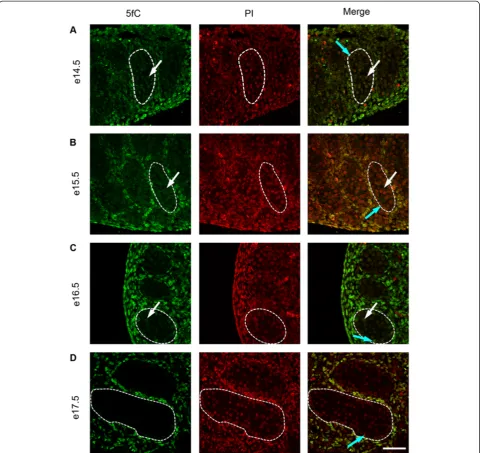
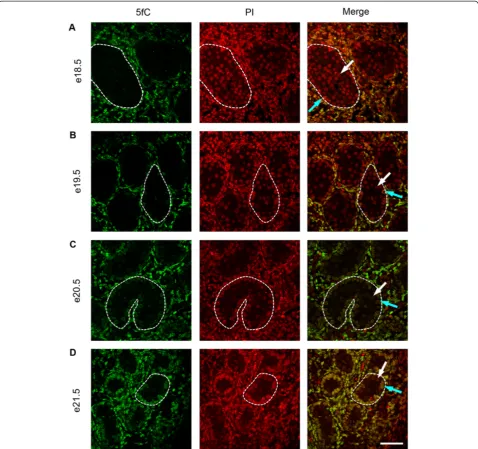
To investigate the temporal expression patterns for germ cell 5mC, 5hmC, 5fC and 5caC, immunofluorescence was used. Consistent with the IHC findings, little 5mC was detectable in germ cells between e14.5 and e18.5 (Figure 3A,B,C,D and Figure 4A), but 5mC was detectable in some germ cells at e19.5, with enhanced staining at e21.5 (Figure 4B,C,D). In contrast, 5hmC showed some germ cell localisation between e14.5–e16.5 (Figure 3A,B,C)
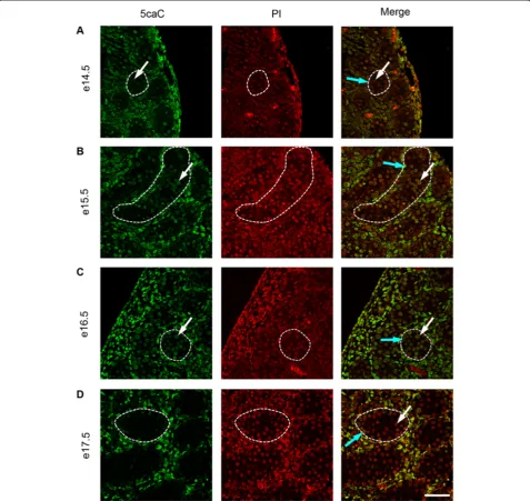
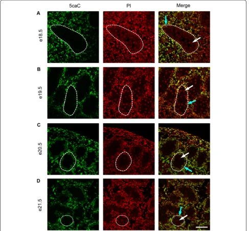
but had become undetectable by e17.5 and remained un-detectable up to e21.5 (Figure 3D and Figure 4A,B,C,D). Semi-quantitative analysis of the images confirmed a significant decrease in 5mC and an increase in 5hmC be-tween e16.5 and e21.5 (Additional file 1: Figure S1). Similarly, 5fC was detectable in germ cells at e14.5–e16.5 (Figure 5A,B,C) but not after this age (Figure 5D and Figure 6A,B,C,D). 5caC was also detectable in germ cells only at e14.5–e16.5 (Figure 7A,B,C) but not at later ages (Figure 7D and Figure 8A,B,C,D). In contrast to the germ
cells, 5mC and 5hmC in somatic cells was omnipresent and appeared stable throughout late gestation. The findings are summarised in Table 1. The absence of detect-able 5mC from e14.5 onwards until e19.5 and the limited appearance (between e14.5–e16.5) of 5hmC, 5fC and 5caC might suggest that the oxidised forms of 5mC are part of a demethylation process that is complete by e16.5. It is unclear if this is indicative of an active or a passive de-methylation pathway during rat male germ cell develop-ment. We therefore investigated the pattern of TDG
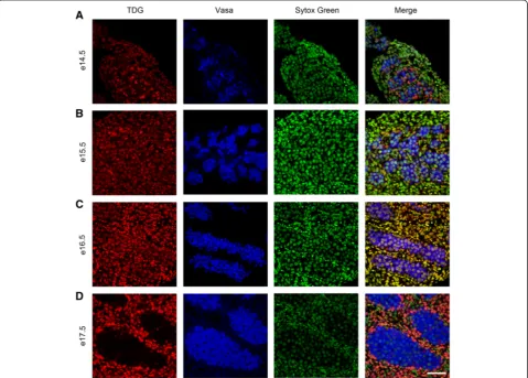
staining in developing fetal germ cells. The protocol for TDG staining was compatible with protocols for counter-staining for germ cell cytoplasm (VASA) and nucleus (sytox green) (Figure 9). TDG was clearly detectable in VASA positive germ cell nuclei between e14.5 and e16.5 (Figure 9A,B,C) but was largely undetectable at later ages (Figure 9D and Figure 10A,B,C,D). It is noteworthy that TDG expression in e14.5–e16.5 germ cells is coincident with the presence of 5hmC, 5fC and 5caC. In somatic cells, TDG was detectable at all stages examined.
Discussion
Our study represents a novel characterisation of the dy-namics of DNA modification of fetal germ cells in the male rat during the latter half of gestation. The lack of detectable 5mC in germ cells at e14.5 suggests that the process of 5mC erasure is already complete by this time. This is consistent with data in mice showing that DNA methylation is erased during PGC migration [5,14,42] such that by e13.5, the overall level of DNA methylation is reduced by more than 90% [7].
One proposed mechanism for DNA demethylation in fetal germ cells is TET-mediated 5mC to 5hmC
conver-sion [13]. In vitro PGC (iPGC) formation and
genome-wide DNA demethylation are unaffected by the absence of 5hmC mediated by TET1 and TET2 [46]. However, this leads to hypermethylation in many target loci in mutant iPGCs, consistent with a role for 5hmC as an intermediate in locus-specific demethylation. In the mouse germline, the loss of 5mC in exons, at promoters and imprinted regions is associated with an increase in 5hmC which is mostly completed by e11.5, followed by a progressive
decline over several cell cycles between e11.5 and e13.5, suggesting a replication-dependent mechanism for 5hmC loss [13]. Our study, showing the stable persistence of 5hmC in fetal rat germ cells until e16.5, suggests that the removal of 5mC through conversion to 5hmC might also occur in the rat. 5fC and 5caC were also present in fetal germ cells between e14.5 and e16.5, but, as with 5hmC, were no longer present at e17.5. Recent data has shown enrichment of 5hmC in mouse PGCs during the phase of DNA demethylation [13,42], with a progressive later decline; however, these studies were undertaken using
Oct4-GFP transgenic mice which allow the isolation of migratory germ cells from urogenital ridges at an early stage and such studies are not yet possible in the Wistar rat. We did not observe enrichment of 5hmC in rat germ cells relative to the surrounding somatic cells between e14.5 and e16.5, and this may indicate that this occurs be-fore e14.5 in the rat and that 5hmC levels have already begun to decline by this time, as in the mouse [13,42]. The lack of enrichment of germ cell 5fC and 5caC and the presence of these modifications in surrounding somatic
cells has previously been reported, at least up until e12.5 in the mouse [13,42].
Given their proposed role in the TET-mediated de-methylation process, the presence of 5fC and 5caC in fetal germ cells could reflect an accumulation of uncon-verted intermediates during global demethylation before rapid removal between e16.5 and e17.5. Although little is known regarding the function of 5fC and 5caC, a re-cent study showing that the levels of these modifications gradually decrease with cell division in the mouse zygote,
rather than exhibiting the rapid removal that might be ex-pected if they existed solely as part of a demethylation mechanism, indicates that they may also have a functional role in chromatin remodelling [37]. Thus, the stable pres-ence of 5hmC, 5fC and 5caC in rat germ cells between e14.5 and e16.5, and in testicular somatic cells throughout late gestation, may indicate potential functionality of these modifications during germ cell development.
TET-mediated iterative oxidation of 5mC and 5hmC to 5fC and/or 5caC may provide suitable substrates for demethylation to non-modified cytosine via the rapid
excision of 5fC and 5caC by TDG, followed by base exci-sion repair (BER) [29,37,45,47]. Support for this comes from comparative analysis of 5hmC/5fC/5caC distribu-tions in wild-type and TDG-deficient mouse ES cells showing that a large number of genomic loci are targeted by TET/TDG activities [48] and from recent work sug-gesting that 5fC is detectably enriched over the bodies of actively transcribing genes as well as over CpG islands and promoters in ES cells, and that these levels increase in the absence of TDG activity [43]. Consistent with a role for TDG in germ cell DNA demethylation in the rat, TDG
was present in germ cells between e14.5 and e16.5, but largely absent thereafter, when 5fC and 5caC were no lon-ger detectable. There are, however, alternative mechanisms for DNA demethylation including through deamination of 5mC to thymine by an activation-induced deaminase/
apolipoprotein B editing complex (AID/APOBEC), result-ing in a G/T mismatch that is subsequently converted to G/C by TDG and subsequent BER. Support for this comes from data showing that AID-deficient PGCs have substan-tially delayed demethylation (although loss of 5mC still occurs) [49,50]. Recent studies in the mouse zygote show that there are two phases of active DNA demethylation with involvement of TET3 and, independently, AID-mediated cytosine deamination and subsequent BER in the second phase whilst the mechanisms accounting for DNA demethylation in the first phase remain unclear [41]. Further clarification of the roles of AID- and/or TDG-mediated demethylation in PGCs is still required.
The re-establishment of 5mC in male rat germ cells ap-pears to begin at e19.5. The remethylation phase has been less well characterised in mouse germ cells, although gene-targeted studies suggest that in male germ cells, the onset of DNA remethylation occurs at some imprinted loci and at repetitive elements from e15.5, with global remethyla-tion re-established by e18.5 [51,52] which may be further augmented perinatally [17,18]. Our demonstration of the Table 1 Detection of 5mC, 5hmC, 5fC, 5caC and TDG
during mid to late gestation (e14.5–e21.5) in fetal rat germ cells
Day 5mC 5hmC 5fC 5caC TDG
e14.5 − + + + +
e15.5 − + + + +
e16.5 − + + + +
e17.5 − − − − −
e18.5 − − − − −
e19.5 + in a subset of germ cells − − − −
e20.5 + − − − −
e21.5 + − − − −
5mC, 5hmC, 5fC and 5caC are detectable in somatic cells throughout this time-course. + indicates presence, and−indicates absence of staining.
Figure 9Immunofluorescence showing localisation of TDG between e14.5 and e17.5.TDG is stained in red, the cytoplasm of germ cells is stained for VASA in blue and nuclei are stained using sytox green (green). Images show TDG is present in germ cells between e14.5–e16.5(AtoC)
absence of 5hmC, 5fC and 5caC in rat germ cells during the re-establishment of DNA methylation between e19.5 and e21.5 further supports the hypothesis that these vari-ants form part of the initial demethylation pathway in the male germline [29].
Conclusions
This study provides the first characterisation of global patterns of DNA methylation in the fetal rat testis for all currently recognised forms of cytosine methylation dur-ing mid to late gestation and provides an insight into the dynamics of remethylation in male rat fetal germ cells. The timing of both erasure and re-establishment of DNA methylation is later than that in the mouse, consistent with the longer gestation period in the rat (22 days in Wistar rats, compared to approximately 19 days in the mouse [53]). The extent to which germline reprogram-ming operates in other species is currently poorly under-stood. However, studies for example in pigs indicate that epigenetic reprogramming in germ cells follows the same dynamics as in mice, although the phase of demethylation
occurs over a period of 20 days [54]. There are few pub-lished studies in humans, but data suggests that in human males, germ cell DNA is hypomethylated in mid-gestation and 5mC levels increase from around 20 weeks to term [55,56]. Levels of 5hmC, 5fC and 5caC decrease as human spermatogenesis proceeds, whilst 5mC levels remain con-stant [57]. As in mouse, this suggests that during sperm-atogenesis active DNA demethylation mechanisms are down-regulated, stabilising methylation profiles in mature sperm, which can be subsequently reprogrammed in ferti-lised zygotes [58]. Our study, demonstrating the dynamics of epigenetic reprogramming in fetal germ cells in the rat, provides further evidence that this is conserved across species. The stable presence of 5hmC, 5fC and 5caC in germ cells for several days following the loss of 5mC also support data from the pig suggesting that epigenetic repro-gramming may occur over an extended period in species with longer gestation [54]. Although altered epigenetic re-programming early in gestation during the period of DNA methylation erasure has been proposed as one mechanism accounting for transgenerational inheritance in mammals
Figure 10Immunofluorescence showing localisation of TDG between e18.5 and e21.5.TDG is stained in red, the cytoplasm of germ cells is stained for VASA in blue and nuclei are stained using sytox green (green). Images show TDG is largely undetectable in germ cells between e18.5–e21.5
[14], we and others have proposed that disruption of epi-genetic reprogramming in the second half of gestation could also be important in the transmission of environ-mentally induced effects across generations through the male line [59-64]. Our study therefore also provides a base-line for investigating the susceptibility of this process to disruption, for example as a consequence of exposure to environmental factors.
Methods
Animals
Ethics statement
These studies were specifically approved by the UK Home Office and were conducted under an approved Project Licence (PPL 60/3914) in accordance with the UK Animals (Scientific Procedures) Act 1986 following re-view by the University of Edinburgh Animal Research Ethics Committee. Wistar rats were maintained in our own facility in an environment of controlled humid-ity, temperature (22°C), lighting (artificial light between 7.00 a.m.–7.00 p.m.), and constant access to breeding diet (RM3(E) soya free; Special Diets Services, Witham, Essex, UK) and water. Females were time mated and killed by CO2 asphyxiation and subsequent cervical dislocation at
experimental time points between e14.5 and e21.5 (gesta-tion is approximately 22 days in our colony). Fetuses were removed, decapitated, and placed in ice cold phosphate-buffered saline (PBS, Sigma-Aldrich, Dorset, England, UK). Testes were microdissected and incubated in Bouin's fixative for 1 h at room temperature. Tissues were then embedded in paraffin wax following standard procedures and 5μm sections prepared.
5mC and DAZL immunohistochemistry
Tissue sections were de-waxed and rehydrated before sub-mersion in Novocastra Epitope Retrieval Solution (pH9, Leica, Milton Keynes, UK) and pressure cooking for 5 min at 125°C. Endogenous peroxidase activity was then blocked by incubation in 3% (v/v) hydrogen peroxide/methanol. All subsequent washes were in TBS, and antibody/serum incubations were conducted in a humidity chamber (Fisher Scientific, Loughborough, Leicestershire, UK). Tissues were blocked with normal goat serum (NGS, Biosera, East Sussex, UK) diluted 1:5 with 5% (w/v) BSA in TBS (NGS/ TBS/BSA) before incubation with DAZL antibody (1:500, mouse, AbD Serotec, Oxford, UK) overnight at 4°C. Slides were then washed and incubated with goat anti-mouse bio-tinylated antibody (1:500, Vector Laboratories, Burlingame, CA, USA) for 30 min. Slides were washed before incuba-tion with Streptavidin-Alkaline Phosphatase (1:200, Vector Laboratories) for 30 min. Following further washing, anti-bodies were detected using PermaBlue Plus/AP (Diagnostic Biosystems, Pleasanton, CA, USA) following manufactur-er's instructions. Tissues were incubated for 15 min in
4 M hydrochloric acid (HCl)/TBS, preheated to 37°C, then washed with 0.1% Tween in TBS. Tissue was permeabi-lised with 1% Triton X-100 (Sigma-Aldrich, St. Louis, MO, USA) in TBS for 30 min before blocking with normal horse serum (NHS, Biosera) diluted 1:5 with 5% (w/v) BSA in TBS (NHS/TBS/BSA). Slides were incubated with 5mC antibody (1:300, mouse, Eurogentec, London, UK) overnight at 4°C. Slides were then washed and antibody detected using the ImmPress anti-mouse Ig (peroxidase) Polymer Detection Kit (Vector Laboratories) and ImmPress Diaminobenzidine (DAB, Vector Laboratories) following manufacturers' instructions. Mounting was conducted with PermaFluor Aqueous Mounting Medium (Thermo Fisher Scientific, Waltham, MA, USA).
Immunofluorescence for 5mC and 5hmC
For immunofluorescence, following initial preparation as above, tissues were blocked with NGS (Biosera) diluted
1:5 with 5% (w/v) BSA in TBS (NGS/TBS/BSA) before
incubation with 5mC antibody (1:100, mouse, Eurogentec) overnight at 4°C. Slides were washed before the addition of goat anti-mouse biotinylated secondary antibody (1:500, Dako, Berkshire, UK), subsequently detected during in-cubation for 60 min with Alexa Fluor 488 streptavidin (1:200, Invitrogen, Paisley, UK). For detection of 5hmC, following further washing, tissues were again blocked with NGS/TBS/BSA and incubated with 5hmC antibody (1:50, rabbit, Active Motif, Rixensart, Belgium) overnight at 4°C. Goat anti-rabbit Alexa Fluor 555 (1:200 in TBS, Invitrogen) was used to detect the primary antibody. All slides were mounted in PermaFluor Aqueous Mounting Medium (Thermo Fisher Scientific). Analysis was per-formed on three to ten fetal testes from three to six differ-ent litters for each modification at each time point and representative images captured as specified below. Semi-quantification of immunofluorescence was obtained using Image J Software (National Institute of Health). Each complete tubule in the image was analysed, identifying the region of interest as being within the seminiferous tu-bule, inside the ring of Sertoli cell nuclei. Intensity was expressed as mean pixel intensity for this region, normal-ised to the mean pixel intensity for somatic cells within the same image.
Immunofluorescence for 5fC and 5caC
at each time point and representative images captured as specified below.
Immunofluorescence for TDG
Following re-hydration, tissues were submerged in 0.01 M citric acid (sigma, pH6) and pressure cooked for 5 min at 125°C. Endogenous peroxidase activity was then blocked by incubation in 3% (v/v) hydrogen peroxide/methanol for 30 min. Tissues were blocked with normal chicken serum (NChS, Biosera) diluted 1:5 with 5% (w/v) BSA in TBS (NChS/TBS/BSA), before incubation with anti-TDG (1:500, rabbit, Sigma) antibody overnight at 4°C. Following washing, tissues were incubated with chicken anti-rabbit peroxidase-conjugated secondary antibody (1:200, Santa Cruz, CA, USA) in NChS/TBS/BSA for 30 min. Following further washing, slides were incubated with Tyramide-Cy3 (Perkin Elmer-TSA-Plus Cyanine 3 System, Perkin Elmer Life Sciences, Waltham, MA, USA) (1:50 in kit diluent) for 10 min. Following further washing, slides were micro-waved at full power for 2.5 min in boiling 0.01 M citric acid before further blocking in NGS (Biosera) diluted 1:5 with 5% (w/v) BSA in TBS (NGS/TBS/BSA), then incuba-tion with anti-Vasa (1:150, rabbit, Abcam, Cambridge, UK) antibody overnight at 4°C. Following further washing, tissue was incubated with goat anti-rabbit peroxidase-conjugated secondary antibody (1:200, Dako) in NGS/TBS/BSA for 30 min. Following further washing, slides were incubated with Tyramide-Cy5 (Perkin Elmer-TSA-Plus Cyanine 5 System, Perkin Elmer Life Sciences) (1:50 in kit diluent) for 10 min. Slides were washed and counterstained with sytox green (1:500 in TBS) for 30 min before mount-ing in Permafluor as previously.
Image capture and processing
DAB immunohistochemistry was imaged using a Provis AX70 microscope (Olympus Optical, Southend-on-Sea, Essex, UK) and AxioCam HRc (Carl Zeiss Ltd., Cambridge, UK). An LSM 510 Meta confocal microscope (Carl Zeiss Ltd.) was used to image immunofluorescence, and all fig-ures were produced using Photoshop CS5.1 (Adobe, San Jose, CA, USA).
Additional file
Additional file 1: Figure S1.Semi-quantification of immunofluorescence for 5mC and 5hmC at e16.5 and e21.5. Semi quantification of immunofluorescence was obtained using Image J Software and intensity expressed as mean pixel intensity for this region, normalised to the mean pixel intensity for somatic cells within the same image. There was a significant decrease in 5mC and an increase in 5hmC between e16.5 and e21.5.
Abbreviations
5caC:5-carboxylcytosine; 5fC: 5-formylcytosine; 5hmC: 5-hydroxymethylcytosine; 5mC: 5-methylcytosine; (e): embryonic day; AID/APOBEC: activation-induced deaminase/apolipoprotein B editing complex; BER: base excision repair;
DAB: diaminobenzamine; IHC: immunohistochemistry; iPGC:in vitroprimordial germ cells; PGC: primordial germ cells; TDG: Thymine DNA Glycosylase; TET: ten-eleven translocase.
Competing interests
The authors declare that they have no competing interests.
Authors' contributions
CR, RM and AD generated the main idea of the work and developed the study design. CR and SvdD acquired and analysed the data. AD, RM and RS contributed to analysis and interpretation of the data. RS and SvdD contributed materials. CM, RM and AD wrote the manuscript. SvdD and RS made comments, suggested appropriate modifications and made corrections. All authors read and approved the final manuscript.
Acknowledgements
CR is supported by the British Heart Foundation. RMS and SvdD are supported by the Medical Research Council (Grant G1100358). Work in RM's lab is supported by the MRC, IMI-MARCAR (under grant agreement number [115001]) and the BBSRC. Work in the AD lab is supported by a Scottish Senior Clinical Fellowship (SCD/09) and the MRC (MR/K018310/1). We thank Sheila MacPherson, Gael Sugano and William Mungall for expert technical assistance.
Author details
1Endocrinology Unit, University/BHF Centre for Cardiovascular Science,
University of Edinburgh, The Queen's Medical Research Institute, 47 Little France Crescent, Edinburgh EH16 4TJ, UK.2MRC Centre for Reproductive
Health, University of Edinburgh, The Queen's Medical Research Institute, 47 Little France Crescent, Edinburgh EH16 4TJ, UK.3MRC Human Genetics Unit,
IGMM, Western General Hospital, Crewe Road, Edinburgh EH4 2XU, UK.
Received: 8 April 2014 Accepted: 23 July 2014 Published: 7 August 2014
References
1. Reddington J, Perricone S, Nestor C, Reichmann J, Youngson N, Suzuki M, Reinhardt D, Dunican D, Prendergast J, Mjoseng H, Ramsahoye B, Whitelaw E, Greally J, Adams I, Bickmore W, Meehan RR:Redistribution of H3K27me3 upon DNA hypomethylation results in de-repression of Polycomb target genes.Genome Biol2013,14:R25.
2. Reddington JP, Pennings S, Meehan RR:Non-canonical functions of the DNA methylome in gene regulation.Biochem J2013,451:13–23. 3. Bird A:DNA methylation patterns and epigenetic memory.Genes Dev
2002,16:6–21.
4. Hackett JA, Reddington J, Nestor C, Dunican DS, Branco M, Reichmann J, Reik W, Surani MA, Adams IR, Meehan RR:Promoter DNA methylation couples genome-defence mechanisms to epigenetic reprogramming in the mouse germline.Development2012,139:3623–3632.
5. Hajkova P, Erhardt S, Lane N, Haaf T, El-Maarri O, Reik W, Walter J, Surani MA:Epigenetic reprogramming in mouse primordial germ cells.Mech Dev2002,117:15–23.
6. De Felici M:Nuclear reprogramming in mouse primordial germ cells: epigenetic contribution.Stem Cells Int2011,2011:425863.
7. Hajkova P, Jeffries SJ, Lee C, Miller N, Jackson SP, Surani MA:Genome-wide reprogramming in the mouse germ line entails the base excision repair pathway.Science2010,329:78–82.
8. Hajkova P:Epigenetic reprogramming in the germline: towards the ground state of the epigenome.Philos Trans R Soc Lond B Biol Sci2011, 366:2266–2273.
9. Culty M:Gonocytes, the forgotten cells of the germ cell lineage.Birth Defects Research Part C2009,87:1–26.
10. Borgel J, Guibert S, Li Y, Chiba H, Schubeler D, Sasaki H, Forne T, Weber M: Targets and dynamics of promoter DNA methylation during early mouse development.Nat Genet2010,42:1093–1100.
11. Meissner A, Mikkelsen TS, Gu H, Wernig M, Hanna J, Sivachenko A, Zhang X, Bernstein BE, Nusbaum C, Jaffe DB, Gnirke A, Jaenisch R, Lander ES:Genome-scale DNA methylation maps of pluripotent and differentiated cells.Nature2008,454:766–770.
methylation define restriction and potential of neuronal progenitors.
Mol Cell2008,30:755–766.
13. Hackett JA, Sengupta R, Zylicz JJ, Murakami K, Lee C, Down TA, Surani MA: Germline DNA demethylation dynamics and imprint erasure through 5-hydroxymethylcytosine.Science2013,339:448–452.
14. Seisenberger S, Andrews S, Krueger F, Arand J, Walter J, Santos F, Popp C, Thienpont B, Dean W, Reik W:The dynamics of genome-wide DNA methylation reprogramming in mouse primordial germ cells.Mol Cell
2012,48:849–862.
15. Guibert S, Forne T, Weber M:Global profiling of DNA methylation erasure in mouse primordial germ cells.Genome Res2012,22:633–641. 16. Crichton J, Dunican D, MacLennan M, Meehan R, Adams I:Defending the
genome from the enemy within: mechanisms of retrotransposon suppression in the mouse germline.Cell Mol Life Sci2014,71:1581–1605. 17. Lees-Murdock DJ, De Felici M, Walsh CP:Methylation dynamics of
repetitive DNA elements in the mouse germ cell lineage.Genomics2003, 82:230–237.
18. Li JY, Lees-Murdock DJ, Xu GL, Walsh CP:Timing of establishment of paternal methylation imprints in the mouse.Genomics2004, 84:952–960.
19. Smallwood SA, Tomizawa S, Krueger F, Ruf N, Carli N, Segonds-Pichon A, Sato S, Hata K, Andrews SR, Kelsey G:Dynamic CpG island methylation landscape in oocytes and preimplantation embryos.Nat Genet2011, 43:811–814.
20. Coffigny H, Bourgeois C, Ricoul M, Bernardino J, Vilain A, Niveleau A, Malfoy B, Dutrillaux B:Alterations of DNA methylation patterns in germ cells and Sertoli cells from developing mouse testis.Cytogenet Cell Genet1999, 87:175–181.
21. Lucifero D, Mertineit C, Clarke HJ, Bestor TH, Trasler JM:Methylation dynamics of imprinted genes in mouse germ cells.Genomics2002, 79:530–538.
22. Beaujean N, Hartshorne G, Cavilla J, Taylor J, Gardner J, Wilmut I, Meehan R, Young L:Non-conservation of mammalian preimplantation methylation dynamics.Curr Biol2004,14:R266–R267.
23. Beaujean N, Taylor JE, McGarry M, Gardner JO, Wilmut I, Loi P, Ptak G, Galli C, Lazzari G, Bird A, Young LE, Meehan RR:The effect of interspecific oocytes on demethylation of sperm DNA.Proc Natl Acad Sci U S A2004, 101:7636–7640.
24. Young LE, Beaujean N:DNA methylation in the preimplantation embryo: the differing stories of the mouse and sheep.Anim Reprod Sci2004, 82:61–78.
25. Tahiliani M, Koh KP, Shen Y, Pastor WA, Bandukwala H, Brudno Y, Agarwal S, Iyer LM, Liu DR, Aravind L, Rao A:Conversion of 5-methylcytosine to 5-hydroxymethylcytosine in mammalian DNA by MLL partner TET1.
Science2009,324:930–935.
26. Ito S, D'Alessio AC, Taranova OV, Hong K, Sowers LC, Zhang Y:Role of Tet proteins in 5mC to 5hmC conversion, ES-cell self-renewal and inner cell mass specification.Nature2010,466:1129–1133.
27. Koh KP, Yabuuchi A, Rao S, Huang Y, Cunniff K, Nardone J, Laiho A, Tahiliani M, Sommer CA, Mostoslavsky G, Lahesmaa R, Orkin SH, Rodig SJ, Daley GQ, Rao A:Tet1 and Tet2 regulate 5-hydroxymethylcytosine production and cell lineage specification in mouse embryonic stem cells.Cell Stem Cell
2011,8:200–213.
28. Wossidlo M, Nakamura T, Lepikhov K, Marques CJ, Zakhartchenko V, Boiani M, Arand J, Nakano T, Reik W, Walter J:5-Hydroxymethylcytosine in the mammalian zygote is linked with epigenetic reprogramming.
Nat Commun2011,2:241.
29. Ito S, Shen L, Dai Q, Wu SC, Collins LB, Swenberg JA, He C, Zhang Y: Tet proteins can convert 5-methylcytosine to 5-formylcytosine and 5-carboxylcytosine.Science2011,333:1300–1303.
30. Song CX, Szulwach KE, Fu Y, Dai Q, Yi C, Li X, Li Y, Chen CH, Zhang W, Jian X, Wang J, Zhang L, Looney TJ, Zhang B, Godley LA, Hicks LM, Lahn BT, Jin P, Chuan H:Selective chemical labeling reveals the genome-wide distribution of 5-hydroxymethylcytosine.Nat Biotechnol2011,29:68–72. 31. Stroud H, Feng S, Morey Kinney S, Pradhan S, Jacobsen SE:
5-Hydroxymethylcytosine is associated with enhancers and gene bodies in human embryonic stem cells.Genome Biol2011,12:R54. 32. Nestor CE, Ottaviano R, Reddington J, Sproul D, Reinhardt D, Dunican D,
Katz E, Dixon JM, Harrison DJ, Meehan RR:Tissue type is a major modifier of the 5-hydroxymethylcytosine content of human genes.Genome Res
2012,22:467–477.
33. Ficz G, Branco MR, Seisenberger S, Santos F, Krueger F, Hore TA, Marques CJ, Andrews S, Reik W:Dynamic regulation of 5-hydroxymethylcytosine in mouse ES cells and during differentiation.Nature2011,473:398–402. 34. Liu Y, Liu P, Yang C, Cowley AW, Liang M:Base-resolution maps of
5-methylcytosine and 5-hydroxymethylcytosine in Dahl S rats: Effect of salt and genomic sequence.Hypertension2014,63:827–838. 35. Kellinger MW, Song CX, Chong J, Lu XY, He C, Wang D:5-formylcytosine
and 5-carboxylcytosine reduce the rate and substrate specificity of RNA polymerase II transcription.Nat Struct Mol Biol2012,19:831–833. 36. Almeida RD, Sottile V, Loose M, De Sousa PA, Johnson AD, Ruzov A:
Semi-quantitative immunohistochemical detection of 5-hydroxymethyl-cytosine reveals conservation of its tissue distribution between amphibians and mammals.Epigenetics2012,7:137–140.
37. Inoue A, Shen L, Dai Q, He C, Zhang Y:Generation and replication-dependent dilution of 5fC and 5caC during mouse preimplantation development.
Cell Res2011,21:1670–1676.
38. Iqbal K, Jin SG, Pfeifer GP, Szabo PE:Reprogramming of the paternal genome upon fertilization involves genome-wide oxidation of 5-methylcytosine.Proc Natl Acad Sci U S A2011,108:3642–3647. 39. Gu TP, Guo F, Yang H, Wu HP, Xu GF, Liu W, Xie ZG, Shi LY, He XY, Jin SG,
Iqbal K, Shi YG, Deng Z, Szabo G, Pfeifer GP, Li J, Xu GL:The role of Tet3 DNA dioxygenase in epigenetic reprogramming by oocytes.Nature2011, 477:606–U136.
40. Inoue A, Zhang Y:Replication-dependent loss of 5-hydroxymethylcytosine in mouse preimplantation embryos.Science2011,334:194.
41. Santos F, Peat J, Burgess H, Rada C, Reik W, Dean W:Active demethylation in mouse zygotes involves cytosine deamination and base excision repair.Epigenetics Chromatin2013,6:39.
42. Yamaguchi S, Hong K, Liu R, Inoue A, Shen L, Zhang K, Zhang Y:Dynamics of 5-methylcytosine and 5-hydroxymethylcytosine during germ cell reprogramming.Cell Res2013,23:329–339.
43. Raiber E-A, Beraldi D, Ficz G, Burgess H, Branco M, Murat P, Oxley D, Booth M, Reik W, Balasubramanian S:Genome-wide distribution of 5-formylcytosine in ES cells is associated with transcription and depends on thymine DNA glycosylase.Genome Biol2012,13:R69.
44. Song C-X, Szulwach Keith E, Dai Q, Fu Y, Mao S-Q, Lin L, Street C, Li Y, Poidevin M, Wu H, Gao J, Liu P, Li L, Xu GL, Jin P, He C:Genome-wide profiling of 5-formylcytosine reveals its roles in epigenetic priming.
Cell2013,153:678–691.
45. He YF, Li B-Z, Li Z, Liu P, Wang Y, Tang Q, Ding J, Jia Y, Chen Z, Li L, Sun Y, Li X, Dai Q, Song C-X, Zhang K, He C, Xu G-L:Tet-mediated formation of 5-carboxylcytosine and its excision by TDG in mammalian DNA.
Science2011,333:1303–1307.
46. Vincent JJ, Huang Y, Chen P-Y, Feng S, Calvopiña Joseph H, Nee K, Lee Serena A, Le T, Yoon Alexander J, Faull K, Fan G, Rao A, Jacobsen SE, Pellegrini M, Clark AT:Stage-specific roles for Tet1 and Tet2 in DNA demethylation in primordial germ cells.Cell Stem Cell2013,12:470–478. 47. Maiti A, Drohat AC:Thymine DNA glycosylase can rapidly excise
5-formylcytosine and 5-carboxylcytosine: potential implications for active demethylation of CpG sites.J Biol Chem2011, 286:35334–35338.
48. Shen L, Wu H, Diep D, Yamaguchi S, D’Alessio Ana C, Fung H-L, Zhang K, Zhang Y:Genome-wide analysis reveals TET- and TDG-dependent 5-methylcytosine oxidation dynamics.Cell2013,153:692–706. 49. Hackett JA, Zylicz JJ, Surani MA:Parallel mechanisms of epigenetic
reprogramming in the germline.Trends Genet2012,28:164–174. 50. Popp C, Dean W, Feng S, Cokus SJ, Andrews S, Pellegrini M, Jacobsen SE,
Reik W:Genome-wide erasure of DNA methylation in mouse primordial germ cells is affected by AID deficiency.Nature2010,463:1101–1105. 51. Yamazaki Y, Mann MR, Lee SS, Marh J, McCarrey JR, Yanagimachi R,
Bartolomei MS:Reprogramming of primordial germ cells begins before migration into the genital ridge, making these cells inadequate donors for reproductive cloning.Proc Natl Acad Sci U S A2003, 100:12207–12212.
52. Kafri T, Ariel M, Brandeis M, Shemer R, Urven L, McCarrey J, Cedar H, Razin A: Developmental pattern of gene-specific DNA methylation in the mouse embryo and germ line.Genes Dev1992,6:705–714.
54. Hyldig S, Croxall N, Contreras D, Thomsen P, Alberio R:Epigenetic reprogramming in the porcine germ line.BMC Dev Biol2011,11:11. 55. Almstrup K, Nielsen JE, Mlynarska O, Jansen MT, Jorgensen A, Skakkebak NE,
Rajpert-De Meyts E:Carcinoma in situ testis displays permissive chromatin modifications similar to immature foetal germ cells.Br J Cancer2010, 103:1269–1276.
56. Wermann H, Stoop H, Gillis AJM, Honecker F, van Gurp RJ, Ammerpohl O, Richter J, Oosterhuis JW, Bokemeyer C, Looijenga LHJ:Global DNA methylation in fetal human germ cells and germ cell tumours: association with differentiation and cisplatin resistance.J Pathol2010, 221:433–442.
57. Nettersheim D, Heukamp LC, Fronhoffs F, Grewe MJ, Haas N, Waha A, Honecker F, Waha A, Kristiansen G, Schorle H:Analysis of TET expression/ activity and 5mC oxidation during normal and malignant germ cell development.PLoS One2013,8:e82881.
58. Salvaing J, Aguirre-Lavin T, Boulesteix C, Lehmann G, Debey P, Beaujean N: 5-methylcytosine and 5-hydroxymethylcytosine spatiotemporal profiles in the mouse zygote.PLoS One2012,7:e38156.
59. Drake AJ, Liu L, Kerrigan D, Meehan RR, Seckl JR:Multigenerational programming in the glucocorticoid programmed rat is associated with generation-specific and parent of origin effects.Epigenetics2011, 6:1334–1343.
60. Drake AJ, Walker BR, Seckl JR:Intergenerational consequences of fetal programming by in utero exposure to glucocorticoids in rats.Am J Physiol2005,288:R34–R38.
61. Radford EJ, Isganaitis E, Jimenez-Chillaron J, Schroeder J, Molla M, Andrews S, Didier N, Charalambous M, McEwen K, Marazzi G, Sassoon D, Patti M-E, Ferguson-Smith AC:An unbiased assessment of the role of imprinted genes in an intergenerational model of developmental programming.
PLoS Genet2012,8:e1002605.
62. Drake AJ, Liu L:Intergenerational transmission of programmed effects: public health consequences.Trends Endocrinol Metab2010,21:206–213. 63. Daxinger L, Whitelaw E:Transgenerational epigenetic inheritance: more
questions than answers.Genome Res2010,20:1623–1628.
64. Radford EJ, Ito M, Shi H, Corish JA, Yamazawa K, Isganaitis E, Seisenberger S, Hore TA, Reik W, Erkek S, Peters AHFM, Patti M-E, Ferguson-Smith AC: In utero undernourishment perturbs the adult sperm methylome and intergenerational metabolism.Science2014, 10.1126/ science.1255903.
doi:10.1186/1756-8935-7-19
Cite this article as:Roseet al.:Dynamic changes in DNA modification states during late gestation male germ line development in the rat.
Epigenetics & Chromatin20147:19.
Submit your next manuscript to BioMed Central and take full advantage of:
• Convenient online submission
• Thorough peer review
• No space constraints or color figure charges
• Immediate publication on acceptance
• Inclusion in PubMed, CAS, Scopus and Google Scholar
• Research which is freely available for redistribution