PRIMARY RESEARCH
CircHIPK3/miR-876-5p/PIK3R1 axis regulates
regulation proliferation, migration, invasion,
and glutaminolysis in gastric cancer cells
Qingchun Li
1, Yuan Tian
2, Yun Liang
2and Chang Li
1*Abstract
Background: Circular RNAs (circRNAs) are a new group of non-coding RNAs that play vital roles in cancer occurrence, including gastric cancer (GC). Nevertheless, the role and underlying regulatory mechanisms of circHIPK3 in GC remain unclear.
Methods: The expression levels of circHIPK3, miR-876-5p, and phosphoinositide-3-kinase regulatory subunit 1 (PIK3R1) were estimated by real-time quantitative polymerase chain reaction (RT-qPCR) assay. The proliferation, migration, and invasion of GC cells were determined by 3-(4, 5-dimethylthiazol-2-yl)-2, 5-diphenyl-2H-tetrazol-3-ium bromide (MTT) and transwell assay. Glutaminolysis of GC cells was assessed by measuring glutamine, glutamate, and α-ketoglutarate levels. The western blot was employed to examine the related-protein expression. The association between miR-876-5p and circHIPK3 or PIK3R1 was predicted and affirmed by bioinformatics database starBase v2.0 and dual-luciferase reporter assay, respectively. Eventually, the xenograft experiment was used to assess the role of circHIPK3 silencing in vivo.
Results: CircHIPK3 was upregulated in GC tissues and cells compared with controls, and circHIPK3 was more resist-ance to RNase R than linear homeodomain interacting protein kinase 3 (HIPK3) mRNA. Silencing of circHIPK3 inhibited GC cells proliferation, migration, invasion, and glutaminolysis as well as tumor tumorigenic ability. Moreover, we also found that miR-876-5p, interacted with PIK3R1, was a target gene of circHIPK3. CircHIPK3 silencing induced effects on GC cells were abolished by silencing of miR-876-5p. In addition, upregulation of PIK3R1 inversed miR-876-5p overex-pression-induced effects on GC cells.
Conclusion: The circHIPK3 mediated the proliferation, migration, invasion, and glutaminolysis of GC cells partly through regulation of miR-876-5p/PIK3R1 axis by the mechanism of competing endogenous RNAs (ceRNA), indicat-ing circHIPK3 was a GC-associated circRNA that promoted GC development.
Keywords: CircRNA, ceRNA, Tumorigenic, Target
© The Author(s) 2020. This article is licensed under a Creative Commons Attribution 4.0 International License, which permits use, sharing, adaptation, distribution and reproduction in any medium or format, as long as you give appropriate credit to the original author(s) and the source, provide a link to the Creative Commons licence, and indicate if changes were made. The images or other third party material in this article are included in the article’s Creative Commons licence, unless indicated otherwise in a credit line to the material. If material is not included in the article’s Creative Commons licence and your intended use is not permitted by statutory regulation or exceeds the permitted use, you will need to obtain permission directly from the copyright holder. To view a copy of this licence, visit http://creat iveco mmons .org/licen ses/by/4.0/. The Creative Commons Public Domain Dedication waiver (http://creat iveco mmons .org/publi cdoma in/ zero/1.0/) applies to the data made available in this article, unless otherwise stated in a credit line to the data.
Highlights
1. CircHIPK3 is obviously overexpressed in gastric can-cer tissues and cells.
2. Knockdown of circHIPK3 inhibits gastric cancer cells proliferation, migration, invasion, and glutaminolysis through miR-876-5p/PIK3R1 axis.
3. CircHIPK3 increases PIK3R1 expression by targeting miR-876-5p.
Open Access
*Correspondence: jewjjx@163.com
1 Department of Gastrointestinal Colorectal and Anal Surgery,
China-Japan Union Hospital of Jilin University, No. 126, Xiantai Street, Changchun 130031, Jilin, China
Background
Gastric cancer (GC) is universal malignant tumor all over the world, ranking as the third leading reason of cancer-associated mortality [1]. According to statistics, there are 1,000,000 new cases and 783,000 mortalities of GC in 2018 [2]. The clinical outcomes of GC patients remain poor in most countries, although many advancements have been achieved in terms of technologic methods. Therefore, it is required to discover new diagnosis bio-markers and comprehend the pathophysiology of GC.
Circular RNAs (circRNAs) are a class of circularly config-ured RNA molecules, lacking 5′ to 3′ polar or polyadenyla-tion tails [3]. Recent data showed that circRNAs were widely expressed in eukaryotes and could act as key regulators in multiple biological processes [4]. Coincidentally, numerous studies revealed that circRNAs were closely associated with the occurrence and progress of malignant tumors, including GC. For example, Rong et al. revealed that circPSMC3 was closely connected with the progression of GC by interacting miRNA-296-5p, indicating that circPSMC3 was novel a tar-get for the therapy of GC [5]. In view of this, it is meaningful to excavate the relevant molecular mechanisms of circH-IPK3 in GC. CircHcircH-IPK3 (hsa_circ_0000284) is derived from the homeodomain interacting protein kinase 3 (HIPK3) gene and located on chr11 (33307958–33309057). Evidence indicated that circHIPK3 facilitated colorectal cancer cells proliferation and metastasis [6]. In addition, circHIPK3 also was overexpressed in epithelial ovarian cancer, which was associated with poor prognosis of patients [7]. Nevertheless, it was uncertain whether circHIPK3 is associated with regu-lation of GC development.
Previously published studies have described that aberrant expression of miRNAs played vital role in tumorigenesis, drug-resistance, and immune response [8–10]. By comple-mentary base pairing with the 3′untranslated region (UTR) of mRNA, miRNA triggered mRNA degradation or trans-lational repression [11]. Furthermore, the tumor inhibition impacts of miR-876-5p were confirmed in many types of tumor cells, including lung cancer [12], hepatocellular car-cinoma [13], and GC [14]. An extensive understanding of the function of miR-876-5p in GC was necessary.
Phosphoinositide-3-kinase (PI3K) regulatory subunit 1 (PIK3R1) was identified as a regulator of PI3K/pro-tein kinase B (AKT) signal pathway that was important and complicated in tumorigenesis [15]. Furthermore, PIK3R1 was overexpressed in endometrial cancer cells, and upregulation of miR-495 impeded endometrial can-cer cells proliferation while induced apoptosis by directly targeting PIK3R1. The further investigation of the molec-ular mechanisms of PIK3R1 in GC was required.
Currently, the study was aimed to explore the biologi-cal function and underlying mechanism of circHIPK3 in GC. We measured circHIPK3 expression in GC tissues
samples and cells. Additionally, functional experiments were used to investigate the regulatory mechanisms of circHIPK3 in regulation proliferation, migration, inva-sion, and glutaminolysis in GC cells.
Materials and methods
Tissues collection
In total of 26 GC patients who had not received any preop-erative treatments were registered in the present study. The GC tissues and contiguous noncancerous tissue samples were harvested from patients with surgery at China-Japan Union Hospital of Jilin University and then transferred to a − 80 °C refrigerator for further preservation until further use. All patients offered the written informed consents, and the research was permitted by the Ethics Committee of China-Japan Union Hospital of Jilin University.
Cell culture
The human normal gastric epithelial cell line GES-1 and GC cells (HGC-27) were bought from China Life Science Academy (Shanghai, China). GC cells (AGS) were gained from the American Type Culture Collection (Rockville, MD, USA). Cells were cultivated in RPMI 1640 medium supplemented with penicillin (100 U/mL), streptomycin (100 mg/mL), and 10% fetal bovine serum (GIBCO BRL, Grand Island, NY, USA) in a suitable environment con-taining 5% CO2 at 37 °C.
Real‑time quantitative polymerase chain reaction (RT‑qPCR)
RNA samples were isolated from GC tissues and cells using Trizol reagent (Takara, Dalian, China) in accordance with the manufacturer’s instructions. The complementary DNA was synthesized using 2 μg of RNA template as tem-plate and reverse transcription kit (Takara) or microRNA Reverse Transcription Kit (Qiagen, Hilden, Germany). All complementary DNA products were quantitatively ana-lyzed using SYBR Green Real-Time PCR Master Mix (Qia-gen) under ABI Prism 7900 real-time PCR system (Applied Biosystems, Foster City, CA, USA). The transcription lev-els of circHIPK3, HIPK3, miR-876-5p, and PIK3R1 were computed based on the 2−ΔΔCt method. Notable, phos-phate dehydrogenase (GAPDH) and small nuclear RNA U6 were used to as housekeeping genes.
The sequences of primers were listed:
circHIPK3 (F-5′-GGG TCG GCC AGT CAT GTA TC-3′; R-5′-ACA CAA CTG CTT GGC TCT ACT-3′);
HIPK3 (F-5′-TCA CAA GTC TTG GTC TAC CCA-3′; R-5′-CAC ATA GGT CCG TGG ATA GTTTC-3′);
miR-876-5p (F-5′-GCC GAG TGG ATT TCT TTG TG-3′; R-5′-CTC AAC TGG TGT CGT GGA -3′);
GAPDH (F-5′-TCC CAT CAC CAT CTT CCA GG-3′; R-5′-GAT GAC CCT TTT GGC TCC C-3′);
U6 (F-5′-AAC GCT TCA CGA ATT TGC GT-3′; R-5′ -CTC GCT TCG GCA GCACA-3′).
Transfection assay
Specific small interfering RNA (siRNA) against circH-IPK3 (si-circHcircH-IPK3) and siRNA scrambled control (si-NC), and short hairpin RNA objecting circHIPK3 (sh-circHIPK3) and shRNA scrambled control (sh-NC) were purchased from GenePharma (Shanghai, China). The miR-876-5p mimic (miR-876-5p) and its negative control (miR-NC), and miR-876-5p inhibitor (anti-miR-876-5p) and its negative control (anti-miR-NC) were designed and acquired from Thermo Fisher Scientific (Waltham, MA, USA). HGC-27 and AGS cells were transfected with plasmids or oligonucleotides using Lipofecamine2000 (Invitrogen, Carlsbad, CA, USA). The sequences for si-circHIPK3, si-NC, sh-circHIPK3 and sh-NC were as fol-lows: si-circHIPK3: 5′-CUA CAG GUA UGG CCU CAC A-3′, si-NC: 5′-UUC UCC GAA CGU GUC ACG UTT-3′, sh-circHIPK3: 5′-ccggCUA CAG GUA UGG CCU CAC AttcaagagaTGT GAG GCC ATA CCT GTA GTT TTT TGG TAC C-3′, sh-NC: 5′- ccggUUC UCC GAA CGU GUC ACG UTTttcaagagaAAT CGT GAC ACG TTC GGA GAA TTT TTT GGT ACC-3′. For overexpression of PIK3R1 (PIK3R1), the primers were used for amplification and then cloned in the mammalian expression pcDNA3.1 vector (Invitrogen), PIK3R1 F-5′-CCG GAA TTC ATG AGT GCT GAG GGG TAC CAG TAC-3′; and R-5′-CCG CTC GAG ATC GCC TCT GCT GTG CAT ATA TA-3′.
RNase R treatment
3 U/mg of RNase R (Epicenter, Madison, WI, USA) was applied to treat RNA for 15 min at 37 °C, using Mock as control.
Cell proliferation assay
HGC-27 and AGS cells were seeded into the 96-well plates with a concentration of 3000 cells per well and incubated overnight. After transfection, 20 μL of 3-(4, 5-dimethylthiazol-2-yl)-2, 5-diphenyl-2H-tetrazol-3-ium bromide (MTT; Beyotime, Shanghai, China) was added into the 96-well plates at the designated time points. After further culturing for 4 h, the supernatants were replaced with 150 μL of dimethyl sulfoxide (DMSO). The cell viability was assessed by detecting optical density at wavelength of 490 under a microplate reader (Applied Biosystems).
Transwell assay
The migrant and invasive capabilities of GC cells were measured using 24-well transwell chamber pro-adhered
without Matrigel (Becton–Dickinson, San Jose, CA, USA) or with Matrigel. HGC-27 and AGS cells (5 × 10⁴) were re-suspended in 200 μL of free-serum medium and then added into the upper chamber. After culturing for 24 h, the migrated and invaded cells on the lower cham-ber were fixed with 4% formaldehyde and then stained by violet crystalline for 1 5 min. Subsequently, each well of transwell was observed and photographed using a micro-scope (Olympus, Tokyo, Japan).
Analyses of glutamine, glutamate, and α‑ketoglutarate (α‑KG) levels
Transfected or non-transfected HGC-27 and AGS (1 × 10⁴/well) were seeded into 6-well plates and cul-tured for 24 h. Glutamine/Glutamate Determination Kit (Sigma-Aldrich, Merck KGaA, Darmstadt, Germany) was used for detection of concentrations of glutamine and glutamate based on user’s guideline. The relative level of α-KG level was assessed by the α-KG Assay Kit (Abcam, Cambridge, MA, USA) in compliance with the specifica-tion of manufacturer’s manuals.
Western blot assay
Proteins were isolated from tissues and cell lysis from HGC-27 and AGS cells by Radio-Immunoprecipitation assay (RIPA) buffer (Beyotime). After that, 40 μg of total protein was segregated by 10% sodium dodecyl sulfate polyacrylamide gel electrophoresis, and then protein blots were transferred onto nitrocellulose membranes (Beyotime). After that, membranes were shaken in 5% skim milk solution, and then incubated with appropri-ate primary antibodies: anti-amino-acid transporter 2 (ASCT2; ab237704; 1:1000 dilution; Abcam), anti-glu-taminase (GLS1; ab156876; 1:1000 dilution; Abcam), or anti-GAPDH (ab181602; 1:1000 dilution; Abcam). After washing, the membranes were reacted with secondary antibody (ab150077; 1:2000 dilution; Abcam). Protein bands were examined using the Western Blotting Detec-tion Kit (Solarbio, Beijing, China) and the intensity of bands was quantified using Image Lab software 5.2 (Bio-Rad, Hercules, CA, USA).
Dual‑luciferase reporter assay
HGC-27 and AGS cells were lysed, and luciferase activity was checked using dual-luciferase reporter assay system (Promega).
In vivo experiment
The lentiviral vectors with sh-circHIPK3 (sh-circHIPK3) or control (sh-NC) were constructed by GenePharma. A total of 12 male BALB/c nude mice were purchased from Shanghai Experimental Animal Center (4–5 weeks old; Shanghai, China) and then were divided into 2 groups (6 mice per group). Sh-circHIPK3 or sh-NC sta-bly infected HGC-27 cells (5 × 10⁶ cells were suspended in 200 μL of FBS-free culture medium) were hypodermi-cally inoculated left axillary region of male BALB/c nude mice. Tumor growth was measured every week based on V = 1/2 × abasedonb2 method [length (a) and width (b) length of the tumor]. At 35 days, subcutaneous tumors were removed for weight detection and further analysis. This study was directed with approval from the Institu-tional Animal Care and Use Committee of China-Japan Union Hospital of Jilin University.
Statistical analysis
Statistical analyses were conducted by Student’s t-test and one-way analysis of variance using the SPSS 21.0 software (IBM, Somers, NY, USA). In addition, P-value less than 0.05 denoted significant. All data were exhib-ited as mean ± standard deviation. Pearson’s correlation assay was carried out to analyze the correlations rela-tionship between miR-876-5p and circHIPK3 or PIK3R1 expression.
Results
CircHIPK3 was overexpressed in GC tissues and cells
Initially, the results of RT-qPCR revealed that circHIPK3 was significantly upregulated in GC tissues and cells compared with neighboring normal tissues and human normal gastric epithelial cell GES-1, respectively (Fig. 1a, b). In addition, we also confirmed that circHIPK3 was more resistance to RNase R than linear HIPK3 mRNA, revealing that circHIPK3 was a circular RNA to some extent (Fig. 1c, d). Therefore, the biological role of circH-IPK3 was investigated in the next experiments.
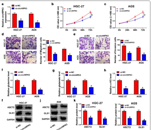
Knockdown of circHIPK3 inhibited proliferation, migration, invasion, and glutaminolysis in GC cells
As revealed in Fig. 2a, the knockdown experiments were successful using si-circHIPK3 in HGC-27 and AGS cells; the expression of circHIPK3 was decreased after trans-fection with si-circHIPK3. Additionally, cell viability was prominently declined at 72 h in HGC-27 and AGS cells infected with si-circHIPK3 than those cells trans-fected with si-NC (Fig. 2b, c). By performing transwell migration and invasion assays, we found that silencing of circHIPK3 decreased migration and invasion in HGC-27
and AGS cells (Fig. 2d, e). Furthermore, the levels of glu-tamine, glutamate, and α-KG were observed to assess glutaminolysis in GC cells. As presented in Fig. 2f–h, glutamine, glutamate, and α-KG were all declined in HGC-27 and AGS cells by transfection with si-circHIPK3 compared with control group. Consistently, ASCT2 and GLS1 were decreased after circHIPK3 silencing in HGC-27 and AGS cells (Fig. 2i–k). The above results revealed that silencing of circHIPK3 participated in proliferation, migration, invasion and glutaminolysis of GC cells, indi-cating the important role of circHIPK3 in GC.
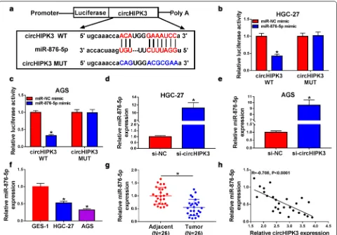
MiR‑876‑5p was a target of circHIPK3 and was decreased in GC tissues and cells
To identify target of circHIPK3, we searched for pos-sible target using bioinformatics software. As presented in Fig. 3a, circHIPK3 had the complementary sequence in miR-876-5p. After that, dual-luciferase reporter vec-tors contained wild type or mutant circHIPK3 were con-structed, and HGC-27 and AGS cells were co-transfected with indicated vectors and 876-5p mimic or miR-NC. The luciferase activity of circHIPK3 WT was inhib-ited in miR-876-5p mimic group, while the luciferase activity of circHIPK3 MUT had no difference between miR-876-5p and miR-NC group (Fig. 3b, c). In addition, knockdown of circHIPK3 enhanced miR-876-5p level in HGC-27 and AGS cells (Fig. 3d, e). Importantly, miR-876-5p showed lower expression in GC cells and tissues with respect to GES-1 cells and adjacent normal tissues, respectively (Fig. 3f, g). An adverse connection between
circHIPK3 and miR-876-5p was observed in GC tissues (Fig. 3h). Therefore, circHIPK3 negatively regulated miR-876-5p expression in GC cells.
Silencing of circHIPK3 impeded proliferation, migration, invasion and glutaminolysis of GC cells by upregulation of miR‑876‑5p
It has been confirmed that knockdown of circHIPK3 enhanced miR-876-5p level in HGC-27 and AGS cells, while it was abolished in HGC-27 and AGS cells by transfection with anti-miR-876-5p (Fig. 4a). Impor-tantly, depletion of miR-876-5p abolished the inhibitory effect on proliferation in HGC-27 and AGS cells caused by si-circHIPK3 (Fig. 4b, c). Transwell analysis suggested that downregulation of miR-876-5p could rescue loss of migration and invasion capabilities caused by circH-IPK3 knockdown (Fig. 4d, e). In addition, the levels of
glutamine, glutamate, and α-KG were increased in circH-IPK3-silencing GC cells after knockdown of miR-876-5p (Fig. 4f–h). Additionally, silencing of circHIPK3 induced the reduction in ASCT2 and GLS1 expression, while co-transfection of si-circHIPK3 and anti-miR-876-5p abol-ished these effects (Fig. 4i–k). Collectively, circHIPK3/ miR-876-5p axis mediated proliferation, migration, inva-sion, and glutaminolysis of GC cells.
PIK3R1 was regulated by circHIPK3/miR‑876‑5p axis in GC cells
As shown in Fig. 5a, we constructed luciferase reporter vectors PIK3R1 3′UTR WT contained complementary sequence to miR-876-5p, with PIK3R1 3′UTR MUT as control. Moreover, miR-876-5p mimic declined luciferase activity of PIK3R1 3′UTR WT instead of PIK3R1 3′UTR MUT in HGC-27 and AGS cells, implying that PIK3R1 was a direct target of miR-876-5p (Fig. 5b, c). The lev-els of PIK3R1 were decreased in HGC-27 and AGS cells
after transfection with miR-876-5p, including mRNA and protein expression (Fig. 5d–f). What’s more, trans-fection with anti-miR-876-5p overturned the decrease in PIK3R1 expression in HGC-27 and AGS cells caused by silencing of circHIPK3 (Fig. 5g–i). We also confirmed that mRNA and protein expression levels of PIK3R1 were upregulated in GC tissues and cells than that in matched controls (Fig. 5j–m). Besides, PIK3R1 was observed to be negatively correlated with miR-876-5p, while positively correlated circHIPK3 expression in GC tissues (Fig. 5n, o). In summary, these results revealed that circHIPK3 regulated PIK3R1 by sponging miR-876-5p in GC.
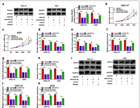
Overexpression of PIK3R1 overturned the inhibitory effects on proliferation, migration, invasion, and glutaminolysis of GC cells caused by overexpression of miR‑876‑5p
The results of western blot assay indicated that the upreg-ulation of miR-876-5p inhibited PIK3R1 expression, which was reversed by transfection with PIK3R1 (Fig. 6a).
Fig. 4 Knockdown of circHIPK3-mediated effects on proliferation, migration, invasion, and glutaminolysis were overturned by depletion of miR-876-5p in gastric cancer cells. a–k HGC-27 and AGS cells were transfected with si-NC, si-circHIPK3, si-circHIPK3 + anti-miR-NC, or
The decrease of cell proliferation in miR-876-5p-overex-pression cells was abrogated by upregulation of PIK3R1 (Fig. 6b, c). Moreover, transwell analysis displayed that co-transfection with miR-876-5p and PIK3R1 rescued loss of migration and invasion abilities induced by miR-876-5p (Fig. 6d, e). The overexpression of PIK3R1 could obviously increase glutamine, glutamate, and α-KG lev-els in HGC-27 and AGS cells with miR-876-5p overex-pression (Fig. 6f–h). Interestingly, miR-876-5p-induced inhibitory effects on ASCT2 and GLS1 expression was abrogated by overexpression of PIK3R1 in both HGC-27
and AGS cells (Fig. 6i–k). Conclusively, our results revealed that miR-876-5p regulated growth, migration, invasion and glutaminolysis by targeting PIK3R1 in GC cells.
Knockdown of circHIPK3 inhibited GC tumor growth
To probe the effect of circHIPK3 on GC cells prolifera-tion in vivo, a xenograft tumor model in nude mice was established. As displayed in Fig. 7a, b, downregulation of circHIPK3 led to the smaller tumor volumes and weight than volumes tissues from control group. Furthermore,
Fig. 5 CircHIPK3 regulated PIK3R1 expression by sponging miR-876-5p. a Binding regions between circHIPK3 and PIK3R1, as well as matched mutation form of PIK3R1 were presented. b, c Dual-luciferase reporter assay was introduced to test the luciferase activity in HGC-27 and AGS cells.
d–f RT-qPCR and western blot assays were applied to measure PIK3R1 levels in HGC-27 and AGS cells transfected with miR-NC or miR-876-5p. g–i
circHIPK3 was decreased, while miR-876-5p was increased in xenografts from sh-circHIPK3 group com-pared with sh-NC group (Fig. 7c, d). The western blot assay results indicated that suppression of circHIPK3 impeded PIK3R1 expression in xenografts (Fig. 7e). Syn-thetically, silencing of circHIPK3 suppressed GC growth in vivo.
Discussion
Conclusively, our study suggested the cancerogenic role of circHIPK3 in GC, and the silencing of circHIPK3 impeded GC cells proliferation, migration, invasion, as well as inhibited the tumor growth in vivo. Impor-tantly, we found that a novel regulatory mechanism of
circHIPK3/miR-876-5p/PIK3R1 axis was involved in GC development. Furthermore, evidence has shown that sur-vive and proliferation of cancer cells were highly depend-ent on glutaminolysis. ASCT2, high-affinity glutamine importer, was involved in amino acid transport and metabolism in cancer [16]. Glutamine was metabolized by rate-limiting enzyme GLS1, and eventually converted to α-KG [17]. Therefore, the levels of ASCT2, GLS1, and α-KG were closely correlated with glutaminolysis. In this paper, knockdown of circHIPK3 inhibited ASCT2, GLS1, and α-KG levels, suggesting silencing of circHIPK3 inhib-ited glutaminolysis in GC cells.
Recently, multiple studies have revealed an extensive landscape of circRNAs with exquisite regulation of the
Fig. 6 Overexpression of miR-876-5p inhibited proliferation, migration, invasion and glutaminolysis of gastric cancer cells by affecting PIK3R1. a–k
HGC-27 and AGS cells were transfected with miR-NC, miR-876-5p, miR-876-5p +vector, or miR-876-5p + PIK3R1. a The protein expression level of PIK3R1 was measured by western blot assay in HGC-27 and AGS cells. b, c The cell proliferation was assessed by MTT assay in HGC-27 and AGS cells.
development of malignant tumors through interaction with miRNAs [18, 19]. The studies had shown that circH-IPK3 functioned as an oncogene in malignant tumors [6, 7, 20]. By the bioinformatics prediction, we noticed that circHIPK3 had the binding sites on miR-876-5p. Subsequently, the direct association between circH-IPK3 and miR-876-5p was verified using dual-luciferase reporter assay. In addition, circHIPK3 was adversely con-nected with miR-876-5p expression in GC tissues. As we expected, knockdown of miR-876-5p could counteract the inhibitory effects on growth, mobility, and glutami-nolysis in GC cells caused by silencing of circHIPK3. Analogously, Sang et al. revealed that ciRS-7 stimulated esophageal squamous cell carcinoma progression by sponging miR-876-5p [21]. In addition, previous study by Xie et al. has unveiled the anti-tumor role of miR-876-5p in osteosarcoma by targeting c-Met [22].
For investigating the downstream target of the circH-IPK3-miR-876-5p axis, dual-luciferase reporter analysis was used and identified that PIK3R1 was a functional target of miR-876-5p. The PI3K family, belong to kinases of inositol and phosphatidylinositol, were oncogenes according pervious report [23]. The regulatory subu-nit (p85a), the composition of PI3K, was encoded by
PIK3R1 [24], while PI3K signaling was involved in mul-tiple human diseases, including, inflammation, malignant tumor, and diabetes [25, 26]. We supposed that PIK3R1 was an oncogene in malignant tumors. Surely, decrease of PIK3R1 suppressed proliferation and invasion in glio-blastoma multiform cells [27]. Moreover, reduction of PIK3R1 induced apoptosis and cycle arrest in colorec-tal cancer cells by repressed cyclin D1 expression [28]. Analogously, Huang et al. confirmed that miR-486-5p played a tumor-inhibitory role by suppressing PI3K/AKT pathway activation via targeting PIK3R1 [29]. However, the potential mechanism of PI3K/AKT regulation in GC by PIK3R1 should be further analyzed According above researches, PIK3R1 might be a disadvantageous index for prediction of GC. Not surprisingly, Xia et al. discov-ered PIK3R1 level was increased by nuclear paraspeckle assembly transcript 1 in GC and afterward stimulated GC progression [30]. In this study, PIK3R1 was increased in GC and adversely correlated with that of miR-876-5p expression in GC tissues. Furthermore, functional cel-lular experiments indicated that the upregulation of PIK3R1 increased the proliferation, migration, invasion, and glutaminolysis in miR-876-5p-overexpression GC cells.
In the current study, circHIPK3 was upregulated in GC tissues and cells after RT-qPCR examination. The following loss-of-function assays revealed that silencing of circHIPK3 impaired proliferation, migration, invasion and glutami-nolysis capacities of GC cells. Mechanistically, circHIPK3 exerted its cancerogenic functions through miR-876-5p/ PIK3R1 axis in GC.
Conclusion
In summary, circHIPK3 was upregulated in GC sam-ples and cells when compared with adjacent noncancer-ous tissues and gastric epithelial cells, correspondingly. Functional experiments indicated that silencing of circHIPK3 impeded GC progression by inhibiting pro-liferation, migration, invasion and glutaminolysis of GC cells through miR-876-5p/PIK3R1 axis, suggesting a potential therapeutic strategy for GC in the future.
Acknowledgements
None.
Authors’ contributions
Conception and design: QL; Development of methodology: YT; Acquisition of data: YL; Analysis and interpretation of data: CL and QL; Writing, review, and revision of article: QL; All coauthors have reviewed the article before submis-sion. All authors read and approved the final manuscript.
Funding
None.
Availability of data and materials
All data generated or analysed during this study are included in this published article.
Ethics approval and consent to participate
All patients offered the written informed consents and the research was permitted by the Ethics Committee of China-Japan Union Hospital of Jilin University. This study was directed with approval from the Institutional Animal Care and Use Committee of China-Japan Union Hospital of Jilin University.
Consent for publication
Informed consent was obtained from all patients.
Competing interests
The authors declare that they have no competing interests.
Author details
1 Department of Gastrointestinal Colorectal and Anal Surgery, China-Japan
Union Hospital of Jilin University, No. 126, Xiantai Street, Changchun 130031, Jilin, China. 2 Center of Physical Examination, China-Japan Union Hospital
of Jilin University, Changchun 130031, Jilin, China.
Received: 1 April 2020 Accepted: 25 July 2020
References
1. Hamashima C. Current issues and future perspectives of gastric cancer screening. World J Gastroenterol WJG. 2014;20(38):13767.
2. Bray F, Ferlay J, Soerjomataram I, Siegel RL, Torre LA, Jemal A. Global cancer statistics 2018: GLOBOCAN estimates of incidence and mor-tality worldwide for 36 cancers in 185 countries. CA Cancer J Clin. 2018;68(6):394–424.
3. Wilusz JE, Sharp PA. A circuitous route to noncoding RNA. Science. 2013;340(6131):440–1.
4. Jens M. Circular RNAs are a large class of animal RNAs with regulatory potency. Dissecting regulatory interactions of RNA and protein. Springer; 2014. p. 69–80.
5. Rong D, Lu C, Zhang B, Fu K, Zhao S, Tang W, Cao H. CircPSMC3 sup-presses the proliferation and metastasis of gastric cancer by acting as a competitive endogenous RNA through sponging miR-296-5p. Mol Cancer. 2019;18(1):25.
6. Zeng K, Chen X, Xu M, Liu X, Hu X, Xu T, Sun H, Pan Y, He B, Wang S. CircH-IPK3 promotes colorectal cancer growth and metastasis by sponging miR-7. Cell Death Dis. 2018;9(4):417.
7. Liu N, Zhang J, Zhang L, Wang L. CircHIPK3 is upregulated and predicts a poor prognosis in epithelial ovarian cancer. Eur Rev Med Pharmacol Sci. 2018;22(12):3713–8.
8. Yin Y, Song M, Gu B, Qi X, Hu Y, Feng Y, Liu H, Zhou L, Bian Z, Zhang J. Systematic analysis of key miRNAs and related signaling pathways in colorectal tumorigenesis. Gene. 2016;578(2):177–84.
9. Riquelme I, Letelier P, Riffo-Campos AL, Brebi P, Roa JC. Emerging role of miRNAs in the drug resistance of gastric cancer. Int J Mol Sci. 2016;17(3):424.
10. Paladini L, Fabris L, Bottai G, Raschioni C, Calin GA, Santarpia L. Targeting microRNAs as key modulators of tumor immune response. J Exp Clin Cancer Res. 2016;35(1):103.
11. Pu M, Chen J, Tao Z, Miao L, Qi X, Wang Y, Ren J. Regulatory network of miRNA on its target: coordination between transcriptional and post-transcriptional regulation of gene expression. Cell Mol Life Sci. 2019;76(3):441–51.
12. Bao L, Lv L, Feng J, Chen Y, Wang X, Han S, Zhao H. MiR-876-5p suppresses epithelial–mesenchymal transition of lung cancer by directly down-regulating bone morphogenetic protein 4. J Biosci. 2017;42(4):671–81. 13. Wang Y, Xie Y, Li X, Lin J, Zhang S, Li Z, Huo L, Gong R. MiR-876-5p acts as an inhibitor in hepatocellular carcinoma progression by targeting DNMT3A. Pathol Res Pract. 2018;214(7):1024–30.
14. Xu Z, Yu Z, Tan Q, Wei C, Tang Q, Wang L, Hong Y. MiR-876-5p regulates gastric cancer cell proliferation, apoptosis and migration through target-ing WNT5A and MITF. Biosci Rep. 2019. https ://doi.org/10.1042/bsr20 19006 6.
15. Hamidi A, Song J, Thakur N, Itoh S, Marcusson A, Bergh A, Heldin C-H, Landström M. TGF-β promotes PI3K-AKT signaling and prostate cancer cell migration through the TRAF6-mediated ubiquitylation of p85α. Sci Signal. 2017;10(486):eaal4186.
16. Van Geldermalsen M, Wang Q, Nagarajah R, Marshall A, Thoeng A, Gao D, Ritchie W, Feng Y, Bailey C, Deng N. ASCT2/SLC1A5 controls glutamine uptake and tumour growth in triple-negative basal-like breast cancer. Oncogene. 2016;35(24):3201–8.
17. Jin L, Alesi G, Kang S. Glutaminolysis as a target for cancer therapy. Onco-gene. 2016;35(28):3619.
18. Li P, Yang X, Yuan W, Yang C, Zhang X, Han J, Wang J, Deng X, Yang H, Li P. CircRNA-Cdr1as exerts anti-oncogenic functions in bladder Cancer by sponging MicroRNA-135a. Cell Physiol Biochem. 2018;46(4):1606–16. 19. Yuan Y, Liu W, Zhang Y, Zhang Y, Sun S. CircRNA circ_0026344 as a
prognostic biomarker suppresses colorectal cancer progression via microRNA-21 and microRNA-31. Biochem Biophys Res Commun. 2018;503(2):870–5.
20. Chen G, Shi Y, Liu M, Sun J. circHIPK3 regulates cell proliferation and migration by sponging miR-124 and regulating AQP3 expression in hepatocellular carcinoma. Cell Death Dis. 2018;9(2):175.
21. Sang M, Meng L, Sang Y, Liu S, Ding P, Ju Y, Liu F, Gu L, Lian Y, Li J. Circular RNA ciRS-7 accelerates ESCC progression through acting as a miR-876-5p sponge to enhance MAGE-A family expression. Cancer Lett. 2018;426:37–46.
22. Xie W, Xiao J, Wang T, Zhang D, Li Z. MicroRNA-876-5p inhibits cell pro-liferation, migration and invasion by targeting c-Met in osteosarcoma. J Cell Mol Med. 2019;23(5):3293–301.
23. Samuels Y, Ericson K. Oncogenic PI3K and its role in cancer. Curr Opin Oncol. 2006;18(1):77–82.
•fast, convenient online submission
•
thorough peer review by experienced researchers in your field
• rapid publication on acceptance
• support for research data, including large and complex data types
•
gold Open Access which fosters wider collaboration and increased citations maximum visibility for your research: over 100M website views per year
•
At BMC, research is always in progress.
Learn more biomedcentral.com/submissions
Ready to submit your research? Choose BMC and benefit from:
25. Rathinaswamy MK, Burke JE. Class I phosphoinositide 3-kinase (PI3K) regulatory subunits and their roles in signaling and disease. Adv Biol Regul. 2019;75:100657.
26. Wong K-K, Engelman JA, Cantley LC. Targeting the PI3K signaling pathway in cancer. Curr Opin Genet Dev. 2010;20(1):87–90.
27. Weber GL, Parat M-O, Binder ZA, Gallia GL, Riggins GJ. Abrogation of PIK3CA or PIK3R1 reduces proliferation, migration, and invasion in glio-blastoma multiforme cells. Oncotarget. 2011;2(11):833.
28. Sun Y, Zhao S, Tian H, Xie X, Xiao F, Li K, Song Y. Depletion of PI3K p85α induces cell cycle arrest and apoptosis in colorectal cancer cells. Oncol Rep. 2009;22(6):1435–41.
29. Huang XP, Hou J, Shen XY, Huang CY, Zhang XH, Xie YA, Luo XL. Micro RNA-486-5p, which is downregulated in hepatocellular carcinoma, sup-presses tumor growth by targeting PIK3R1. FEBS J. 2015;282(3):579–94. 30. Xia T, Chen J, Wu K, Zhang J, Yan Q. Long noncoding RNA NEAT1
promotes the growth of gastric cancer cells by regulating miR-497-5p/ PIK3R1 axis. Eur Rev Med Pharmacol Sci. 2019;23(16):6914–26.
Publisher’s Note