RONALD T. ROLLEY
Department of Surgery, Medical College of Virginia, Richmond 23219
Introduction
The problem of homograft rejec-tion in man remains unsolved. Any organ transplanted from one indi-vidual to another (except in the case of identical twins) will be re-jected, unless the rejection process can be abated by some method. Un-fortunately, homograft rejection still occurs all too frequently, and the toxicity of the drugs used pre-sents problems. However, in other animal systems the rejection pro-cess is directly related to the degree of antigenic difference between the donor and the recipient. Recently, various transplantation centers have tried to achieve antigenic similarity between the donor and the recipient as a means of improving the homo-graft survival rate. In order to do this, the antigens of the potential donors and the recipient are meas-ured in vitro. This is called tissue typing, or histocompatibility match-ing.
Humans have been shown to de-velop antibodies against donor cells following multiple blood transfu-sions, multiple pregnancies, organ homografting and deliberate immu-nization using repeated injections of leukocytes or platelets. Such an-tibodies are cross-reactive in vary-ing percentages of the random population. A panel of such anti-bodies or antisera can be tested against any individual's cells to de-termine his antigenicity. This can
*
Presented at the Fortieth Annual McGuire Lecture Series, October 31 -N ovember 1, 1968, Medical College of Virginia, Richmond.then be compared with the results of typing from other individuals prior to transplantation.
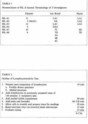
Antibodies have been identified against an antigen system known as HL-A (Table 1). Tissue typing studies performed in families have shown that this system is the result of antigenic determinants present on the same pair of chromosomes at several subloci. However, the exact interrelationship of these de-terminants is not fully known. The use of the HL-A terminology for the antigens has simplified the ex-change of information in this field. The original terminology of vari-ous investigators for these antigens is also shown in Table 1.
Methods
The methods most commonly employed in tissue typing are: 1) lymphocytotoxicity; 2) leukoagglu-tination; 3) platelet complement fixation; 4) mixed cell agglutina-tion; 5) immune adherence; and 6) mixed lymphocyte culture. The lymphocytotoxicity test is the most commonly employed test and re-quires viable lymphocytes, a for-eign complement source (generally, rabbit complement), and a battery of antisera. Cell death occurs if an antigen-antibody-complement reac-tion takes place. The most common method of detecting cell death is a dye exclusion method. Whereas dead cells imbibe the surrounding fluid, including the dye, and ap-pear larger and darker on the in-verted phase microscope, living cells with intact cell membranes exclude the surrounding fluid and
dye. Terasaki and co-workers have developed one form of this tech-nique (Terasaki, Vredevoe and Mickey, 1967; Mittal et al., 1968). An alternate method of determin-ing cell death is to preincubate the lymphocytes with 51Cr and then determine the release of this isotope as an indication of cell death (Ro-gentine and Plocinik, 1967).
Reproducibility of these tests is good, with about a 3% error. How-ever, the tests cannot be done with cells other than lymphocytes, e.g., kidney cells. An assumption is made that lymphocytes react simi-larly to cells from transplantable organs, i.e., kidney, heart. How-ever, this may not be the case. The need for viable lymphocytes to per-form the test precludes the later testing of cadaver donors unless the lymphocytes are specially freeze-preserved or maintained in tissue culture. An outline of the lympho-cytotoxicity test employed at the Medical College of Virginia is shown in Table 2.
TABLE 1
Nomenclature of HL-A System Terminology of 3 Investigators
Dausset van Rood
HL-Al 11 LAl
HL-A2 1 (MAC) SA
HL-A3 12 LA3
HL-A5 5 DAS
HL-A7 10 7C
HL-AS 8 7D
4A 4B 4C
TABLE 2
Outline of Lymphocytotoxicity Test
1 . Prepare pure suspension of lymphocytes a. Freshly drawn specimen
b. Mailed specimen
2. Add lymphocytes to previously prepared trays of 110 antisera + recipient's sera
3. Add pooled rabbit complement 4. Add eosin and formalin
5. Allow cells to resettle and prepare trays for readings 6. Read microtest trays on inverted phase microscope 7. Evaluate typing
MIXED AGGLUTINATION
Rh (D)• RBC
Payne
LAI LA2 LA3
4D 7D
45 min
60 min 60-120 min 30 min 15 min 15 min
4-5 hr
·
w~
~
[><ANTIl~;G~NTIBOD'
GOAT
ANTI~
-.f\~
id--
~SERUM
ABHUMAN lgG .-:;;__,_._ ~
KIDNEY CELLS
Fig. 1-The mixed cell agglutination test. See text.
are not recognizing important trans-plant antigens.
The platelet complement fixation test requires platelets, complement, and a battery of antisera (Shulman et al., 1964). This test is no longer widely employed. Dausset (1969) recently described new antigen-antibody reactions-related to the HL-A system-which occurred with this test but were not demonstrable in the lymphocytotoxic or leukoag -glutination tests.
All three of these tests require a large panel of antisera. Whereas erythrocyte typing requires only a single anti-A serum and a single anti-B serum. This is because the transplantation antigens as well as the antisera employed for detecting these antigens are poorly defined.
The three other techniques of tissue typing are not widely em-ployed at present but may have wider application in the future.
Mixed cell agglutination requires
that the target cells be grown in tissue culture monolayers (Milgrom et al., 1966). However, any cell line capable of being grown in tis-sue culture may be used. Attempts to employ this test with dispersed, fresh cells, cryostat sections, or fresh tissue slices have been unsuc-cessful. This test requires a special indicator cell system consisting of human erythocytes precoated with an antibody directed against human erythrocytes and an anti-human an-tibody capable of reacting with both the first antibody and the test anti-sera. At the Medical College of Virginia this indicator system con-sists of Rh+ erythrocytes precoated with anti-Rh antibody and a goat anti-human antibody. The test anti-serum is layered over the mono-layer of target cells, followed by thorough washing and the addition of the coated indicator cell system. Agglutination of the indicator cell system indicates that the test anti-serum has reacted with the target cell (Fig. 1).
The immune adherence test
erythrocytes (Nelson, 1956). It can be performed with various types of cells-dead or alive. If an antigen-antibody-complement reac-tion takes place, the erythrocyte in-dicator cells will adhere to the tar-get cell. The immune adherence and mixed cell agglutination tests are much more sensitive than the previously described tests. The mixed lymphocyte culture technique has been described in full by Bach
(1966).
In the immune adherence test a comparison of reactivity of lympho-cytes and kidney cells from one individual against the same anti-sera revealed these results in the five individuals studied: 37 nega-tive reactions against lymphocytes and kidney cells, 15 positive re-actions against lymphocytes and kidney cells, 29 reactions in which the antisera reacted positively with kidney cells and negatively with lymphocytes, and four instances in which the reverse relationship existed. This is presumptive evi-dence for an antigenic difference between kidney cells and lympho-cytes. Whether or not it is a quali-tative or quantiquali-tative difference is not shown by these data. At any rate, the data are against the as-sumption that, by typing leuko-cytes, one can determine the full antigenicity of an organ to be trans-planted.
A similar discrepancy has been demonstrated by testing lympho-cytes and kidney cells from the same individual-through use of the lymphocytotoxicity and mixed cell agglutination tests, respectively. In retesting an individual's cells against an antiserum in which the lymphocytotoxicity reaction was negative and the mixed cell agglu-tination reaction positive, the dif-ference in reactivity appeared to be quantitative, as prior absorption with the individual's leukocytes re-moved the reactivity against kidney cells, despite there originally being no reactivity demonstrable by lym-phocytotoxicity testing.
Since the antigenic determinants
TABLE 3
Examples of Lymphocyte Typing Results "A" Match
Example 1
HL-Al HL-A2 HL-A3 HL-A7 HL-AS 4A 4B 4C
Donoc
+
+
+
+
+
Recipient
+
+
+
+
+
4 of 100 sera "killed" donor's but not recipient's cells
Example 2
HL-AI HL-A2 HL-A3 HL-A7 HL-AS 4A 4B 4C
Donor
+
Recipient
+
+
+
+
+
1 of 100 sera "killed" donor's but not recipient's cells. All of above differences in antigenic groups are in the "minor direction."
for the HL-A system occur on the same pair of chromosomes, there is a 25 % chance that a sibling will be identical with any other sibling for the HL-A system. By tissue typ-ing a family, it can be determined which individuals share 0, 1, or both chromosomes. Amos et al. ( 1969) have shown that skin grafts between nonimmunosuppressed sib-lings known to be identical for the HL-A system have a mean sur-vival of about 25 days. The failure of permanent graft take might in-dicate that there are other antigenic systems present on other chromo-somes which are not yet defined.
Detailed absorption studies have been done in our laboratory to demonstrate the presence of HL-A antigens in various human tissues, i.e., kidney, liver, heart, blood ves-sels, lung, skin, pancreas, ureter and fat. Kidney and liver tissue
Histocompatibility Matching
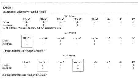
Of the six tissue typing tech-niques discussed above, only mixed lymphocyte culture involves direct interaction between donor and re-cipient cells. The other five tests all use a comparison of results against a panel of antisera. A histocompat-ibility match may be reported on an A, B, C, or D scale. The terms A and B are comparable to the terms matched or compatible; the terms C and D are comparable to the terms mismatched or incom-patible. Table 3 shows two exam-ples of an A match. In the upper example of that table, the antigenic-ity of the donor and the recipient for these eight antigens is the same. However, a great deal of attention is also given to individual serum reactions, since not all sera have been classified for one of these par-ticular antigen groups. In this ex-ample 4% of the sera reacted with antigens of the donor but not of the recipient. Theoretically, the recipient is capable of reacting to such foreign antigens.
TABLE 4
Examples of Lymphocyte Typing Results
HL-Al HL-A2
Donor
+
+
Recipient
+
+
In the second example of an A
match in Table 3, the recipient bas more antigen groups than the donor. At our present level of knowledge, this is assumed not to affect graft survival and therefore, the example is not considered a mismatch; but rather an A match. Table 4 shows an example of a B match in which no recognized antigen groups are present on donor cells but absent on recipient cells. However, more than 5 % of the individual sera detected antigens on donor but not recipient cells; hence, this match is rated B rather than A. Table 4 also shows an example of a C match in which a well rec-ognized antigen group is present on donor cells but absent on recipient cells. A D match (Table 4) indi-cates a donor-to-recipient mismatch of two antigen groups.
Preformed Antibodies
Tissue typing tests determine neither the presence of preformed antibodies against donor antigens, nor the presence of organ-specific
"B" Match
auto-antibodies, such as the anti-glomerular basement membrane antibodies. To look for preformed antibodies against histocompatibil-ity antigens, it is necessary to per-form a cross match test using the recipient's serum and donor's cells.
This can be done by any one of the five tissue typing methods dis-cussed in this paper. If preformed antibodies are found and a tran s-plant performed, there is a great probability that the transplant will be rejected very quickly. A dis-cussion of the detection of aut o-antibodies is beyond the scope of this paper.
At present, bistocompatibility matching is an inexact science, and there are undoubtedly many histo-compatibility antigens either poorly defined or undefined. With further definition of such antigens, im-provement can be expected in the correlation of tissue typing with homograft function and survival. Further work also needs to be done to determine which method of tis-sue typing is best.
HL-A3 HL-A7 HL-AS 4A 4B 4C
+
+
12 of 100 sera "killed" donor's but not recipient's sera."C" Match
-I
HL-Al HL-A2 HL-A3 HL-A7 HL-A8 4A 4B 4CDonor
+
+
+
Recipient
l
+
+
+
+
1 group mismatch in "major direction."
"D" Match
HL-Al
ff
HL-A3ITJ
HL-AS 4A 4B 4CDonor
+
+
+
Recipient
+
+
+
References
AMOS, D. B., H. F. SEIGLER, J. G. SOUTHWORTH AND F. E. WARD. Skin graft rejection between subjects genotyped for HL-A. Transplanta-tion Proc. 1: 342-346, 1969. BACH, F. H. Lymphocyte reactivity
in mixed leukocyte cultures-its assay and genetics. In in vitro.
Vol. 2. Phenotypic Expression-Im-munological, Biochemical and Morphological. Baltimore: Williams and Wilkins, 1966, pp. 32-39. DAUSSET, J., R. L. WALFORD, J.
Co-LOMBANI, L. LEGRAND, N. FEIN-GOLD, A. BARGE AND F. T. RAPA-PORT. The HL-A system sub-loci and their importance in transplanta-tion. Transplantation Proc. 1: 331
-338, 1969.
MILGROM, F., B. I. LITVAK, K. KANO AND E. WITEBSKY. Humoral anti-bodies in renal homograft. JAMA
198:226-230, 1966.
MITTAL, K. K., M. R. MICKEY, D. P. SINGAL ANO P. I. TERASAKI. Sero-typing for homotransplantation. XVIII. Refinement of microdroplet lymphocyte cytotoxicity test. Trans-plantation 6:913-927, 1968. NELSON, R. A., JR. The
immune-adherence phenomenon. A hypo-thetical role of erythrocytes in de-fence against bacteria and viruses.
Proc. Royal Soc. Med. 49:55-58, 1956.
ROGENTINE, G. N., JR. AND B. A. PLOCINIK. Application of the 51Cr cytotoxicity technique to the analy-sis of human lymphocyte isoanti-gens. Transplantation 5: 1323-1333,
1967.
ROLLEY, R. T., G. M. WILLIAMS AND D. M. HUME. Distribution of leu-kocyte antigens in human tissues.
Surg. Forum 19:241-243, 1968. SHULMAN, N. R., V. R. NARDER,
M. C. HILLER ANOE. M. COLLIER.
Platelet and leukocyte isoantigens and their antibodies: Serologic, physiologic and clinical studies. In
Progress in Hematology. Vol. 4.
C. V. Moore and E. B. Brown (eds.). New York/London: Grune and Stratton, 1964, pp. 222-304.
TERASAKI, P. I., D. L. VREDEVOE AND M. R. MICKEY. Serotyping for ho-motransplantation. X. Survival of 196 grafted kidneys subsequent to typing. Transplantation 5: 1057-1070, 1967.
ZMIJEWSKI,
c
.
M., R. L. ST. PIERRE, J. L. FLETCHER, S. F. WILSON, W. CANNADY AND H. E. ZMIJEWSKI.A comparison of two micro-leu koagglutination test. In
Histo-compatibility Testing. E. S. Curtoni, P. L. Mattiuz and R. M. Tost (eds.). Copenhagen: Munksgaard,