HTTPS://WWW.HEIGHPUBS.ORG ISSN
2573-6191
Summary
Several articles have discussed mandibular condyle position after bilateral sagittal split osteotomies (BSSO). However, previous studies have sought to establish a relationship between orthognathic surgery and the improvement of temporomandibular joint (TMJ) signs and symptoms. Also, they have been limited to two-dimensional evaluation of condylar position considering only the condylar “sag” or improper inferior seating of the condyle. The purpose of this study was to investigate a relationship between changes in condylar position using three-dimensional computed tomography and the negative clinical impact of BSSO on the temporomandibular joint.
Research Article
Correlation between mandibular
condylar position and clinical
dysfunction index after bilateral
sagittal split osteotomies
Kalach Mussali Alberto J
1* Gonzalez-Magana Fernando
2and
Malagon Hidalgo Hector O
31Resident, Plastic & Reconstructive Surgery, ISSEMyM Medical Center, Toluca, State of Mexico,
Mexico
2Clinical Assistant Professor, Maxillofacial Surgery, ISSEMyM Medical Center, Toluca, State of
Mexico, Mexico
3Program Director, Plastic & Reconstructive Surgery, ISSEMyM Medical Center, Toluca, State of
Mexico, Mexico
*Address for Correspondence: Alberto J Kalach Mussali, Avenida Baja Velocidad Speed 284, Colonia San Jerónimo Chicahualco, Metepec, State of Mexico, Mexico, C.P. 52170, Tel: +52-722-275-6300; Email: [email protected]
Submitted: 05 December 2018 Approved: 03 June 2019 Published: 04 June 2019
Copyright:© 2019 Kalach Mussali AJ, et al. This is an open access article distributed under the Creative Commons Attribution License, which permits unrestricted use, distribution, and reproduction in any medium, provided the original work is properly cited
Keywords: Bilateral sagittal splitosteotomy; Mandibular condyle position; Temporomandibular joint; Clinical dysfunction index; Orthognathic surgery
How to cite this article: Kalach Mussali AJ, Gonzalez-Magana F, Malagon Hidalgo HO. Correlation between mandibular condylar position and clinical dysfunction index after bilateral sagittal split osteotomies. J Oral Health Craniofac Sci. 2019; 4: 001-007. https://doi.org/10.29328/journal.johcs.1001026
Introduction
Dentofacial deformities can be de ined as signi icant deviations from normal pro-portions of the maxillo-mandibular complex that also negatively affect the relationship of the teeth within each arch and the relationship of the arches with one another (oc-clusion). These deformities may be minimal, such as a slight projection of the chin, or extreme, such as a severe vertical maxillary excess or hemifacial microsomia [1].This condition may be evident at birth or appear during growth and development, creating functional, degenerative, aesthetic and psychosocial problems [2]. Serious problems of dental and skeletal malocclusion require combined treatment with orthodontics and orthognathic surgery, intending to achieve facial, dental and functional harmony [3].
Surgery to reposition the jaws (i.e., an orthognathic procedure) as part of an interdisciplinary approach is often recommended to manage the related skeletal, dental, and soft-tissue dysfunctions and concerns [4]. Orthognathic surgery is a treatment option that provides stability, function and aesthetics [5].
Mandibular topographic changes have a displacement of the mandibular condyle within the temporomandibular joint, leading to temporomandibular disorders [9]. The potential effects of BSSO on signs and symptoms of temporomandibular joint disorder are still controversial. The surgeon must be aware of the potential implication of BSSO advancement on the onset and progression of temporomandibular disorders in patients with Class II malocclusion. Interestingly, counterclockwise rotation of the mandible appears to be associated with more muscle tenderness, especially in patients undergoing greater BSSO advancements [10].Mandibular condyle displacements after BSSO may cause temporomandibular joint dysfunction; the aim of this study is to investigate the relationship between the clinical dysfunction index and the mandibular condyle position after BSSO [10].
Material and Methods
A population of 47 patients who underwent BSSO in the Maxillofacial / Plastic & Reconstructive Surgery Department in ISSEMyM Medical Center were studied retro-spectively. All of the patients had preoperative orthodontics and they were operated between December 2012 and January 2016. During preoperative and postoperative examination, Helkimo’s Anamnestic and Dysfunction Index (HADI) [11], were deter-mined. Three-dimensional computed tomography were acquired with 1.0mm-thick slices using a Siemens SOMATOM Emotion scanner, data acquisition occurred in the preoperative surgical planning and >6 months postoperatively for both the Helkimo’s Anamnestic and Dysfunction Index and tomographic measurements using Pullinger & Hollender modi ied by Pereira method (P&HmP) [12]. (to determine antero-poste-rior condylar displacement) and determination of condylar rotation as seen in igure 1 (ConRot) (determined in the axial plane parallel to Frankfort’s plane, tracing a line between the anterior and posterior nasal spine determining the midsagittal plane; and a line on the mayor condylar axis, resulting in an angle) [13,14].
Each examination included a written anamnestic questionnaire and both a clinical and radiological examination. In the questionnaire the patient answered yes or no to questions concerning joint noises, locking, joint pain, pain in the facial musculature, stiffness/dif iculties in mouth opening, clenching of the teeth and ear pain. The basic data from the questionnaire and clinical examination were calculated and classi ied according to Helkimo’s Anamnestic Index and clinical Dysfunction Index.
Statistical analysis was performed using SPSS 24.0 software measuring Pearson’s correlation coef icient to establish a relationship between tomographic measurements and Helkimo’s Anamnestic and Dysfunction Index in the preoperative and postoperative setting.
This study received approval from the institutional review of boards and ethics committee approval.
Results
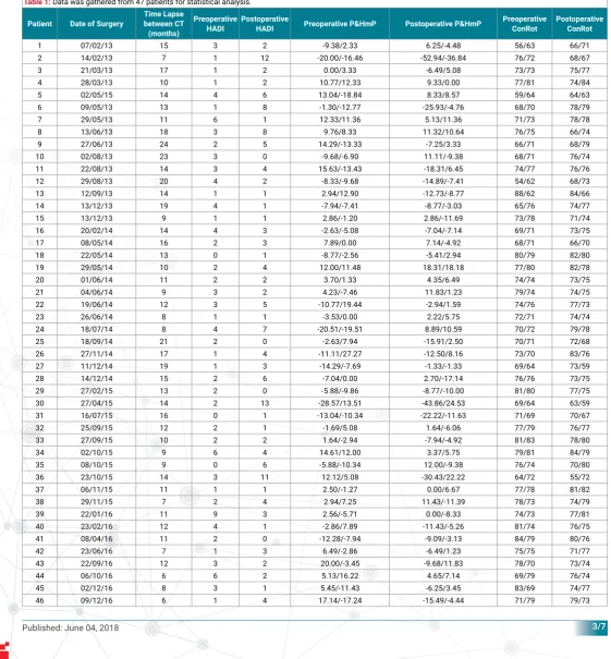
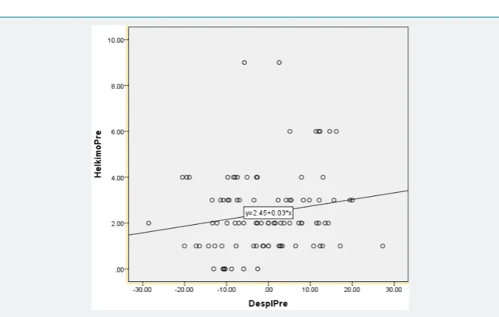
Data was gathered from 47 patients for statistical analysis shown in table 1. There were no signi icant differences between the Helkimo’s Anamnestic and Dysfunction Index and the tomographic measurements in the preoperative setting. A weak correlation was found between condylar displacement (P&HmP) and HADI with a Pearson’s correlation coef icient of 0.175 (p=0.091) as seen in igure 2; nor condylar rotation and HADI with a Pearson’s correlation coef icient of 0.158 (p=0.129) as seen in igure 3.
During the postoperative period, patients with a posterior mandibular condyle displacement in the temporomandibular joint showed a higher HADI score, proving
Table 1:Data was gathered from 47 patients for statistical analysis.
Patient Date of Surgery
Time Lapse between CT (months) Preoperative HADI Postoperative
HADI Preoperative P&HmP Postoperative P&HmP
Preoperative ConRot
Postoperative ConRot
1 07/02/13 15 3 2 -9.38/2.33 6.25/-4.48 56/63 66/71
2 14/02/13 7 1 12 -20.00/-16.46 -52.94/-36.84 76/72 68/67
3 21/03/13 17 1 2 0.00/3.33 -6.49/5.08 73/73 75/77
4 28/03/13 10 1 2 10.77/12.33 9.33/0.00 77/81 74/84
5 02/05/15 14 4 6 13.04/-18.84 8.33/8.57 59/64 64/63
6 09/05/13 13 1 8 -1.30/-12.77 -25.93/-4.76 68/70 78/79
7 29/05/13 11 6 1 12.33/11.36 5.13/11.36 71/73 78/78
8 13/06/13 18 3 8 9.76/8.33 11.32/10.64 76/75 66/74
9 27/06/13 24 2 5 14.29/-13.33 -7.25/3.33 66/71 68/79
10 02/08/13 23 3 0 -9.68/-6.90 11.11/-9.38 68/71 76/74
11 22/08/13 14 3 4 15.63/-13.43 -18.31/6.45 74/77 76/76
12 29/08/13 20 4 2 -8.33/-9.68 -14.89/-7.41 54/62 68/73
13 12/09/13 14 1 1 2.94/12.90 -12.73/-8.77 88/62 84/66
14 13/12/13 19 4 1 -7.94/-7.41 -8.77/-3.03 65/76 74/77
15 13/12/13 9 1 1 2.86/-1.20 2.86/-11.69 73/78 71/74
16 20/02/14 14 4 3 -2.63/-5.08 -7.04/-7.14 69/71 73/75
17 08/05/14 16 2 3 7.89/0.00 7.14/-4.92 68/71 66/70
18 22/05/14 13 0 1 -8.77/-2.56 -5.41/2.94 80/79 82/80
19 29/05/14 10 2 4 12.00/11.48 18.31/18.18 77/80 82/78
20 01/06/14 11 2 2 3.70/1.33 4.35/6.49 74/74 73/75
21 04/06/14 9 3 2 4.23/-7.46 11.83/1.23 79/74 74/75
22 19/06/14 12 3 5 -10.77/19.44 -2.94/1.59 74/76 77/73
23 26/06/14 8 1 1 -3.53/0.00 2.22/5.75 72/71 74/74
24 18/07/14 8 4 7 -20.51/-19.51 8.89/10.59 70/72 79/78
25 18/09/14 21 2 0 -2.63/7.94 -15.91/2.50 70/71 72/68
26 27/11/14 17 1 4 -11.11/27.27 -12.50/8.16 73/70 83/76
27 11/12/14 19 1 3 -14.29/-7.69 -1.33/-1.33 69/64 73/59
28 14/12/14 15 2 6 -7.04/0.00 2.70/-17.14 76/76 73/75
29 27/02/15 13 2 0 -5.88/-9.86 -8.77/-10.00 81/80 77/75
30 27/04/15 14 2 13 -28.57/13.51 -43.86/24.53 69/64 63/59
31 16/07/15 16 0 1 -13.04/-10.34 -22.22/-11.63 71/69 70/67
32 25/09/15 12 2 1 -1.69/5.08 1.64/-6.06 77/79 76/77
33 27/09/15 10 2 2 1.64/-2.94 -7.94/-4.92 81/83 78/80
34 02/10/15 9 6 4 14.61/12.00 3.37/5.75 79/81 84/79
35 08/10/15 9 0 6 -5.88/-10.34 12.00/-9.38 76/74 70/80
36 23/10/15 14 3 11 12.12/5.08 -30.43/22.22 64/72 55/72
37 06/11/15 11 1 1 2.50/-1.27 0.00/6.67 77/78 81/82
38 29/11/15 7 2 4 2.94/7.25 11.43/-11.39 78/73 74/79
39 22/01/16 11 9 3 2.56/-5.71 0.00/-8.33 74/73 77/81
40 23/02/16 12 4 1 -2.86/7.89 -11.43/-5.26 81/74 76/75
41 08/04/16 11 2 0 -12.28/-7.94 -9.09/-3.13 84/79 80/76
42 23/06/16 7 1 3 6.49/-2.86 -6.49/1.23 75/75 71/77
43 22/09/16 12 3 2 20.00/-3.45 -9.68/11.83 78/70 73/74
44 06/10/16 6 6 2 5.13/16.22 4.65/7.14 69/79 76/74
45 02/12/16 8 3 1 5.45/-11.43 -6.25/3.45 83/69 74/77
Figure 2: A weak correlation was found between condylar displacement (P&HmP) and HADI with a Pearson’s correlation coeffi cient of 0.175 (p=0.091).
Figure 3: Nor condylar rotation and HADI with a Pearson’s correlation coeffi cient of 0.158 (p=0.129).
a strong Pearson’s correlation coef icient with a score of -0.217 (p<0.036) as seen in igure 4.
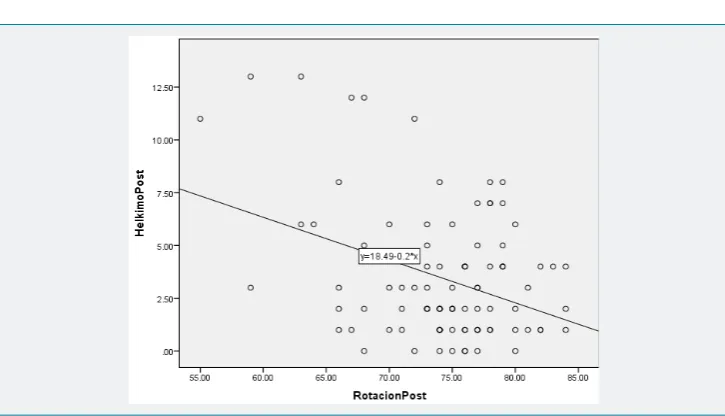
A stronger Pearson’s correlation coef icient was found between mandibular condyle rotation (ConRot) and HADI -0.367 (p<0.01) as seen in igure 5.
No association could be found between HADI and anterior condyle displacement measured with P&HmP or with lateral condylar rotation (ConRot).
Discussion
The main goal of most patients who undergo bilateral sagittal split osteotomies is to achieve a better aesthetic facial result. During examination and planning, it’s fundamental to perform cephalometric measurements for an adequate prediction of inal results and prevent causing evident clinical dysfunction. Orthognathic surgery displaces and modi ies mandibular topography in planned predictable sites for which surgery is intended, but secondarily disturbs the anatomy of the mandibular condyle causing clinical dysfunction in the temporomandibular joint [5]. Surgery also changes the facial osseous and muscle / soft tissue balance) which will elicit signs and symptoms observed and described in HADI [11].
In the tomographic sagittal plane, antero-posterior displacement of the mandibular condyle is observed after BSSO, for which Pullinger & Hollender designed a measuring method to establish a condylar relationship with the temporomandibular joint [15]. Later Pereira modi ied this method in which he did the same measurements using a resting position centered bite instead of a maximal intercuspation bite [12]. In the tomographic axial plane rotational condylar movements are observed after surgery despite of condylar transoperative repositioning, most condylar position studies have omitted this measurement and have measured the condyle bi-dimensionally only. Also in the axial tomographic plane lateromedial condylar movements and “sag” can be observed, but have been negligible during postoperative clinical examination [8,16].
Other studies have focused on remodeling and displacement of the mandibular condyles, and established this changes as a physiological adaptation process of the TMJ, allowing major consequences related to occlusion to be avoided. Sometimes the adaptive capacity of the joint is exceeded, leading to the onset of a resorption process of the condyle, characterized by bone remodeling with a negative balance in the volume of the condyle [17].These studies have shown no correlation between changes in condylar volume and joint spaces. Therefore, changes in joint spaces are more likely to
be related to factors other than remodeling, such as condylar displacement. Superior, posterior and medial joint spaces are reduced after orthognathic surgery, changes not related to the amount of the mandibular advancement that could possibly relate to TMJ dysfunction. However the measurements were performed in 2-D examinations, making it impossible to evaluate medial-lateral and rotational alterations [18-20].
In this study we observed that posterior displacement and medial condylar rota-tion are the most representative and valuable, since they will evoke a negative clinical impact in BSSO patients [21]. Despite the latter, this study could not establish a move-ment prediction method for the mandibular condyle. Follow-up examinations after at least 6 months, allowing symptoms that could be related directly to the postsurgical recovery period, showed that even with a protocolized technique, a broad range of displacement and rotation of the condyles was observed.
Also, the aim of this study was to establish a relationship between condylar dis-placement/rotation and the clinical dysfunction index after BSSO, thus other variables such as ixation method, type of ixation, osteosynthesis material position and distribu-tion, osteotomy types, association to bimaxillary surgery, and others were not consid-ered, and could be subject to future studies as they could also modify the position of the mandibular condyles after surgery.
Conclusion
Condylar displacements are a consequence of BSSO and mandibular advancement. These displacements represent a relationship with a negative clinical impact, mostly medial rotation and posterior condylar displacement.
This study could not establish a quantitative relationship between condylar dis-placements and the presentation of signs and symptoms in the orthognathic surgery patient. Mandibular advancement, setback or rotation planned in cephalometry are predictable and reproducible, but condylar displacements secondary to these are not, being in luenced also by masticatory muscles and soft tissue, that after changing posi-tion are subject to elongaposi-tion and compression forces evoking signs and symptoms, sometimes worsening the dysfunction of the patients.
References
1. Fish LC, Epker BN, Sullivan CR. Orthognathic surgery: the correction of dentofacial deformities. J Oral Maxillofac Surg. 1993; 51(Suppl 1): 28-41. Ref.: https://tinyurl.com/y2m6l7js
2. American Association of Oral and Maxillofacial Surgeons. Parameters of care: clinical practice guidelines for oral and maxillofacial surgery (AAOMS Par Care 2012). J Oral Maxillofac Surg. 2012; 70: 107-136. Ref.: https://tinyurl.com/y28tl8ra
3. Sinko K, Jagsch B, Benes G, Millesi F, Fischmeister R. Facial aesthetics and the assignment of personality traits before and after orthognathic surgery. Int J Oral Maxillofac Surg. 2012; 41: 469-476. Ref.: https://tinyurl.com/yyxjtdfs
4. Zins JE, Bruno J, Moreira-Gonzalez A, Bena J. Orthognathic surgery: is there a future? Plast Reconstr Surg. 2005; 116: 1442–1450. Ref.: https://tinyurl.com/y6qgmw9q
5. Posnick JC. Orthognathic Surgery, Principles and Practice. Elsevier Health Sciences. 2013; 61–68.
Ref.: https://tinyurl.com/y2abqzmg
6. Bell WH, Schendel SA. Biologic basis for modifi cation of the sagittal ramus split operation. J Oral Surg. 1977; 35: 362-369. Ref.: https://tinyurl.com/yys2sccm
7. Onizawa K, Schmelzen R, Vogt S. Alteration of temporomandibular joint symptoms after orthognathic surgery comparison with healthy volunteers. J Oral Maxillofac Surg. 1995; 53: 117-121. Ref.:
https://tinyurl.com/y35vnktq
9. Kundert M, Hadjianghelou O. Condylar displacement after sagittal splitting of the mandibular rami. J Maxillofac Surg. 1980; 8: 278-287. Ref.: https://tinyurl.com/yyfr426z
10. Bagheri SC, Bell RB, Khan HA. Current Therapy in Oral and Maxillofacial Surgery. Elsevier Saunders; 2012. Mandibular Defi ciency: Bilateral Sagittal Split Osteotomy. 617-626. Ref.: https://tinyurl.com/y4av8wkx 11. Helkimo M. Studies on function and dysfunction of the masticatory system. II. Index for anamnestic and
clinical dysfunction and occlusal state. Swed Dent J. 1974: 67: 101–121. Ref.: https://tinyurl.com/y3tdjrc2 12. Pereira LJ, Gavião MB. Tomographic evaluation of TMJ in adolescents with temporomandibular
disorders. Braz. Oral Res. 2004; 18: 208-214. Ref.: https://tinyurl.com/y6g43qzx
13. Pérez Garcidueñas CM, Malagón Hidalgo HO. Medición Tomográfi ca del ángulo cóndilo-mandibular en pacientes del servicio de cirugía plástica del Centro Médico ISSEMyM “Arturo Montiel Rojas” de 1 de enero de 2013 al 31 de diciembre de 2013. Trabajo de Tesis, Reg 009/14, Universidad Autónoma del Estado de México. 2014.
14. An SB, Park SB, Kim YI, Son WS. Effect of post-orthognathic surgery condylar axis changes on condylar morphology as deter- mined by 3-dimensional surface reconstruction. Angle Orthod. 2014; 84: 316–321. Ref.: https://tinyurl.com/y4ubab52
15. Pullinger AG, Hollender L, Solberg WK, Petersson A. A tomographic study of mandibular condyle position in an asymptomatic population. J Prosthet Dent. 1985; 53: 706-713. Ref.: https://tinyurl.com/yxfk22ek 16. Kadesjö N, Benchimol D, Falahat B, Näsström K, Shi XQ. Evaluation of the effective dose of cone beam CT and multislice CT for temporomandibular joint examinations at optimized exposure levels. Dentomaxillofac Radiol. 2015; 44: 20150041. Ref.: https://tinyurl.com/yxnrgpgu
17. Ferri J, Nicor R, Màes JM, Raoul G, Lauwers L. Condylar resorptions and orthodontic surgical treatment: state of the art. Int Orthod. 2016; 14: 503-527. Ref.: https://tinyurl.com/y5447neg 18. Kawamata A, Fujishita M, Nagahara K, Kanematu N, Niwa K, et al. Three-dimensional computed
tomography evaluation of postsurgical condylar displacement after mandibular osteotomy. Oral Surg Oral Med Oral Pathol Oral Radiol Endod. 1998; 85: 371-376. Ref.: https://tinyurl.com/y2dmcchr 19. Alder ME, Deahl ST, Matteson SR, Van Sickels JE, Tiner BD, et al. Short-term changes of condylar position after sagittal split osteotomy for mandibular advancement. Oral Surg Oral Med Oral Pathol Oral Radiol Endod. 1999; 87: 159-165. Ref.: https://tinyurl.com/y2y6bbjn
20. Méndez-Manjón I, Guijarro-Martínez R, Valls-Ontañón A, Hernández-Alfaro F. Early changes in condylar position after mandibular advancement: a three-dimensional analysis. Int J Oral Maxillofac Surg. 2016; 45: 787–792. Ref.: https://tinyurl.com/y5uscdgy