R E S E A R C H
Open Access
Epigenetic change in kidney tumor:
downregulation of histone acetyltransferase
MYST1 in human renal cell carcinoma
Yong Wang
1,2†, Rui Zhang
3,4†, Donglu Wu
3,4, Zhihua Lu
1, Wentao Sun
1, Yong Cai
3, Chunxi Wang
1*and Jingji Jin
3*Abstract
Background:MYST1 (also known as hMOF), a member of the MYST family of histone acetyltransferases (HATs) as an epigenetic mark of active genes, is mainly responsible for histone H4K16 acetylation in the cells. Recent studies have shown that the abnormal gene expression of hMOF is involved in certain primary cancers. Here we examined the involvement of hMOF expression and histone H4K16 acetylation in primary renal cell carcinoma (RCC).
Simultaneously, we investigated the correlation between the expression of hMOF and clear cell RCC (ccRCC) biomarker carbohydrase IX (CA9) in RCC.
Materials and methods:The frozen RCC tissues and RCC cell lines as materials, the reverse transcription polymerase chain reaction (RT-PCR), western blotting and immunohistochemical staining approaches were used. Results:RT-PCR results indicate that hMOF gene expression levels frequently downregulated in 90.5% of patients (19/21) with RCC. The reduction of hMOF protein in both RCC tissues and RCC cell lines is tightly correlated with acetylation of histone H4K16. In addition, overexpression of CA9 was detected in 100% of ccRCC patients (21/21). However, transient transfection of hMOF in ccRCC 786–0 cells did not affect both the gene and protein expression of CA9.
Conclusion:hMOF as an acetyltransferase of H4K16 might be involved in the pathogenesis of kidney cancer, and this epigenetic changes might be a new CA9-independent RCC diagnostic maker.
Keywords:Renal cell carcinoma, hMOF, Carbonate anhydrase IX
Introduction
Changes of chromatin structure are mainly regulated by epigenetic regulations including ATP-dependent remode-ling of nucleosomes, the incorporation of variants histones into nucleosomes and posttranslational modifications of histones [1]. Post-translational modifications of the N-terminal tails of histones include acetylation, methylation, phosphorylation, ubiquitination, sumoylation, and ADP-ribosylation [2,3]. Histone acetylation as one of the best characterized epigenetic modifications is controlled by his-tone acetyltransferases (HATs) and hishis-tone deacetylases (HDAC). The balance between histone acetylation and
deacetylation serves as a key epigenetic mechanism for gene expression, DNA repair, developmental processes and tumorigenesis [4-6]. Thus, any reason to make this imbalance can lead to abnormal cell function, even cancer. MYST1 (also known as hMOF), is the human ortholog
of theDrosophilaMOF protein containing chromodomain
and acetyl-CoA binding motif which is one of the key components of the dosage compensation complex (DCC) or the male specific lethal (dMSL) complex [7]. Recent biochemical purifications revealed that hMOF forms at least two distinct multi-protein complexes in mammalian cells. One complex is the evolutionary conserved human MSL complex which is responsible for the majority of his-tone H4 acetylation at lysine 16 [8,9]. The other hMOF-containing complex is the human non-specific lethal (NSL) complex which is recently characterized by Cai Y et al. [10]. hNSL complex can also acetylate histone H4 at * Correspondence:chunxi_wang@126.com;jjjin@jlu.edu.cn
†Equal contributors
1Urology Department, The First Clinical Hospital, Jilin University,
Changchun City, Jilin 130021, China
3College of Life Science, Jilin University, Changchun City, Jilin 130012, China
Full list of author information is available at the end of the article
lysine 5 and 8 on the recombinant polynucleosomes with the exception of histone H4K16. Although the functions of hMSL and hNSL complexes in human cells are not very clear, both complexes can acetylate histone H4 at lysine 16, suggesting the importance of acetylation of H4K16 in cells. Except for acetylation of H4K16, NSL complex was found to be able to acetylate the tumor suppressor protein p53, and this acetylation is able to affect the behavior of p53 in response to DNA damage [11]. It has been reported that depletion of hMOF in human cells leads to genomic instability, spontaneous chromosomal aberrations, cell cycle defects, reduced transcription of certain genes, and defect-ive DNA damage repair and early embryonic lethality [4-7]. This suggests a critical role for hMOF in fundamental pro-cesses such as gene transcription, cell proliferation, differ-entiation and DNA repair response. It is worth mentioning that depletion of hMOF also leads to global reduction of histone H4K16 acetylation in human cells [8,12]. However, recent studies suggest that the global modification status of H4K16Ac is also affected by Gcn-5-containing HAT and SIRT-LSD1 HDAC complexes [13,14], indicating hMOF might not be the only HAT fulfilling acetylation of H4K16 in cells. Although the role of histone H4K16 acetylation in transcription regulation is not completely understood, loss of H4K16 acetylation has been found in certain cancers. Pfister et al. [15] found that frequent downregulation of hMOF in large series of primary breast carcinomas and medulloblastomas and hMOF protein expression tightly correlated with acetylation of H4K16 in both cancers. In addition, analysis of the tissue microarray slides revealed low or absent histone H4K16 acetylation in majority of breast cancer tissues [16].
Renal cell carcinoma (RCC) is one of the most com-mon genitourinary malignancies, accounting for about 3% of all cancers worldwide [17]. With the improved im-aging diagnostic technology, more RCC cases have been diagnosed at an early stage. However, there is a consider-able number of RCC patients at the time of diagnosis has been transferred [18]. Research efforts have found various biomarkers of diagnostic and prognostic of RCC such as hypoxia-induced factor 1alpha (HIF1α), vascular endothelial growth factor (VEGF), and carbonic anhy-drase IX (CA9), but they are not specific and sensitive enough to accurately predict the survival of RCC patients [19-21]. Recent studies indicate that epigenetic alterations play an important role in carcinogenesis, and global histone modifications as predictors of cancer re-currence in various tumor entities has begun to study. Patients with RCC have been found that total acetylation levels of histone H3 were inversely correlated with pT-stage, distant metastasis, Fuhrman grading and RCC pro-gression, whereas total histone H4Ac deacetylation was correlated with pT-stage and grading [22]. All the above observations strongly suggest that histone modifications
might be involved in the development and progression of RCC. However, it is not clear which particular enzyme or specific modified lysine residue is responsible for tumori-genesis in RCC. This study aims to assess hMOF expres-sion and its corresponding acetylation of histone H4K16 in the RCC via qRT-PCR, western blotting and immuno-histochemistry. Simultaneously, we also investigated the correlation between the expression of hMOF and CA9.
Materials and methods Materials
Anti-H4K16 (Cat# H9164) polyclonal antibody was pur-chased from Sigma. Anti-MYST1 (Cat# A300-992A) was obtained from Bethyl Laboratories. Anti-CA9 (Cat# sc-25599) was from Santa Cruz Biotechnology. Anti-GAPDH and anti-hMOF rabbit polyclonal antibodies were raised against bacterially expressed proteins (Jilin University).
Tissue collection
Human paired clinical RCC tissues and matched adja-cent tissues were collected from patients with primary RCC between March 2011 and May 2012, who under-went kidney tumor radical surgery at the First Hospital of Jilin University. The study was approved by the Ethics Committee of the First Hospital of Jilin University and all patients gave informed consent. All removed tissues during the surgery were frozen immediately in liquid ni-trogen and then stored at−80°C. Patient medical records including tumor staging, pathological diagnosis, and sur-gical records were reviewed. The pathologic diagnosis of the resected tumors was based on the American Joint Committee on Cancer [23]. All patients did not receive chemotherapy or radiotherapy before surgery.
Cell culture and maintenance
Human embryonic kidney cell line HEK293T, human clear cell renal cell carcinoma (ccRCC) cell lines 786–0 (TCHu3) and MN/CA9 positive human renal cell car-cinoma cell line OS-RC-2 (TCHu40) were obtained from Cell Resource Center of Shanghai Institute of Life Science, Chinese Academy of Science. Cells were
cul-tured in Dulbecco’s Modified Eagle’s Medium (DMEM,
Sigma) with 5% glucose and 10% fetal bovine serum, 100 U/mL penicillin, 100 mg/mL streptomycin in 10 cm dishes at 37°C in a humidified atmosphere of 5% CO2. Cultured cells were harvested from 1 well of 6-well plate and lysed using ice-cold RIPA lysis buffer (50 mM Tris HCl (pH7.4), 150 mM NaCl, 1% Nonidet P-40, 0.25% Na-deoxycholate, 1 mM EDTA and protease in-hibitor cocktail). Following centrifugation at 12,000 × g for 15 min at 4°C, total proteins in resulting supernatant was quantified using the Bradford assay following the manufacturer’s instruction (BioRad).
Western blotting
Aliquot of whole cell extract from cultured cells was mixed
with 4xSDS sample buffer (0.25 M Tris–HCl pH 6.8, 8%
SDS, 30% Glycerol, 0.02% Bromophenol Blue containing 10% BME). Denatured proteins were separated by SDS polyacrylamide gel (SDS-PAGE) and specific proteins were analyzed by western blotting. 200 mg of kidney tissue sam-ples were homogenized with liquid nitrogen and
solubi-lized in 200 μl cold PBS containing 1.0% Nonidet P-40,
0.5% Na- deoxycholate, 0.1% SDS, 0.05 mM PMSF and protease inhibitor cocktail. The homogenate was swirled and kept on ice for 30 minutes. Whole cell extracts were prepared by sonication (SCIENTZ-IID, China) for 10 sec-onds with 50% duty cycle and centrifugation at 12,000 rpm for 15 min. Spectrophotometer used to measure protein concentrations in a solution using a Bradford assay kit. Equal total amounts of denatured proteins were separated by SDS-PAGE. Specific proteins were detected by im-munoblotting using hMOF, H4K16Ac, CA9 and GAPDH polyclonal antibodies. Immunoblotted proteins were vi-sualized using the chemiluminescent detection system (PierceTechnology).
Reverse transcription PCR (RT-PCR)
Cells were harvested from 1 well of a 6-well plate and total RNA was isolated using TRIzolWLS Reagent (Invitrogen). Total RNA from kidney tissues (normal/adjacent or tumor) were also isolated using TRIzolWLS Reagent. 1μg of RNA from each sample was used as a template to pro-duce cDNA with PrimeScript 1st Strand cDNA Synthesis Kit (TAKARA). hMOF, CA9 and GAPDH mRNA levels were analyzed by Polymerase chain reaction (PCR) with
C1000™Thermal Cycler (BIO-RAD) and quantitative real
time PCR with Real Time PCR Detector Chromo 4 (BIO-RAD). All PCR reactions were finished under following program: initial denaturation step was 95°C for 3 min, fol-lowed by 35 cycles of denaturation at 95°C for 30 seconds, annealing at 60°C for 30 seconds and extension at 72°C for 30 seconds. The primer sets used for PCR were as
follows: GAPDH, 50-ATCACTGCCACCCAGAAGAC-30
(forward) and 50-ATGAGGTCCACCACCCTGTT-30
(re-verse), yielding a 460 bp product; CA9, 50–GCAGGAG
GATTCCCCCTTG-3 (Forward) and 50-GGAGCCTC
AACAGTAGGTAGAT-30 (Reverse), yielding a 185 bp
product; hMOF, 50
-GGCTGGACGAGTGGGTAGACAA-Figure 1hMOF is downregulated in human ccRCC. A.Clinical informations of four newly diagnosed patients with ccRCC.B.hMOF mRNA levels are dropped down in 4 random cases of ccRCC tissues. Total RNA from tissue was isolated using trizol. mRNA levels of hMOF, CA9, VEGF and HIF1αin paired human clinical ccRCC and adjacent kidney tissue was analyzed by RT-PCR (upper panel). mRNA levels were quantified by densitometry using Quantity One Basic software (Bio- Rad) (lower panel).C.Total hMOF protein expression and the acetylation of histone H4K16 levels are decreased in selected ccRCC tumor tissue. Aliquots of whole cell extracts from four selected ccRCC tumor samples and its
30(forward) and 50-TGGTGATCGCCTCATGCTCCTT-30 (reverse), yielding a 227 bp product.
Immunohistochemical staining
Formalin-fixed and paraffin-embedded clear cell renal car-cinoma tissue blocks were from the The First Clinical Hospital of Jilin University. Tissue blocks were sectioned and deparaffinized in xylene and rehydrated through a graded ethanol series. Tissue slides were then subjected to antigen retrieval by boiling in 0.01 M sodium citrate buffer (pH 6) in a microwave oven for 10 min. Endogenous per-oxidase was blocked by incubation for 10 min in 3% hydrogen peroxide in methanol. Finally, the reactions were detected using the DAB detection kit (Dako). Anti-MYST1 and acetylated H4K16 polyclonal antibodies were used at a 1:500 dilution. MYST1 protein expression status and the histone H4K16 acetylation levels were estimated in a four-step scale (none, weak, moderate, strong). The determination criteria are shown below: score 0 = none, no staining or nuclear staining <10% of tumor cells; score 1 = weak, partial or weak complete nuclear staining >10% of tumor cells; score 2 = moderate complete nuclear stain-ing >10% of tumor cells; score 3 = strong and complete nuclear staining in >10% of tumor cells [24].
Transient transfection
Human embryonic kidney (HEK) 293T cells, renal cell
carcinomas 786–0 and OS-RC-2 cells were cultured in 6
well tissue culture plates (~2 × 10⁵cells/well) in DMEM containing 10% fetal bovine serum and antibiotics. The
cells were transiently transfected with 0.25~2 μg of
hMOF cDNAs using polyethylenimine (PEI). At 48 hrs post-transfection, cells were harvested and lysed for immunoblot and RT-PCR analysis.
Statistical analysis
The expression difference of genes and proteins between ccRCC and normal tissues were statistically analyzed. Statistical analysis was completed with SPSS 17.0 (SPSS, Inc., Chicago IL). Statistical comparisons were analyzed using the student’st-test. Values ofP< 0.05 were consid-ered to be statistically significant.
Results
Downregulation of hMOF mRNA in primary renal cell carcinoma tissues
In order to know whether the hMOF is involved in the pathogenesis of primary RCC or not, we first examined the mRNA levels of hMOF and other hypoxia signature
genes including CA9, VEGF and HIF1α in 4 random
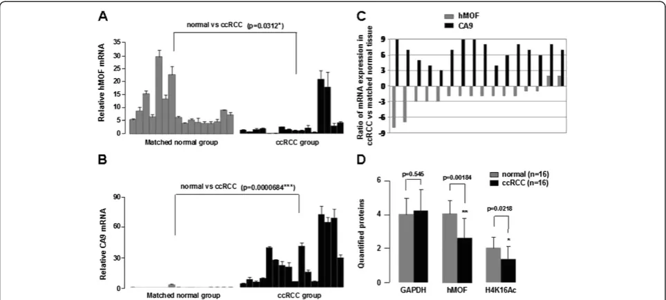
Figure 2Downregulation of hMOF is accompanied by increased CA9 in ccRCC. A-B.Relative mRNA expression levels of hMOF and CA9 in ccRCC. Total RNA was isolated from sixteen paired clinical ccRCC and adjacent kidney tissues. Relative mRNA expression levels of hMOF and CA9 were analized by quantitative RT-PCR. Error bars represent the standard error of the mean of 3 independent experiments. Student’st-test was performed to compare the difference between ccRCC and normal tissues.C.Expression patterns of hMOF and CA9 mRNAs in ccRCC and its corresponding adjacent kidney tissues. Expression is displayed as a ratio of expression of hMOF or CA9 gene in ccRCC versus matched normal tissues. Each bar is the log2 value of the ratio of hMOF or CA9 expression levels between ccRCC and matched normal tissues from the same patients. Bar value >1 represents >2-fold increased, whereas bar value <−1, represents >2-fold decreased.D.Protein expression levels of hMOF and its H4K16Ac status in ccRCC. Aliquots of whole cell extracts from sixteen selected ccRCC tumor samples and its corresponding adjacent tissues were analyzed by western blotting. The blots were then scanned and quantified with Quantity One software. The significant difference is expressed as *p<0.05, **p<0.01.
cases of newly diagnosed ccRCC (Figure 1A) by reverse transcription PCR (RT-PCR) and quantitative real-time PCR (qPCR). As shown in Figure 1B, the gene expres-sion levels of hMOF were markedly decreased in all ccRCC tissues compared to matched normal tissues (p<0.001). In contrast, CA9 expression levels were sig-nificantly increased in all ccRCC tissues (p<0.01). How-ever, no significant difference was observed in VEGF and
HIF1α expression. Additional 16 paired clinical ccRCC
and matched normal tissues were used to further valid-ate the frequent downregulation of hMOF mRNA ex-pression in primary ccRCC. Analysis of performed mRNA expression of 16 samples revealed significant (>2-fold decreased) downregulation of hMOF mRNA in 87.5% (14/16) of patients (Figure 2A and C), whereas 12.5% (2/16) of patients showed significant (>2-fold increased) upregulation of hMOF (Figure 2A and C). However, less relationship between hMOF expression and tumor size, stage and grading was detected in our limited number of cases (data not shown). To examine the gene expression status of hMOF in other types of
RCC, four kidney cancer patients with pathologically daignosed ccRCC, chRCC (chromophobe RCC), paRCC (papillary RCC) and unRCC (unclassified RCC), respect-ively, were selected. Analysis of qRT-PCR results showed that the gene expression of hMOF significantly downre-gulated in all types of RCC (>2-fold) (Figure 3A and B).
Reduction of hMOF protein in human primary renal cell carcinoma tissues
acetylation status of histone H4K16 was also signifi-cantly reduced or lost (p<0.05). To further confirm these results, we performed immunohistochemical staining for hMOF and histone H4K16 acetylation in the formalin fixed paraffin embedded tissue sections of same four selected ccRCC patients. The results revealed that both the hMOF protein levels and the histone H4K16 acetyl-ation status were markedly reduced (score 1 to 2 for
hMOF staining, and score 0–1 for H4K16Ac staining) in
all ccRCC tissues compared to adjacent tissues. For ex-ample, the results of immunohistochemical staining for hMOF and H4K16Ac are presented in Figure 1D. Weak staining of hMOF and no staining of H4K16Ac in the ccRCC paraffin embedded tissue sections were detected. In the additional 16 paired clinical ccRCC and matched normal tissues, hMOF protein expression and H4K16Ac status were detected by western blotting. The quantified protein levels (Quantity One software) were analyzed by
t-test. As shown in Figure 2D, hMOF protein expression
levels were significantly reduced in ccRCC tissues (p<0.01), and the expression of hMOF was tightly corre-lated with H4K16 acetylation (p<0.05). Furthermore, in the four different pathologically diagnosed ccRCC, chRCC, paRCC and unRCC, hMOF protein expression was significantly decreased in ccRCC, chRCC and
unclassified RCC, whereas less changes were detected in paRCC (Figure 3C).
Elevation of CA9 gene expression is accompanied by frequent reduction of hMOF mRNA in ccRCC
CA9 is not expressed in healthy renal tissue but is
expressed in most ccRCC through HIF1α accumulation
driven by hypoxia [25]. In our study, the gene expression of CA9 was significantly increased (>2-fold) in 100% of ccRCC patients (21/21; Figure 3D) including four initial selected ccRCC, sixteen additional ccRCC (Figure 2B) and one case (Figure 3A and B) used in comparing experi-ment. Among these cases, reduction of hMOF mRNA ex-pression was detected in 90.5% of cases (19/21). There were only 2 cases presenting elevation of hMOF mRNA expression in ccRCC (Figure 2A and C). However, no ele-vation of CA9 gene expression was detected in different pathologically diagnosed RCC including chRCC, paRCC and unRCC, although the mRNA levels of hMOF were significantly decreased in those RCC (Figure 3B).
Non-correlation between hMOF and CA9 is found in renal cell carcinoma cells
To extend these observations and to know whether there is a correlation regulatory relationship between Figure 4Non-correlation between hMOF and CA9 is found in renal cell carcinoma cells. A.hMOF protein expression was correlated with acetylation of H4K16 in RCC cell 786–0 and OSRC-2. 293T, 786–0 or OSRC-2 cells were cultured in 6-well tissue culture plates (~2x10⁵cells/well) in
DMEM medium containing 10% fetal bovine serum. Whole cell extracts were subjected to immunoblotting using indicated antibodies (right panel). 293T, 786–0 or OSRC-2 cells from 1 well of a 6 well plate were lysed and total RNA was isolated using Trizol. hMOF and CA9 gene expressions were measured by RT-PCR (left panel) and qRT-PCR (B).C.Effect of hMOF on CA9 mRNA expression levels in RCC cells. RCC 786–0 cells were cultured in 6-well tissue culture plates (~2x10⁵cells/well) in DMEM medium containing 10% fetal bovine serum. The cells were transfected with 0.25, 0.5, 1 and 2μg of hMOF cDNAs. 48 hours after transfection, cells were lysed and total RNA was isolated using Trizol. Indicated gene expressions were analyzed by qRT-PCR.D.Effect of hMOF on CA9 protein expression in RCC cells. RCC 786–0 cells were
transfected with 0.25, 0.5, 1 and 2μg of hMOF cDNAs. 48 hours after transfection, cells were harvested and lysed in RIPA buffer. Aliquots of whole cell extracts were subjected to 12% SDS-PAGE, and specific proteins were detected by indicated antibodies.
hMOF and CA9 in cells, we performed experiments
using RCC cell 786–0 and OS-RC-2 as model. We first
examined both the protein levels and mRNA expression
levels of the hMOF and CA9 in 293T, 786–0 and
OS-RC-2 cells. The results as shown in Figure 4A indicate the opposing gene expression patterns between hMOF and CA9 were observed. The expression of hMOF was
reduced in both 786–0 and OS-RC-2 cells compared to
293T cells, and the log2 ratio changes are−0.84 and−1.9, respectively. Western blotting analysis revealed that the hMOF proteins were markedly decreased in both renal cell carcinoma cells. In addition, the reduction of hMOF proteins resulted in loss of the acetylation of histone H4K16 in RCC cells. In contrast with hMOF, the gene ex-pression of CA9 was increased in both 786–0 (log2=6.2) and OS-RC-2 cells (log2=12.3) compared to 293T cells. To determine whether the CA9 gene expression was
regu-lated by hMOF, renal cell carcinoma 786–0 cells were
transiently transfected with 0.25 to 2μg of hMOF cDNAs. The results are shown in Figure 4C and D, both the gene and protein expression levels of hMOF were dose-dependently increased. However, neither the gene nor pro-tein expression of CA9 levels were affected by transient transfection RCC 786–0 cells with hMOF cDNAs.
Discussion
The HAT hMOF belongs to the MYST (Moz-Ybf2/Sas3-Sas2-Tip60) family, and is believed to be responsible for
histone H4 acetylation at lysine 16 in both Drosophila
and human cells [7,8,12]. Abnormal expression of the hMOF and its corresponding modification of H4K16 have been found in certain primary cancer tissues. The expression behavior of hMOF in different primary can-cers was observed to be different. Frequent downregula-tion of hMOF expression was found in primary breast cancer and medulloblastoma [15]. On the contrary, hMOF was overexpressed in non-small cell lung carcin-oma tissues [26]. Regardless of what type of situation, hMOF protein expression tightly correlated with acetyl-ation of histone H4K16. In this study, we investigated the expression of histone acetyltransferase hMOF and its corresponding H4K16 acetylation in a series of primary kidney tumor tissues by qRT-PCR, western blotting, and immunohistochemistry. The results revealed that either hMOF mRNA expression or hMOF protein expression was frequently downregulated in human RCC (19/21 cases; >90%), and hMOF protein expression was corre-lated with acetylation of histone H4K16 in parallel. In addition, low protein expression levels of hMOF and loss of histone H4K16 acetylation were detected in renal
car-cinoma cells 786–0 and OS-RC-2 compared to human
embryonic kidney cell HEK293T. Together this, HAT hMOF might have an important role in primary renal cell carcinoma tumorigenesis.
CA9 is a transmembrane, zinc-containing metalloen-zyme that catalyzes reversible reactions of the bicarbonate buffer system to regulate pH in hypoxic conditions [27]. Overexpression of CA9 has been shown in a wide variety of malignant cell lines and tumors [28-30]. It is worth mentioning that CA9 has been well described as a diag-nostic marker for clear cell renal carcinoma (ccRCC), es-pecially by showing high expression in metastastic ccRCC (mccRCC) [31,32]. Therefore, the inhibitor or regulatory proteins of hypoxic tumor-associated CA9 possesses the potential therapeutic possibility for those tumors in which CA9 is involved in perturbing the extra- or intra- tumoral acidification process. In our experiments, although the
ex-pression of VEGF and HIF1αwhich are hypoxia signature
genes were not observed significant difference between ccRCC and normal tissues, overexpression of CA9 was observed in 100% of ccRCC cases and in both renal car-cinoma cell lines. Interestingly, in four different diagnostic RCCs, downregulation of hMOF was detected in all types of RCCs, but the overexpression of CA9 was only pre-sented in ccRCC, suggesting that hMOF might be a new common diagnostic marker for human different diagnostic RCC. Although frequent downregulation of hMOF and overexpression of CA9 were detected in both RCC clinical tissues and RCC cell lines, non-correlation between
hMOF and CA9 was found in RCC 786–0 cells, suggesting
hMOF and its corresponding modifications might be a new CA9-independent RCC diagnosis biomarker. Al-though large series of clinical cases and analyses of overall survival need to be investigated, the molecular mechanism linking loss of hMOF expression to renal cell carcinoma, especially mechanism of hMOF on renal cell carcinomas, will be an exciting avenue for further research.
Conclusion
In conclusion, hMOF as an acetyltransferase of H4K16 might be involved in the pathogenesis of renal cell car-cinoma, and this epigenetic change might be a new CA9-independent RCC diagnostic marker. In addition, our results suggest that a novel molecular mechanism of hMOF might serve as a lead to new therapeutics target in human renal cell carcinoma.
Competing interests
The authors declare that they have no competing interests.
Authors’contributions
YW, RZ, DW and ZL carried out the experiments and data analyses. WS and CW collected the clinical samples and completed immunohistochemistry. YC and JJ drafted the manuscript. All authors read and approved the final manuscript.
Acknowledgements
Author details
1
Urology Department, The First Clinical Hospital, Jilin University, Changchun City, Jilin 130021, China.2Urology Department, Jilin province People’s
Hospital, Changchun City, Jilin 130021, China.3College of Life Science, Jilin University, Changchun City, Jilin 130012, China.4Graduate School of Jilin
University, Changchun City, Jilin 130012, China.
Received: 11 December 2012 Accepted: 2 February 2013 Published: 9 February 2013
References
1. Jin J, Cai Y, Li B, Conaway RC, Workman JL, Conaway JW, Kusch T:In and out: histone variant exchange in chromatin.Trends Biochem Sci2005,30:680–687. 2. Berger SL:The complex languige of chromatin regulation during
transcription.Nature2007,447:407–412.
3. Bhaumik SR, Smith E, Shilatifard A:Covalent modifications of histones during development and disease pathogenesis.Nat Struct Mol Biol2007,
14:1008–1016.
4. Carrouzza MJ, Utley RT, Workman JL, Cote J:The divers functions of histone acetyltransferase complexes.Trends Genet2003,19:321–329. 5. Gupta A, Guerin-Peyrou TG, Sharma GG, Park C, Agarwal M, Ganju RK, Pandita S,
Choi K, Sukumar S, Pandita RK, Ludwig T, Pandita TK:The mammalian ortholog ofDrosophlaMOF that acetylates histone H4 lysine 16 is essential for embryogenesis and oncogenesis.Mol Cell Biol2008,28:397–409. 6. Sharma GG, So S, Gupta A, Kumar R, Cayrou C, Avvakumov N, Bhadra U,
Pandita RK, Porteus MH, Chen DJ, Cote J, Pandita TK:MOF and histone H4 acetylation at lysine 16 are critical for DNA damage response and double-strand break repair.Mol Cell Biol2010,30:3582–3595. 7. Rea S, Xouri G, Akhtar A:Males absent on the first (MOF): from flies to
humans.Oncogene2007,26:5385–5394.
8. Smith ER, Cayrou C, Huang R, Lane WS, Côtê J, Lucchesi JC:A human protein complex homologus to the Drosophila MSL complex is responsible for the majority of histone H4 acetylation at lysine 16. Mol Cell Biol2005,25:9175–9188.
9. Mendjan S, Taipale M, Kind J, Holz H, Gebhardt P, Schelder M, Vermeulen M, Buscaino A, Duncan K, Mueller J, Wilm M, Stunnenberg HG, Saumweber H, Akhtar A:Nuclear pore components are involved in the transcriptional regulation of dosage compensation in Drosophila.Mol Cell2006,21:811–823. 10. Cai Y, Jin J, Swanson SK, Cole MD, Choi SH, Florens L, Washburn MP,
Conaway JW, Conaway RC:Subunit composition and substrate specificity of a MOF-containing histone acetyltransferase distinct from the male-specific lethal (MSL) complex.J Biol Chem2010,285:4268–4272. 11. Sykes SM, Mellert HS, Holbert MA, Li K, Marmorstein R, Lane WS, McMahon SB:
Acetylation of the p53 DNA-binding domain regulates apoptosis induction. Mol Cell2006,24:841–851.
12. Taiple M, Rea S, Richter K, Vilar A, Lichter P, Imhof A, Akhtar A:hMOF histone acetyltransferase is required for histone H4 lysine 16 acetylation in mammalian cells.Mol Cell Biol2005,25:6798–6810.
13. Mulligan P, Yang F, Di Stefano L, Ji JY, Ouyang J, Nishikawa JL, Toiber D, Kulkarni M, Wang Q, Najafi-Shoushtari SH, Mostoslavsky R, Gygi SP, Gill G, Dyson NJ, Näär AM:A SIRT-LSD1 Co-repressor complex regulates notch target gene expression and development.Mol Cell2011,42:689–699. 14. Orpinell M, Fournier M, Riss A, Nagy Z, Krebs AR, Frontini M, Tora L:The
ATAC acetyl transferase complex controls mitotic progression by targeting non-histone substrates.EMBO J2010,29:2381–2394.
15. Pfister S, Rea S, Taipale M, Mendrzyk F, Straub B, Ittrich C, Thuerigen O, Sinn HP, Akhtar A, Lichter P:The histone acetyltransferase hMOF is frequently downregulated in primary breast carcinoma and medulloblastoma and constitutes a biomarker for clinical outcome in medulloblastoma.Int J Cancer2008,122:1207–1213.
16. Elsheikh S, Green AR, Rakha EA, Powe DG, Ahmed RA, Collins HM, Soria D, Garibaldi JM, Paish CE, Ammar AA, Grainge MJ, Ball GR, Abdelghany MK, Martinez-Pomares L, Heery DM, Ellis IO:Globle histone modifications in breast cancer correlate with tumor phenotypes, prognostic factors, and patient outcome.Cancer Res2009,69:3802–3809.
17. Jemal A, Siegel E, Ward E, Murray T, Xu J, Thun MJ:Cancer stastistics.CA Cancer J Clin2007,57:43–66.
18. Janzen NK, Kim HL, Figlin RA, Belldegrun AS:Surveillance after radical or partial nephrectomy for localized renal cell carcinoma and management of recruitment disease.Urol Clin North Am2003,30:843–852.
19. Eichelberg C, Junker K, Ljungberg B, Moch H:Diagnostic and prognostic molecular markers for renal cell carcinoma: a critical appraisal of the current state of research and clinical applicability.Eur Urol2009,55:851–863. 20. Belldegrun AS:Renal cell carcinoma: prognostic factors and patient
selection.Eur Urol Suppl2007,6:477–483.
21. Wu XR, Sha JJ, Liu DM, Chen YH, Yang GL, Zhang J, Xhen YY, Bo JJ, Huang YR:
High expression of p53-induced ring-h2 protein is associated with poor prognosis in clear cell renal cell carcinoma.Eur J Sur Oncol2012,39:100–106. 22. Mosashvilli D, Kahl P, Mertens C, Holzapfel S, Rogenhofer S, Hauser S,
Büttner R, Von Ruecker A, Müller SC, Ellinger J:Globle histone acetylation levels: prognostic relevance in patients with renal cell carcinoma.Cancer Sci2010,101:2664–2669.
23. Edge SB, Byrd DR, Compton CC, Fritz AG, Greene FL, Trotti A:AJCC Cancer Staging Manual. 7th edition. Chicago, IL: Springer; 2010.
24. Choschzick M, Oosterwijl R, Muller V, Woelber L, Simon R, Moch H, Tennstedt P:Overexpression of carbonic anhydrase IX (CAIX) is an independent unfavorable prognostic marker in endometrioid ovarian cancer.Virchows Arch2011,459:193–200.
25. Tostain J, Li G, Gentil-Perret A, Gigante M:Carbonate anhydrase 9 in clear cell renal cell carcinoma: A marker for diagnosis, prognosis and treatment.Eur J Cancer2010,46:3141–3148.
26. Song JS, Chun SM, Lee JY, Kim DK, Kim YH, Jang SJ:The histone acetyltransferase hMOF is overexpressed in non-small cell lung carcinoma.Korean J Pathol2011,45:386–396.
27. Stillebroer AB, Mulders PF, Boerman OC, Oyen WJ, Oosterwijk E:Carbonic anhydrase IX in renal cell carcinoma: implications for prognosis, daignosis, and therapy.Eur Urol2010,58:75–83.
28. Hussain SA, Ganesan R, Reynolds G, Gross L, Stevens A, Pastorek J, Murray PG, Perunovic B, Anwar MS, Billingham L, James ND, Spooner D, Poole CJ, Rea DW, Palmer DH:Hypoxia-regulated carbonic anhydrase IX expression is associated with poor survival in patients with invasive breast cancer. Br J Cancer2007,96:104–109.
29. Klatte T, Seligson DB, Rao JY, Yu H, de Martino M, Kawaoka K, Wong SG, Belldegrun AS, Pantuck AJ:Carbonic anhydrase IX in bladder cancer: a diagnostic, prognostic, and therapeutic molecular marker.Cancer2009,
115:1448–1458.
30. Swinson DE, Jones JL, Richardson D, Wykoff C, Turley H, Pastorek J, Harris AL, O’Byrne KJ:Carbonic anhydrase IX expression, a novel surrogate marker of tumor hypoxia, is associated with a poor prognosis in non-small-cell lung cancer.J Clin Oncol2003,21:473–482.
31. Leibovich BC, Sheinin Y, Lohse CM, Thompson RH, Cheville JC, Zavada J, Kwon ED:Carbonic anhydrase IX is not an independent predictor of outcome for patients with clear cell renal cell carcinoma.J Clin Oncol2007,25:4757–4764. 32. Liao SY, Aurelio ON, Jan K, Zavada J, Stanbridge EJ:Identification of the
MN/CA9 protein as a reliable diagnostic biomarker of clear cell carcinoma of the kidney.Cancer Res1997,57:2827–2831.
doi:10.1186/1756-9966-32-8
Cite this article as:Wanget al.:Epigenetic change in kidney tumor: downregulation of histone acetyltransferase MYST1 in human renal cell
carcinoma.Journal of Experimental & Clinical Cancer Research201332:8.
Submit your next manuscript to BioMed Central and take full advantage of:
• Convenient online submission
• Thorough peer review
• No space constraints or color figure charges
• Immediate publication on acceptance
• Inclusion in PubMed, CAS, Scopus and Google Scholar
• Research which is freely available for redistribution
Submit your manuscript at www.biomedcentral.com/submit