C A S E R E P O R T
Open Access
Development of anti-glomerular basement
membrane glomerulonephritis during the
course of IgA nephropathy: a case report
Tadasu Kojima, Go Hirose, Shuuhei Komatsu, Taito Oshima, Kentaro Sugisaki, Tomohiro Tomiyasu,
Noriko Yoshikawa, Muneharu Yamada and Takashi Oda
*Abstract
Background:Anti-glomerular basement membrane (GBM) glomerulonephritis does not usually coexist with another
glomerulonephritis such as IgA nephropathy. We present a rare case having a combination of these two diseases, and furthermore, histological evaluation could be performed before and after the development of anti-GBM glomerulonephritis over a period of only10 months.
Case presentation: A 66-year-old woman was admitted with complaints of microscopic hematuria and mild
proteinuria for the past 3 years. Serum creatinine level was normal at that time. The first renal biopsy was performed. Light microscopy revealed mesangial proliferative glomerulonephritis with fibro-cellular crescents in one out of 18 glomeruli, excluding one global sclerotic glomerulus. Immunofluorescence (IF) showed IgA and C3 deposition in the mesangium. Therefore, the diagnosis was IgA nephropathy. Eight months later, the patient’s serum creatinine suddenly rose to 4.53 mg/dL and urinalysis showed 100 red blood cells per high power field with nephrotic range proteinuria (12.3 g/gCr). The serological tests revealed the presence of anti-GBM antibody at the titer of 116 IU/mL. Treatments were begun after admission, consisting of hemodialysis, plasma exchange, and intravenous methylprednisolone pulse therapy. At 4 weeks after admission, the second renal biopsy was performed. Light microscopy revealed crescents in 18 of 25 glomeruli, excluding six global sclerotic glomeruli. IF showed linear IgG deposition along the GBM in addition to granular IgA and C3 deposition. Based on these findings, the diagnosis of anti-GBM glomerulonephritis and IgA nephropathy was confirmed. Renal function was not restored despite treatment, but alveolar hemorrhage was prevented. Conclusions: We report a patient with a diagnosis of anti-GBM disease during the course of IgA nephropathy. This case strongly suggests that the presence of autoantibodies should be checked to rule out overlapping autoimmune conditions even in patient who have previously been diagnosed with chronic glomerulonephritis, such as IgA nephropathy, who present an unusually rapid clinical course.
Keywords:Anti-glomerular basement membrane (GBM) glomerulonephritis, Rapidly progressive glomerulonephritis
(RPGN), IgA nephropathy, Crescentic glomerulonephritis
* Correspondence:takashio@tokyo-med.ac.jp
Department of Nephrology and Blood Purification, Kidney Disease Center, Tokyo Medical University Hachioji Medical Center, 1163 Tatemachi, Hachioji, Tokyo 193-0998, Japan
Anti-glomerular basement membrane (GBM) glomer-ulonephritis is an autoimmune glomerular disease that is characterized by linear deposition of IgG along the GBM. The main target of anti-GBM antibody had been shown to lie in the NC1 domain of α3 chains of type IV collagen on the GBM. Histo-logically, it is associated with extensive crescent for-mation and clinically with rapidly progressive glomerulonephritis (RPGN). There are well-known associations between anti-GBM glomerulonephritis and the presence of antineutrophil cytoplasmic antibody (ANCA) and between anti-GBM glomeru lonephritis and membranous nephropathy [1–3]. In a limited number of cases, anti-GBM glomeruloneph-ritis has been associated with IgA nephropathy and other immune complex glomerulonephritis [4–6].
Herein, we report a patient with a diagnosis of anti-GBM disease during the course of IgA nephropathy.
Case presentation
A 66-year-old woman with a significant past medical history of well-controlled hypertension was admitted with complaints of microscopic hematuria and mild proteinuria for the past 3 years. Serum creatinine level was within normal range at that time and therefore the anti-GBM antibody was not tested. The first renal biopsy revealed mesangial proliferative glomerulonephritis with fibro-cellular crescents in one out of 18 glomeruli, excluding one global scler-otic glomerulus (Fig. 1a), and deposition of IgA and C3 in mesangial areas by immunofluorescence mi-croscopy (Fig. 2a). Weak but significant IgG depos-ition was also observed in glomeruli in the distribution somewhat different from IgA or C3 (Fig. 2a). The electron-dense deposits were observed in mesangial areas by electron microscopy.
hypertensive therapy was initiated, mainly with an RAS inhibitor. Eight months later, the patient’s serum creatinine suddenly rose to 4.53 mg/dL (it was 1.04 mg/dL from the routine blood test 1 month be-fore). Urinalysis showed 100 red blood cells per high power field and urinary protein excretion of 12.3 g/ gCr (Fig. 3). The serological tests that were
per-formed to differentiate the cause of rapidly progres-sive glomerulonephritis revealed the presence of anti-GBM antibody at the titer of 116 IU/mL and the absence of anti-nuclear antibody and anti-neutrophil cytoplasmic antibody. Laboratory findings on admis-sion are summarized in the Table 1.
After admission, treatments with hemodialysis, plasma exchange, and intravenous methylpredniso-lone pulse therapy followed by oral prednisomethylpredniso-lone at the dose of 50 mg/day were initiated. The second renal biopsy was performed at 4 weeks after admis-sion in order to assess the probability of renal recov-ery and to make the final diagnosis. It revealed cellular to fibrocellular crescents in 18 of 25 glom-eruli, excluding six global sclerotic glomeruli by light microscopy. By immunofluorescence study, linear IgG deposition along the glomerular capillary walls and mesangial staining for IgA were observed. On the other hand, C3 deposition was observed in the mesangium as well as in the glomerular capillary walls (Fig. 2b). Electron-dense deposits were ob-served in mesangial areas, similarly as in the first bi-opsy, by electron microscopy (Fig. 4). Based on the aforementioned findings, the diagnosis of anti-GBM glomerulonephritis and IgA nephropathy was con-firmed. Plasmapheresis was performed eight times, anti-GBM antibody gradually decreased, and alveolar hemorrhage was prevented. However, her renal func-tion could not be restored and she underwent main-tenance hemodialysis (Fig. 5).
a
b
Additional immunosuppressant was not given be-cause the patient did not show any sign of pulmon-ary involvement and because the renal recovery was quite unlikely from clinical (continuous oliguria and hemodialysis dependence) as well as histological (crescent formation in most of non-sclerotic glom-eruli) point of view.
Clinical and histological presentations from IgA ne-phropathy (at the time of first renal biopsy) and from
anti-GBM disease (at the time of second renal biopsy) were summarized in the Table2.
Discussion and conclusions
IgA nephropathy is an immune complex-mediated glomerulonephritis defined immunohistologically by the presence of glomerular mesangial IgA deposits accompanied by a variety of histopathologic lesions, including mesangial proliferation [7]. Anti-GBM
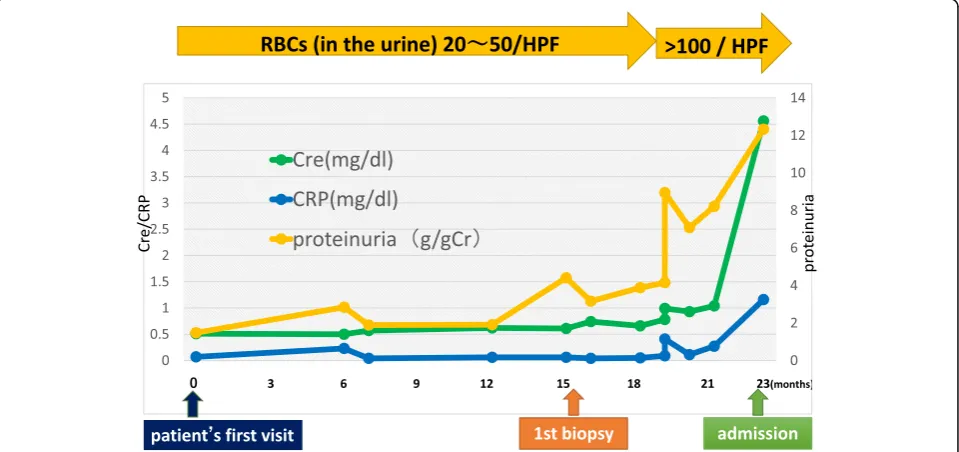
Fig. 3Clinical course before admission. Cre: serum creatinine level
a
I g A IgG C 3
b
I g A IgG C 3
Fig. 2Representative photographs of immunofluorescence staining (Scale bars = 20.0μm).aFirst renal biopsy showing positive staining of IgA, IgG, and C3. The staining pattern was similar in IgA and C3 (granular deposition probably in the mesangial area), but was rather different in IgG.
disease is caused by antibodies reactive to the glom-erular and alveolar basement membrane.
The causal relationship of anti-GBM glomerulo-nephritis and IgA nephropathy is unclear. There was one hypothesis that the IgA-related immune complex might promote immunologic and inflammatory events, resulting in conformational changes and ex-posure of the GBM antigens leading to development
of anti-GBM antibody [4]. However, it is difficult to prove whether anti-GBM disease in this patient de-veloped as an incidental complication or was sec-ondary to IgA nephropathy because there is still no established marker to distinguish primary from sec-ondary anti-GBM disease. In this regard, we per-formed immunofluorescence staining for IgG subclasses on the second renal biopsy, and found that IgG4 was the main subclass of IgG bound to GBM in this patient (Fig. 6). The main subclass of pathogenic IgG in anti-GBM disease was reported to be usually IgG1 [8]. Whether predominance of IgG4 relates with anti-GBM disease developed secondary to IgA nephropathy deserves future study.
The pathophysiological condition of anti-GBM dis-ease before clinical presentation is unknown. In this regard, Olson et al. conducted a case-control study involving 30 patients diagnosed with anti-GBM dis-ease using serum samples from the Department of Defense Serum Repository. In the report, 13% (4 of 30) of study subjects had an elevated GBM anti-body level 2–10 months prior to diagnosis [9]. In the present case, multiple immunofluorescence labelling on the first biopsy showed partial linear IgG depos-ition along the glomerular capillary walls (Fig. 7). Al-though it may be only speculation because serum anti-GBM antibody was not tested at the time of the first biopsy, the patient might have had complica-tions from asymptomatic (subclinical) anti-GBM dis-ease at that point.
In the case of IgA nephropathy complicated by anti-GBM disease, Yamaguchi et al. speculated that the pathological features of IgA nephropathy may not be observed because the number of glomeruli free from destruction is very limited [10]. Therefore, the coexistence of anti-GBM glomerulonephritis and
Fig. 4Electron microscopic photograph of the second renal biopsy, showing the electron-dense deposits in mesangial areas
WBC (4000–8000) 10,500/μL
RBC (390–510) 379 × 104/μL
Hb (12.0–16.0) 11.3 g/dL
PLT (15–35) 36.9 × 10⁴/μL
Chemistry
TP (6.4–8.0) 6.4 g/dL
Alb (3.4–5.0) 2.8 g/dL
AST (8–40) 30 U/L
ALT (4–43) 21 U/L
LDH (106–220) 345 mg/dL
BUN (8–20) 35.6 mg/dL
Cr (0.5–0.8) 5.53 mg/dL
Na (135–147) 136 mEq/L
K (3.4–4.9) 3.9 mEq/L
Cl (98–108) 103 mEq/L
Ca (8.0–10.5) 7.9 mg/dL
P (2.7–4.5) 6.1 mg/dL
CRP (< 0.02) 1.16 mg/dL
Immune-related
C3 (86–160) 145 mg/dL
C4 (17–45) 51.3 g/dL
CH50 (30–45) 76.8 U/mL
IgG (380–1620) 881 mg/dL
IgA (84–438) 186 mg/dL
IgM (57–288) 33 mg/dL
ASO (< 166) 22 IU/mL
ANA (< 40) < 40
Anti–GBM antibody (< 3.0) 116 IU/mL
PR3-ANCA (< 3.5) <1.0 IU/mL
MPO-ANCA (< 3.5) <1.0 IU/mL
Urinalysis
Occult blood 3+
RBCs >100/HPF
Protein 4 + 12.3 g/gCr
WBC 1–4/HPF
Cast Granular cast(+)
RBCcast(+)
WBCwhite blood cells,RBCred blood cells,Hbhemoglobin,Pltplatelets,HPF high-power field,TPtotal protein,Albalbumin,ASTaspartate aminotransferase,ALT
alanine aminotransferase,LDHlactate dehydrogenase,BUNblood urea nitrogen,Cr creatinine,Nasodium,Kpotassium,Clchloride,Cacalcium,Pphosphate,CRP C-reactive protein,ANAanti-nuclear antibody,GBMglomerular basement membrane,
Fig. 5Clinical course after admission. Cre: serum creatinine level, Anti-GBM antibody: anti-glomerular basement membrane antibody, PEX: plasma exchange, mPSL: methylprednisolone, PSL: prednisolone, HD: hemodialysis, RBX: renal biopsy
Table 2Clinical and histological presentation at the time of first and second renal biopsy
First biopsy Second biopsy
Clinical presentation CGN RPGN
U-P 3.2 g/g·Cr U-P 12.3 g/g·Cr
U-RBC 50–99/HPF U-RBC > 100/HPF
sCr 0.74 mg/dL sCr 5.5 mg/dL
Anti-GBM antibody Not tested 116 IU/ml
Light microscopy Mesangial proliferative GN Crescentic GN
Global sclerosis (1/19) Global sclerosis (6/31)
Fibrocellular crescent (1/19) Cellular~fibrocellular crecent (18/31)
Immunofluorescence IgA: mes ++ IgA: mes +
C3: mes ++ C3: mes +
IgG: mes +, peripheral linear +
peripheral linear ± IgG: peripheral linear ++
(focal segmental) (diffuse global)
IgA nephropathy may be more frequent than is be-ing reported.
In summary, we reported a patient with a diagnosis of anti-GBM disease during the course of IgA ne-phropathy. Histological evaluation could be performed before and after the development of anti-GBM disease over a period of 10 months. Even if the patient had already received a diagnosis of a chronic glomerulo-nephritis such as IgA nephropathy, we should check autoantibodies to rule out overlapping autoimmune conditions in case the patient showed an unusually rapid clinical course.
Abbreviations
ANCA:Antineutrophil cytoplasmic antibody; GBM: Glomerular basement membrane; IF: Immunofluorescence; RPGN: Rapidly progressive glomerulonephritis
Acknowledgements
We are grateful to our colleague Sachiko Iwama for excellent technical assistance in histological analysis. Part of this case report was previously presented at the 18th International Vasculitis & ANCA Workshop and was published in abstract form (Rheumatology 56, suppl 3, iii 81, 2017).
Funding
No funding supports.
Availability of data and materials
Not applicable.
Authors’contributions
TK and GH diagnosed, treated, and clinically monitored the patient. TK performed the literature search and wrote the paper. SK, TOshima, KS, TT, NY, and MY participated in treatment and diagnosis of the patient during the hospitalization period. TOda performed the multiple immunofluorescence staining on the renal biopsy and helped to write the manuscript. All authors read and approved the final manuscript.
Ethics approval and consent to participate
Not applicable.
Consent for publication
Written informed consent was obtained from the patient for publication of clinical information including symptom, laboratory data, histological findings and any accompanying images as a case report. A copy of the written consent is available for review by the Editor of this journal.
Competing interests
The authors declare that they have no competing interests.
Publisher’s Note
Springer Nature remains neutral with regard to jurisdictional claims in published maps and institutional affiliations.
IgG 1
IgG 2
IgG 3
IgG 4
Fig. 6Immunofluorescence staining for IgG subclasses on the second renal biopsy (Scale bars = 20.0μm). The main subclass of IgG bound to GBM was IgG4
Received: 11 October 2017 Accepted: 8 January 2019
References
1. Hellmark T, Niels JL, Collins AB, McCluskey RT, Brunmark C. Comparison of anti-GBM antibodies in sera with or without ANCA. J Am Soc Nephrol. 1997; 8:376–85.
2. Petterson E, Törnroth T, Miettinen A. Simultaneous anti-glomerular basement membrane and membrounous glomerulonephritis: case report and literature review. Clin Immunol Immunopathol. 1984;31:171–80. 3. Kielstein JT, Helmchen U, Netzer KO, Weber M, Haller H, Floege J.
Conversion of Goodpasture’s syndrome into membranous glomerulonephritis. Nephrol Dial Transplant. 2001;16:2082–5.
4. Trpkov K, Abdulkareem F, Jim K, Solez K. Recurrence of antiGBM antibody disease twelve years after transplantation associated with de novo IgA nephropathy. Clin Nephrol. 1998;49:124–8.
5. Xu D, Wu J, Wu J, Xu C, Zhang Y, Mei C, Gao X. Novel therapy for anti-glomerular basement membrane disease with IgA nephropathy: a case report. Exp Ther Med. 2016;11:1889–92.
6. Cui Z, Zhao MH, Wang SX, Liu G, Zou WX, Wang HY. Concurrent antiglomerular basement membrane disease and immune complex glomerulonephritis. Ren Fail. 2006;28:7–14.
7. Emancipator SN. IgA nephropathy:morphologic expression and pathogenesis. Am J Kidney Dis. 1994;23:461–2.
8. Hemminger J, Nadasdy G, Satoskar A, Brodsky SV, Nadasdy T. IgG subclass staining in routine renal biopsy material. Am J Surg Pathol. 2016;40(5):617–26. 9. Olson SW, Arbogast CB, Baker TP, Owshalimpur D, Oliver DK, Abbott KC, Yuan
CM. Asymptomatic autoantibodies associate with future anti-glomerular basement membrane disease. J Am Soc Nephrol. 2011;22:1946–52. 10. Yamaguchi H, Takizawa H, Ogawa Y. A case report of the anti-glomerular