R E S E A R C H
Open Access
Small dense LDL particles - a predictor of
coronary artery disease evaluated by invasive
and CT-based techniques: a case-control study
Anne P Toft-Petersen
1, Hans H Tilsted
1, Jens Aarøe
1, Klaus Rasmussen
1, Thorkil Christensen
2, Bruce A Griffin
3,
Inge V Aardestrup
1, Annette Andreasen
1, Erik B Schmidt
1*Abstract
Background:Coronary angiography is the current standard method to evaluate coronary atherosclerosis in patients with suspected angina pectoris, but non-invasive CT scanning of the coronaries are increasingly used for the same purpose.
Low-density lipoprotein (LDL) cholesterol and other lipid and lipoprotein variables are major risk factors for coronary artery disease. Small dense LDL particles may be of particular importance, but clinical studies evaluating their predictive value for coronary atherosclerosis are few.
Methods:We performed a study of 194 consecutive patients with chest pain, a priori considered of low to intermediate risk for significant coronary stenosis (>50% lumen obstruction) who were referred for elective coronary angiography. Plasma lipids and lipoproteins were measured including the subtype pattern of LDL particles, and all patients were examined by coronary CT scanning before coronary angiography.
Results:The proportion of small dense LDL was a strong univariate predictor of significant coronary artery stenosis evaluated by both methods. After adjustment for age, gender, smoking, and waist circumference only results obtained by traditional coronary angiography remained statistically significant.
Conclusion:Small dense LDL particles may add to risk stratification of patients with suspected angina pectoris.
Introduction
Coronary artery disease (CAD) is a common cause of morbidity and mortality in the industrialised world [1]. The diagnosis of coronary atherosclerosis is usually made by invasive coronary angiography (CAG) during which percutaneous coronary intervention (PCI) proce-dures can be applied simultaneously. CAG is invasive, exposes the patient to a moderate amount of radiation and iodinated contrast agent, and depicts only athero-sclerotic lesions that bulge into the lumen. Among the currently available non-invasive alternatives, CT-based coronary angiography (CT CAG) provides a view of the vessel lumen as well as the architecture of the vessel wall. Additionally, the degree of calcification, which has
been shown to be a prognostic marker for CAD [2], can be accessed. Disadvantages of CT CAG include the rela-tively large dose of radiation, the inability to perform intervention, and inconclusive scans. Equipment for CT CAG is, however, being improved, and presumably this will reduce the failure rate and lead to a lower dose of radiation. CT CAG, measured with invasive CAG as the golden standard, offers a high sensitivity for coronary stenoses, whereas the specificity is moderate [3]. It has therefore been argued that patients at low or moderate risk of CAD should undergo a“screening”CT CAG that could select patients for possible intervention.
An elevated concentration of LDL cholesterol is a major risk factor for CAD [4-6]. However, LDL consists of a heterogeneous spectrum of particles with highly variable atherogenic potential [7]. Small dense LDL par-ticles (sdLDL) are believed to be particularly atherogenic due to increased susceptibility to oxidation [8,9], high * Correspondence: ebs@rn.dk
1
Department of Cardiology, Center for Cardiovascular Research, Aalborg Hospital, Aarhus University Hospital, Aalborg, Denmark
Full list of author information is available at the end of the article
endothelial permeability [10], decreased LDL receptor affinity [11], and an increased interaction with matrix components [5].
The main hypothesis to be tested was that sdLDL might be a better predictor of coronary atherosclerosis than standard lipids and lipoproteins. Furthermore, we evaluated levels of apoprotein (apo) B and lipoprotein (a) (Lp(a)) in patients with chest pain considered at low or intermediate risk for CAD and investigated by CAG and CT CAG.
Methods
We performed a case-control study of 194 consecutive patients with low or intermediate risk of CAD, referred for elective CAG (invasive CAG) at Aalborg Hospital between June 2007 and December 2008. Causes of refer-ral were angina pectoris or angina equivalent symptoms. Based on information of previous history, current symp-toms, and risk factors patients were categorized clini-cally as having a high risk or a low to intermediate risk of CAD. We carried out a CT CAG as well as an inva-sive CAG in all patients. Based on this, we grouped the patients in two parallel analyses, i.e. CAD/no CAD on CAG and CAD/no CAD on CT CAG, thus allowing parallel comparisons of sdLDL as a risk factor for CAD measured by different, yet clinically relevant, diagnostic modalities. Patients underwent a CT CAG and later, but within the same week, an invasive CAG, unless they met any of the following exclusion criteria:
•A prior diagnosis of CAD, verified by:
○elevated myocardial enzymes
○ signs of myocardial ischemia in ECG either
spontaneously or during a stress test
○ significant stenoses in the coronary arteries
demonstrated in an earlier invasive CAG
○ischemia demonstrated by myocardial
perfu-sion scintigraphy or stress echocardiography •Invasive CAG referral due to defect cardiac valves, cardiomyopathy, or cardiac arrhythmias
•Elevated serum creatinine •Diabetes mellitus
•Known intolerance to contrast agents
•Inability to hold the breath for at least 10 s in con-nection with the CT CAG
• Contraindications against intravenously
adminis-teredb-adrenoceptor-antagonists •Age less than 40 years
•Lack of contraception in premenopausal women
Patient characteristics are given in Table 1. Of the 194 patients enrolled, 3 dropped out before blood sampling, and 13 were excluded as diabetics. Of the 178 complet-ing, non-diabetic participants, 16 had a technically
unacceptable CT CAG and were excluded from this part of the study. Three patients had serum triglycerides
≥5 mmol/L, and as this interfered with the calculation of LDL cholesterol, they were excluded from the LDL cholesterol comparisons as well as the adjusted analyses.
CAG and CT CAG
On the day of the CT scan, venous blood samples (50 mL) were drawn. Serum lipids (total cholesterol, high-density lipoprotein (HDL) cholesterol, and triglycer-ides) were measured by routine methods at the central laboratory, and LDL cholesterol was calculated according to the Friedewald formula. Serum samples for the mea-surement of apo B, Lp(a), and highly sensitive (hs)CRP were frozen at -80°C and analysed after all patients had been included. Apo B was measured using antibody from DAKO, Glostrup, Denmark, on an Advia 1650 from Bayer Diagnostics, NY, US. Lp(a) was measured using an ELISA kit from Mercodia, Uppsala, Sweden, while hsCRP was measured by an immunoturbidimetric assay from Randox Laboratories LtD, UK, on an Advia 1650.
EDTA plasma (K3-EDTA 1.6 mg/mL blood) for the separation of LDL subfractions was stored at -80°C. Table 1 Basic characteristics of the patients included in the study
Variable n = 178
Age (mean and SD) 62.4
± 9.6
Gender (male) (%) 49.4%
Family disposition to ischemic heart disease (%) 52.8%
Current smoking (%) 21.3%
Lipid-lowering medication (%) 59.0%
Antihypertensive medication (%) 67.4%
Systolic blood pressure (mean and SD) 146.7
± 20.2
Diastolic blood pressure (mean and SD) 79.3
± 10.5
BMI (kg/m2
) (mean and SD) 26.9
4.0
Waist circumference (cm) (mean and SD) 96.3
11.8
Triglycerides (mmol/L) (mean and SD) 1.5
1.0
HDL cholesterol (mmol/L serum) (mean and SD) 1.6 0.5
LDL cholesterol (mmol/L serum) (mean and SD) (n = 175) 2.8 0.9
High sensitive CRP (mean and SD) 2.8
4.7
ApoB (g/L) (mean and SD) 0.9
0.2
Lp(a) (arb. units) (mean and SD) 361.1
Plasma adjusted to a density of 1.067 g/L by iodixanol (Optiprep 60%, Axis-Schield PoC As, Oslo, Norway) was prestained with Coomassie blue, underlayered beneath 9% iodixanol and subjected to ultracentrifugation (2 1/2 h, 65,000 rpm 16ºC, (341,000g) in a near vertical rotor (Beckmann NVT65). A digital photograph of LDL subclass profiles was analysed using Total Lab 1D gel-scan software (Pharmacis, UK). The LDL subfraction pattern was char-acterised based on the fractional (percentage) occurrence of small dense particles (density >1.031 g/mL) and large, buoyant particles (density <1.031 g/mL)[12]. The method has previously been described in detail [13,14].
The technique applied for CT CAG has previously been described by Achenbach et al [15]. The scans were con-ducted on a General Electric 64 slice scanner (LightSpeed VCT). The contrast agent was Jomeron®, 400 mg iod/mL. Scans were performed mainly as helical scans. Only arteries with an estimated luminal diameter >1.5 cm were examined, and stenoses were, as with CAG, considered significant if they obstructed at least 50% of the lumen. CAD was considered present if one or more vessels had a significant luminal obstruction.
Invasive CAG as well as analyses of blood samples were performed after the CT CAG examination and blinded to the evaluation of this.
CAG was conducted according to the routine protocol applied by our department and with standard equip-ment. Only arteries with an estimated luminal diameter >1.5 cm were examined, and stenoses were considered significant if they obstructed at least 50% of the lumen. CAD was considered present if one or more vessels had a significant luminal obstruction.
All participants gave an informed, written statement of consent. The study was approved by the regional ethical committee and conducted according to the Declaration of Helsinki.
Statistics
All statistical analyses were conducted in “R”, version 2.9.1. T-tests were used for comparisons of continuous data. Equality of variances was tested and allowed for in the t-tests, and Fisher’s exact test was applied for com-parisons of binary exposures. Parallel analyses were con-ducted based on CAG and CT CAG, respectively, i.e. the patients were stratified into“CAD” and“no CAD” according to either and then compared with respect to risk factors. To access the impact of sdLDL on presence of CAD binary, logistic regression analyses were made based on 10% increments in sdLDL, and a number of possible confounders, i.e. age, gender, current smoking, waist circumference, and LDL cholesterol, were inter-changeably adjusted for. To ensure that no important differences were overlooked all data were analysed as
median values as well and compared with the Wilcoxon rank sum test (data not shown).
Results
Invasive CAG
When stratified by invasive CAG (Table 2), patients with CAD were significantly more likely to be of male gender and to receive lipid-lowering drugs. They had a larger waist circumference, but no differences were observed as to age, family disposition, antihypertensive medication, BMI, cur-rent smoking, or blood pressure. Patients with CAD had significantly higher triglyceride and lower HDL cholesterol values, but did not significantly differ with respect to total cholesterol, LDL cholesterol, hsCRP, Lp(a), or apoB.
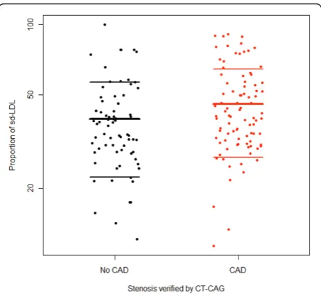
As depicted in Figure 1, patients with CAD had a sig-nificantly higher proportion of small dense LDL (means 50.1% and 40.0%, respectively; p < 0.001).
Unadjusted sdLDL was a significant predictor of pre-sence of CAD verified by invasive CAG in a logistic regression analysis (Table 3; Crude). Adjustment for age, gender, current smoking, and waist circumference (Adjustment 1) or adjustment for LDL cholesterol (Adjustment 2) did not materially reduce the estimate.
CT CAG
Stratified according to CT CAG, the risk profiles of the patients were slightly different (Table 2). Patients with CAD were older, more likely to receive lipid-lowering drugs, and had higher BMI and larger waist circumference. No lipid parameters, total cholesterol, LDL cholesterol, and triglycerides differed between patients with and without CAD, apart from patients with CAD having higher HDL cholesterol. Patients without and with CAD verified by CT CAG differed significantly in proportions of sdLDL (means 39.5% and 45.8%, respectively; p = 0.029) (Figure 2).
Crude sdLDL was a significant predictor of presence of CAD verified by invasive CAG in a logistic regression (Table 4; Crude). Adjustment for age, gender, current smoking, and waist circumference (Adjustment 1) reduced the estimate and rendered the prediction insig-nificant. Additional adjustment for LDL cholesterol (Adjustment 2) reduced the estimate and abolished the significance.
Discussion
The importance of sdLDL in CAD, measured by dif-ferent techniques, has been emphasised in several inves-tigations, among those the Quebec Cardiovascular Study [16] in which men with an elevated sdLDL cholesterol had a significantly higher risk of coronary heart disease (CHD) on follow-up. Also in the EPIC-Norfolk study [17], patients with coronary disease presented with a smaller LDL peak particle size, a higher proportion of sdLDL, and an increased plasma concentration of sdLDL cholesterol than matched, healthy controls. Simi-lar conclusions were drawn in a coinciding evaluation of the same study population by El Harchaoui [18] who reported a stronger association between the number of sdLDL particles and occurrence of CAD, than between serum LDL cholesterol and CAD. In accordance with this finding, we saw no significant difference in LDL
cholesterol in groups with or without CAD, nor did adjustment for LDL cholesterol influence the relation between CAD and sdLDL particles, perhaps because the patients included in the study deviated from the back-ground population by an a priori moderately altered risk profile and the use of statins.
Recognition of an intimate metabolic relationship between triglycerides and other lipids has fuelled debate as to which component is the principal marker of CVD risk and has given rise to the description of a lipid phe-notype consisting of a high proportion of small dense LDL, high levels of triglycerides, and low concentrations of HDL cholesterol [7] typical of patients with the meta-bolic syndrome. Conversely, its degree of covariance with other risk factors may help explain why the strength of associations of these factors with CAD is Table 2 Basic characteristics of patients, stratified according to presence of CAD verified by invasive CAG and
CT CAG, respectively
CAG CT CAG
Variable No CAD
(n = 120)
CAD (n = 58)
p-value No CAD (n = 71)
CAD (n = 91)
p-value
Age (mean and SD)T 61.6
± 9.8 64.1 ± 8.9 0.1 60.4 ± 9.8 64.1 ± 8.9 0.02
Male (number and %)F 46 (38.3) 42 (72.4) <0.0001 40.8 (29) 57.1 (52) 0.06
Family disposition to ischemic heart disease (number and %)F 66 (55) 28 (48.3) 0.43 62.0 (44) 48.4 (44) 0.11
Actual smoking (number and %)F 21 (17.5) 17 (29.3) 0.08 19.7 (14) 24.2 (22) 0.57
Lipid-lowering medication (number and %)F 61 (50.8) 44 (75.9) 0.002 45.1 (32) 70.3 (64) 0.001
Antihypertensive medication (number and %)F 75 (62.5) 45 (77.6) 0.06 62.0 (44) 71.4 (65) 0.24
Systolic blood pressure (mean and SD)T 145.8
± 21.6 148.6 ± 16.8 0.35 144.0 ± 20.1 147.4 ± 19.7 0.29
Diastolic blood pressure (mean and SD)T 78.8
± 11.0 80.4 ± 9.2 0.34 78.2 ± 9.7 79.6 ± 10.6 0.39
BMI (kg/m2)(mean and SD)T 26.7
± 4.3 27.2 ± 3.1 0.4 25.9 ± 4.0 27.4 ± 3.9 0.02
Waist circumference (cm) (mean and SD)T 94.8
± 12.4 99.6 ± 9.7 0.005 92.8 ± 11.9 98.1 ± 11.4 <0.005
Total cholesterol (mmol/L) (mean and SD)T 5.0
± 0.9 5.0 ± 1.0 0.75 5.1 ± 1.0 4.9 ± 0.9 0.35
Triglycerides (mmol/L) (mean and SD)T 1.3
0.7 1.8 1.3 0.02 1.3 0.7 1.2 0.8 0.1
HDL cholesterol (mmol/L ) (mean and SD)T 1.7
0.5 1.4 0.4 <0.001 1.7 0.5 1.5 0.4 0.03
LDL cholesterol (mmol/L) (mean and SD)T(n
total= 175) 2.7
± 0.9 (n = 119)
2.8 ± 0.8 (n = 56)
0.39 2.8
± 1.0 (n = 70)
2.7 ± 0.8 (n = 89)
0.46
High sensitive CRP (mean and SD)T 2.2
2.6 4.0 7.2 0.06 2.2 3.2 3.4 5.8 0.09
ApoB (g/L) (mean and SD)T 0.9
± 0.2 1.0 ± 0.2 0.09 0.9 ± 0.2 0.9 ± 0.2 0.91
Lp(a) (arb. units) (mean and SD)T 315.1
387.9 456.4 511.0 0.06 276.5 346.2 412.9 487.4 0.07
sdLDL (mean and SD percentages)T 40.0
16.8 50.1 19.5 <0.001 39.5 17.1 45.8 18.7 0.03
diminished through statistical adjustment. In accordance with this, Austin et al. [19] found a significant, univariate negative correlation between LDL size and CAD that van-ished upon adjustment for HDL cholesterol, triglycerides, and other traditional risk factors. In the Women’s Health study [20], where LDL particle concentration was com-pared to LDL particle size as risk predictors of coronary mortality and morbidity, even the stronger of the two, the particle concentration, was strongly attenuated by adjust-ment. That sdLDL and LDL particle numbers are by no means independent parameters has been illustrated by Griffin et al. who reported that a predominance of sdLDL, moderately elevated triglycerides, and a low HDL choles-terol were all inversely associated with the number of LDL particles [13].
In a case-control study of 225 middle-aged Japanese CAD patients [21], Koba et al. found that the coronary stenosis was highly correlated with an LDL particle dia-meter less than 255 Å. The CAD patients had LDL
cholesterol and non-HDL cholesterol similar to healthy controls, but sdLDL cholesterol was elevated. In a logistic regression analysis, adjustment for HDL cholesterol did, however, not abolish the effect of sdLDL cholesterol. The present study differs from that study mainly in the measurement of the“sd”parameter (Kola et al. measured cholesterol content whereas we measured the sdLDL proportion) and in the recruitment of patients. Our patients were enrolled based on a clinical suspicion of CAD prior to enrolment, whereas in the study from Japan, confirmed CAD patients were compared to healthy controls who were younger and had a healthier life style.
Kwon et al. [22] used a cross-sectional design to inves-tigate the relation of LDL particle size to CAD. The study population consisted of 504 patients without a prior history of myocardial infarction who underwent a CAG for evaluation of chest pain. Among these, 262 had at least one stenosis by CAG that obstructed 50% of Table 3 Logistic regression analysis of CAD verified by
invasive CAG with proportions of sdLDL as exposure (n = 175)
Odds ratio 95% CI P
Crude 1.36 1.13
1.64
0.001
Adjustment 1 1.26 1.02
1.56
<0.01
Adjustment 2 1.26 1.02
1.56
0.03
Odds ratios are expressed relative to sdLDL increments of 10%. (Adjusted 1: age, gender, current smoking, waist circumference; adjusted 2: age, gender, current smoking, waist circumference, LDL cholesterol).
Figure 1Proportion of small dense LDL stratified according to presence of CAD verified by invasive CAG. (n = 178; lines mean mean ± sd). The mean proportions were 40.0% and 50.1%, respectively.
Figure 2Proportion of small dense LDL, stratified according to presence of CAD verified by CT CAG. (n = 162; lines mean mean ± sd). The proportions were 39.5% and 45.8%, respectively.
Table 4 Logistic regression analysis of CAD verified by CT CAG with proportions of sdLDL as exposure (n = 159)
Odds ratio 95% CI P
Crude 1.26 1.03
1.53
0.02
Adjusted 1 1.20 0.96
1.50
0.11
Adjusted 2 1.21 0.97
1.52
0.09
the lumen, while 242 patients, who showed no or only minimal signs of CAD on a subsequent angiographic examination, served as controls. In accordance with our study, the fraction of sdLDL was significantly higher in the CAD group. In contrast to our data, however, sdLDL remained a significant risk factor upon adjust-ment for traditional risk factors, HDL cholesterol, and LDL cholesterol. Kwon et al. used electrophoresis yield-ing a size scale with a specific nanometre cut-off point between small and large LDL particles, whereas our measurements were based on density and a density cut-off. Indeed, several reports, among those a systematic review [23], emphasise the incongruence between techniques.
Finally, Koz et al. [24] conducted a study in 102 con-secutive young men with chest pain and a low Framing-ham risk score. They all underwent a CAG. LDL size was smaller in patients with CAD (n = 45), but it was not a significant predictor upon adjustment for tradi-tional risk factors. The results are therefore in good agreement with ours. Moreover, the fact that the study populations were quite dissimilar from ours (young men performing military service vs elderly patients) empha-sises the significance of the sdLDL parameter.
The alternative grouping of patients based on CT CAG in our study in general showed a weaker correlation with sdLDL, and its predictive value was lost after adjustment for traditional risk parameters alone. This could in part be explained by a reduced sample size, but it might also reflect the fact that more participants in our study were classified as having CAD on CT examination than on invasive CAG. CT CAG is a fast emerging image modal-ity, but the specificity of the method is suboptimal, and this might readily explain the weaker correlation between sdLDL and CT CAG-evaluated CAD.
The present study has some limitations. The sample size was relatively small, and a priori patients belonged to the low and intermediate CAD risk groups. Likewise, the number of patients with significant stenoses was too limited to enable us to graduate the extent rather than the mere presence of CAD. Identifying patients with CAD on the basis of significant stenoses may underesti-mate the total number of patients with CAD, as visually significant stenoses only represent patients with advanced
disease. Treatment with statins and b-adrenoceptor
antagonists is widespread in Denmark. Apart from a reduction of LDL cholesterol and varying effect on other lipid parameters dependent on the type of dyslipidemia and the specific agent, statins have been shown to induce a significant shift in the sdLDL subtype pattern towards larger and more buoyant particles [25]. Thus, as a larger percentage of the CAD group than of the no CAD group received statins, the connection between sdLDL and CAD may be even stronger than depicted here.
The findings of the present study provide further support to the value of not only cholesterol in the major lipoprotein subfractions (HDL and LDL), but also the distribution and quality of particles within each subfraction in risk assessment, as sdLDL, in con-trast to LDL cholesterol, emerged as a univariate pre-dictor of CAD. HDL cholesterol was, statistically, as closely correlated to the CAD status, but the absolute difference between the mean HDL values in the two groups was small compared to the differences of sdLDL. The study also demonstrates the clinical utility of the iodixanol method which, in contrast to the expensive NMR analysis, can be routinely incorporated into clinical risk assessment. However, the widespread use of statins makes it impossible to exclude patients receiving this treatment and to generalize data to other populations.
Conclusion
A significant difference in the proportion of sdLDL was observed between patients with CAD and otherwise highly similar patients without CAD. Clinical trials are necessary to determine whether this is of importance for stratification or patient outcome.
Abbreviations
CAD: coronary Artery Disease; CAG: invasive coronary angiography; CHD: coronary heart disease; CT: CAG CT-based coronary angiography; HDL: high-density lipoprotein; LDL: low-high-density lipoprotein; PCI: percutaneous coronary intervention; sdLDL: small dense LDL particles
Author details
1Department of Cardiology, Center for Cardiovascular Research, Aalborg
Hospital, Aarhus University Hospital, Aalborg, Denmark.2Department of Radiology, Center for Cardiovascular Research, Aalborg Hospital, Aarhus University Hospital, Aalborg, Denmark.3Faculty of Health & Medical Sciences, University of Surrey, Guildford, Surrey, GU2 7XH UK.
Authors’contributions
APTP compiled the data and wrote the first draft of the paper. HHT and KR evaluated the CT scans (blinded to the invasive coronary angiography results). JA evaluated the results from invasive coronary angiography (blinded to the results of the CT scans). THC was responsible for the technical aspects of the CT scans and helped with the interpretation of the results. BAG set up the analysis of small dense LDL cholesterol particles and interpreted the results together with IVA and AA. HHT, KR, JA, and EBS developed the scientific idea behind the study. All authors contributed to a critical assessment of the manuscript and read and approved the final version.
Competing interests
The authors declare that they have no competing interests.
Received: 1 December 2010 Accepted: 25 January 2011 Published: 25 January 2011
References
1. American Heart Association:Heart attack and angina statistics.2010 [http://www.americanheart.org/presenter.jhtml?identifier=4591]. 2. Greenland P, LaBree L, Azen SP, Doherty TM, Detrano RC:Coronary artery
3. Mowatt G, Cummins E, Waugh N, Walker S, Cook J, Jia X, Hillis GS, Fraser C:
Systematic review of the clinical effectiveness and cost-effectiveness of 64-slice or higher computed tomography angiography as an alternative to invasive coronary angiography in the investigation of coronary artery disease.Health Technol Assess2008,12:iii-143.
4. Wilson PWF, D’Agostino RB, Levy D, Belanger AM, Silbershatz H, Kannel WB:
Prediction of Coronary Heart Disease Using Risk Factor Categories.
Circulation1998,97:1837-47.
5. Tabas I, Williams KJ, Borén J:Subendothelial lipoprotein retention as the initiating process in atherosclerosis: update and therapeutic implications.Circulation2007,116:1832-44.
6. Nordestgaard BG, Benn M, Schnohr P, Tybjaerg-Hansen A:Nonfasting Triglycerides and Risk of Myocardial Infarction, Ischemic Heart Disease, and Death in Men and Women.JAMA2007,298:299-308.
7. Austin MA, King MC, Vranizan KM, Krauss RM:Atherogenic lipoprotein phenotype. A proposed genetic marker for coronary heart disease risk.
Circulation1990,82:495-506.
8. de Graaf J, Hak-Lemmers HL, Hectors MP, Demacker PN, Hendriks JC, Stalenhoef AF:Enhanced susceptibility to in vitro oxidation of the dense low density lipoprotein subfraction in healthy subjects.Arterioscler Thromb1991,11:298-306.
9. Chancharme L, Therond P, Nigon F, Lepage S, Couturier M, Chapman MJ:
Cholesteryl ester hydroperoxide lability is a key feature of the oxidative susceptibility of small, dense LDL.Arterioscler Thromb Vasc Biol1999,
19:810-20.
10. Ip S, Lichtenstein AH, Chung M, Lau J, Balk EM:Systematic review: association of low-density lipoprotein subfractions with cardiovascular outcomes.Ann Intern Med2009,150:474-84.
11. Berneis KK, Krauss RM:Metabolic origins and clinical significance of LDL heterogeneity.J Lipid Res2002,43:1363-79.
12. Davies IG, Graham JM, Griffin BA:Rapid separation of LDL subclasses by iodixanol gradient ultracentrifugation.Clin Chem2003,49:1865-72. 13. Griffin BA, Minihane AM, Furlonger N, Chapman C, Murphy M, Williams D,
Wright JJ, Williams CM:Inter-relationships between small, dense low-density lipoprotein (LDL), plasma triacylglycerol and LDL apoprotein B in an atherogenic lipoprotein phenotype in free-living subjects.Clin Sci (Lond)1999,97:269-76.
14. Rasmussen JG, Eschen RB, Aardestrup IV, Dethlefsen C, Griffin BA, Schmidt EB:Flow-mediated vasodilation: variation and interrelationships with plasma lipids and lipoproteins.Scand J Clin Lab Invest2009,
69:156-60.
15. Achenbach S, Cardiac CT:state of the art for the detection of coronary arterial stenosis.J Cardiovasc Comput Tomogr2007,1:3-20.
16. St-Pierre AC, Cantin B, Dagenais GR, Mauriege P, Bernard PM, Despres JP, Lamarche B:Low-density lipoprotein subfractions and the long-term risk of ischemic heart disease in men: 13-year follow-up data from the Quebec Cardiovascular Study.Arterioscler Thromb Vasc Biol2005,25:553-9. 17. Arsenault BJ, Lemieux I, Despres JP, Wareham NJ, Luben R, Kastelein JJ,
Khaw KT, Boekholdt SM:Cholesterol levels in small LDL particles predict the risk of coronary heart disease in the EPIC-Norfolk prospective population study.Eur Heart J2007,28:2770-7.
18. El Harchaoui KF, van der Steeg WA, Stroes ES, Kuivenhoven JA, Otvos JD, Wareham NJ, Hutten BA, Kastelein JJ, Khaw KT, Boekholdt SM:Value of low-density lipoprotein particle number and size as predictors of coronary artery disease in apparently healthy men and women: the EPIC-Norfolk Prospective Population Study.J Am Coll Cardiol2007,
49:547-53.
19. Austin MA:Low-density lipoprotein particle size, triglycerides, and high-density lipoprotein cholesterol as risk factors for coronary heart disease in older Japanese-American men.Am J Cardiol2000,86:412-6. 20. Blake GJ, Otvos JD, Rifai N, Ridker PM:Low-density lipoprotein particle
concentration and size as determined by nuclear magnetic resonance spectroscopy as predictors of cardiovascular disease in women.
Circulation2002,106:1930-7.
21. Koba S, Hirano T, Ito Y, Tsunoda F, Yokota Y, Ban Y, Iso Y, Suzuki H, Katagiri T:Significance of small dense low-density lipoprotein-cholesterol concentrations in relation to the severity of coronary heart diseases.
Atherosclerosis2006,189:206-14.
22. Kwon SW, Yoon SJ, Kang TS, Kwon HM, Kim JH, Rhee J, Lee SJ, Park JK, Lim JY, Yoon YW, Hong BK:Significance of small dense low-density
lipoprotein as a risk factor for coronary artery disease and acute coronary syndrome.Yonsei Med J2006,47:405-14.
23. Chung M, Lichtenstein AH, Ip S, Lau J, Balk EM:Comparability of methods for LDL subfraction determination: A systematic review.Atherosclerosis 2009,205:342-8.
24. Koz C, Baysan O, Hasimi A, Cihan M, Uzun M, Yokusoglu M, Sag C, Isik E:
Conventional and non-conventional coronary risk factors in male premature coronary artery disease patients already having a low Framingham risk score.Acta Cardiol2008,63:623-8.
25. Backes JM, Gibson CA:Effect of lipid-lowering drug therapy on small-dense low-density lipoprotein.Ann Pharmacother2005,39:523-6.
doi:10.1186/1476-511X-10-21
Cite this article as:Toft-Petersenet al.:Small dense LDL particles - a predictor of coronary artery disease evaluated by invasive and CT-based techniques: a case-control study.Lipids in Health and Disease2011
10:21.
Submit your next manuscript to BioMed Central and take full advantage of:
• Convenient online submission
• Thorough peer review
• No space constraints or color figure charges
• Immediate publication on acceptance
• Inclusion in PubMed, CAS, Scopus and Google Scholar
• Research which is freely available for redistribution