Open Access
Research
Mclk1
+/-
mice are not resistant to the development of
atherosclerosis
Bryan G Hughes and Siegfried Hekimi*
Address: Department of Biology, McGill University, Montreal H3A 1B1, Canada
Email: Bryan G Hughes - [email protected]; Siegfried Hekimi* - [email protected] * Corresponding author
Abstract
Background: Mice with a single copy of Mclk1 (a.k.a. Coq7), a gene that encodes a mitochondrial enzyme required for the biosynthesis of ubiquinone and other functions, live longer than wild-type mice. The prolonged survival implies a decreased mortality from age-dependent lethal pathologies. Atherosclerosis is one of the main age-dependent pathologies in humans and can be modeled in mice that lack Apolipoprotein E (ApoE-/-) or mice that lack the Low Density Lipoprotein Receptor
(LDLr-/-) in addition to being fed an atherosclerosis-inducing diet. We sought to determine if Mclk1
heterozygosity protects against atherosclerosis and dyslipidemia in these models.
Results: We found that Mclk1 heterozygosity did not protect against dyslipidemia, oxidative stress, or atherosclerosis in young (6 or 10 months) or older (18 months) mice. Furthermore, the absence of ApoE suppressed the lifespan-promoting effects of Mclk1 heterozygosity.
Conclusion: These findings indicate that although Mclk1 heterozygosity can extend lifespan of mice, it does not necessarily protect against atherosclerosis. Moreover, in the presence of hyperlipidemia and chronic inflammation, Mclk1 heterozygosity is incapable of extending lifespan.
Introduction
Median lifespan is increased by mutations in the gene clk-1, and by heterozygosity for a null allele of the mamma-lian clk-1 homologue Mclk1, in Caenorhabditis elegans and mice, respectively [1-3]. CLK-1/MCLK1 is a mitochondrial hydroxylase required for the biosynthesis of ubiquinone (UQ or Coenzyme Q) [4,5], a lipid-like molecule that plays a vital role as an electron transporter in the mito-chondrial electron transport chain, is an important lipid-soluble antioxidant, and also performs a variety of other functions [6]. Mclk1-null (Mclk1-/-) cells and embryos make no UQ and the embryos are not viable, while Mclk1
heterozygous (Mclk1+/-) cells and mice are superficially normal and show normal levels of UQ [7,8]. While the biochemical phenotypes associated with Mclk1
heterozy-gosity have begun to be uncovered [3,9], the actual effect on the mouse in terms of resistance to the physical decline associated with age and to age-dependent disease has not yet been examined.
The longest-lived humans display an increased resistance to the common age-dependent diseases (cardiovascular disease, stroke, cancer, etc.), as the majority of them either develop these diseases later than the general population or not at all [10,11]. It is therefore tempting to expect a similar resistance to the development of age-dependent diseases in Mclk1+/- and other lines of long-lived mice. One particularly prevalent age-dependent disease is atherosclerosis, in which the oxidative modification of low-density lipoprotein and the resultant recognition by
Published: 5 May 2009
Lipids in Health and Disease 2009, 8:16 doi:10.1186/1476-511X-8-16
Received: 7 April 2009 Accepted: 5 May 2009
This article is available from: http://www.lipidworld.com/content/8/1/16
© 2009 Hughes and Hekimi; licensee BioMed Central Ltd.
cells of the immune system results in a chronic inflamma-tory state within the wall of blood vessels. This leads to the formation of a plaque containing a mixture of extracellu-lar cholesterol deposits, immune cells and infiltrating smooth muscle cells [12]. Interestingly, manipulations that extend lifespan in mice have been shown to protect against atherosclerosis. Among long-lived mice, calori-cally restricted mice as well as geneticalori-cally modified p66 shc-/- and UCP1 transgenic mice have been evaluated for atherosclerosis susceptibility. The fact that both were found to have an increased resistance to the disease [13-15] suggests that Mclk1+/- mice may also be resistant to the disease.
A further incentive for studying the effect of Mclk1 hetero-zygosity on atherosclerosis is the finding that clk-1 worms have an altered lipid metabolism, consistent with a decrease in oxidatively modified lipoproteins [16]. Fur-thermore, the core non-lipid component of these particles appears to be vitellogenin, a protein that is homologous to apolipoprotein B, the main structural component of atherosclerosis-inducing lipoprotein particles in mam-mals [17,18]. The conservation of lipid metabolism between worms and mammals was further confirmed by the finding that several lipid-lowering drugs developed in mammals are active on lipoprotein metabolism in worms [19].
Atherosclerosis does not appear to develop in mice fed a regular diet, due to the partitioning of most cholesterol into the anti-atherogenic High Density Lipoprotein frac-tion of lipoproteins. Therefore, the lifespan extending effect of Mclk1 heterozygosity cannot be due to a decrease in atherosclerosis-related mortality. However, the under-lying conditions thought to be responsible for atheroscle-rosis, oxidative stress and inflammation, are known to be strongly associated with aging, justifying the study of atherosclerosis in aging mice [20,21]. Mice lacking the Low Density Lipoprotein Receptor and fed a high-fat, high-cholesterol "Western" diet (LDLr-/-), as well as mice lacking apolipoprotein E fed a regular chow diet (ApoE-/-), develop hyperlipidemia and extensive aortic atherosclero-sis, and are commonly used to model atherosclerosis in mice [22,23]. Although the lipid profile of these mice is characteristic of only the more dyslipidemic humans, the histopathological characteristics of the lesions appear similar to those observed in humans and other models, even if they develop over a shorter timeframe [24]. Fur-thermore, although coronary artery disease predomi-nately affects those of middle-age or older, the atherosclerotic lesions that underlie the disease (and which are the focus of this study) begin to develop much earlier in life, with young adults having comparable aortic atherosclerosis to that observed in mice at an equivalent stage in their lives [25].
We elected to use two separate models because, although they theoretically affect the same system (the uptake of lipid from the circulation), they are known to respond dif-ferently to certain interventions [26-28]. Importantly, the
LDLr-/- model develops more severe atherosclerosis than the ApoE-/- model, allowing us to measure the effect of
Mclk1 heterozygosity on atherosclerosis of both medium
and high severity. We also measured atherosclerosis in mice of different ages, in order to detect any possible impact of an age-dependent effect of Mclk1 heterozygosity on atherosclerosis. Here, we demonstrate that Mclk1 het-erozygosity does not consistently affect atherosclerosis or lipid profile in either model. Furthermore, Mclk1 hetero-zygosity fails to extend lifespan in atherosclerosis-suscep-tible ApoE-/- mice.
Methods
Animals and Diet
LDLr-/- [29] and ApoE-\- [30] mice on a C57Bl/6J back-ground were purchased from The Jackson Laboratory (Bar Harbor, ME) and were crossed to Mclk1+/- mice, previously produced by gene targeting [7], to produce LDLr-/-;Mclk1+/ +, LDLr-/-;Mclk1+/-, ApoE-\-; Mclk1+\+ and ApoE-\-; Mclk1 +\-animals. Genotypes were determined by PCR. Mice were maintained on 4.5% fat rodent chow (Charles River diet 5075). At 3 months of age mice in the LDLr-/- groups were placed on a "Western"-type diet (Harlan Teklad, Madison, WI, TD.01444) containing 21% (w/w) anhydrous milk fat and 0.15% cholesterol. Mice in the ApoE-/- groups were fed regular rodent chow throughout the experiments. Mice were housed in a specific pathogen free facility at McGill University, 2–5 animals per cage, and fasted overnight prior to sacrifice by anesthetic overdose (Ketamine/Xyla-zine/Acepromazine).
ApoE-/- mice were sacrificed at 10 and 18 months of age and LDLr-/- mice at 6 and 10 months of age (Table 1). 18 months was judged the greatest age that we could obtain without significant losses due to mortality in the ApoE -/-group. We had originally intended to sacrifice the LDLr -/-mice at 18 months as well, but we found that after 10 months of age on the "Western" diet LDLr-/- mice devel-oped severe health problems. The earlier measurement point for each model is the youngest age where we could
Table 1: Experimental Design
Model Age (months) Gender(s) Background (# Backcrosses)
ApoE-/- 10 M, F CBA × C57BL/6J (4)
10 M, F 129Sv/Balb/c × C57BL/6J (10) 18 F CBA × C57BL/6J (3) Lifespan M, F 129Sv/Balb/c × C57BL/6J (6)
LDLr-/- 6 M, F CBA × C57BL/6J (3)
reliably obtain measurable atherosclerosis in each case. For both groups, male and female mice were sacrificed at the earlier point, and females only at the later time-point.
The genetic backgrounds of the mice varied somewhat between studies, although they were always consistent within a study (Table 1). Mice on the CBA background were backcrossed 3 times into the C57Bl/6J background to create the LDLr-/- mice sacrificed at 6 months of age and mice on a mixed 129Sv × Balb/c background were back-crossed 6 times for LDLr-/- mice sacrificed at 10 months of age. ApoE-/- mice sacrificed at 10 and 18 months were backcrossed 4 and 3 times respectively out of the CBA background, and those used in the aging experiment were backcrossed 6 times out of a mixed 129Sv × Balb/c back-ground. A second group of ApoE-/- mice sacrificed at 10 months of age was backcrossed 10 times from the same background. In all cases, the C57BL/6J strain made the greatest contribution to the background, which is impor-tant because of this strain's relative sensitivity to athero-sclerosis development [31], as well as because the ability
of Mclk1 heterozygosity to extend lifespan in this strain
has been confirmed in our lab [32].
To determine the lifespan of Mclk1+/- mice in the ApoE -/-background, mice were kept until either natural death, or evidence of impending mortality (such as sudden drastic weight loss, lack of movement or a severely distended abdomen) necessitating euthanasia.
All studies were approved by the McGill Faculty of Science Animal Care Committee and conducted according to the guidelines of the Canadian Council on Animal Care.
Atherosclerosis Severity
The surface area of atherosclerotic lesions was measured on the inner surface of the aorta, from the aortic origin to the iliac branch point, as has been previously described [33,34]. Quantification of staining on the acquired images was carried out using UTHSCSA ImageTool v3.0 (University of Texas Health Science Center in San Anto-nio, USA), and results were expressed as percentage sur-face area of the aorta occupied by Oil Red O-staining lesions. The innominate, common carotid and subclavian arteries were excluded from the analysis.
Plasma Measurements
EDTA plasma was collected by cardiac puncture of anaes-thetized mice, flash-frozen in liquid nitrogen and stored at -80°C. Kits for measuring plasma cholesterol were obtained from Wako Chemicals USA and those for triglyc-erides from Sigma-Aldrich. Lipid peroxidation, as quanti-fied by the level of malondialdehyde (MDA), was measured with a Thiobarbituric Acid Reactive Substances
(TBARS) Assay Kit from ZeptoMetrix Corporation. The TBARS method of measuring MDA has received consider-able criticism for being an insufficiently representative measurement of lipid peroxidation [35-37]. Despite this, even some of the harshest critics concede that the assay frequently yields useful results supported by other better-validated assays, although it may be better thought of as a measurement of susceptibility to lipid peroxidation rather than the steady-state level of damage [37].
Statistics
The non-parametric Mann-Whitney test was used to com-pare atherosclerosis surface area in precom-pared aortas. Oth-erwise, the unpaired two-tailed Student's t-test was used. Survival curves were compared using the Log-rank (Man-tel-Cox) test. Two-way ANOVA with Bonferroni posttests was used for comparing body-weights collected over lifespan. Comparisons were always made between geno-types, within genders, using Prism 4.03 (GraphPad Soft-ware, Inc).
Results
Mclk1 heterozygosity does not decrease the severity of atherosclerosis
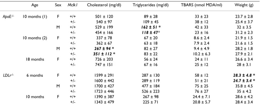
To determine if Mclk1 heterozygosity protected against atherosclerosis, Mclk1+/- mice were crossed into athero-sclerosis-sensitive ApoE-/- and LDLr-/- backgrounds. The proportion of the inner aortic surface occupied by athero-sclerotic lesions was then quantified. Atherosclerosis in both the ApoE-/- and LDLr-/- models of the disease was not inhibited by Mclk1 heterozygosity (Figures 1 and 2). In one condition (ApoE-/- mice sacrificed at 10 months of age), Mclk1+/- females had increased atherosclerosis (6.8 vs. 10 percent of aortic surface area, p = 0.0146). In the same experiment, there was a 37 percent decrease in atherosclerosis in Mclk1+/- males that was not statistically significant (p = 0.21). As the mice in this cohort had only been backcrossed for four generations, it is possible that high variability due to genetic heterogeneity could have been masking an effect of Mclk1 heterozygosity in males. We therefore repeated this study in a second group of
ApoE-/- mice that had been backcrossed ten generations onto the C57BL/6J background. In this second cohort of mice, there was no effect of Mclk1 heterozygosity in either gender.
Heterozygosity for Mclk1 does not prevent atherosclerosis in the LDLr-/- disease model Figure 1
Heterozygosity for Mclk1 does not prevent atherosclerosis in the LDLr-/- disease model. Atherosclerotic lesions that stain with the lipid-sensitive dye Oil Red O were quantified on the inner surface of the aorta in LDLr-/- mice at (A) 6
months of age, with three backcrosses into C57BL/6J and (B) 10 months of age with six backcrosses into C57BL/6J. F and M labels stand for females and males, respectively. The +/+ and +/- labels stand for the Mclk1+/+ and Mclk1+/- genotypes,
Heterozygosity for Mclk1 does not prevent atherosclerosis in the ApoE-/- disease model Figure 2
Heterozygosity for Mclk1 does not prevent atherosclerosis in the ApoE-/- disease model. Atherosclerotic lesions that stain with the lipid-sensitive dye Oil Red O were quantified on the inner surface of the aorta in ApoE-/- mice at (A) 10
weight of female LDLr-/-; Mclk1+/- mice sacrificed at six months of age was slightly decreased, but we did not see an effect on body weight in any other group.
Loss of ApoE suppresses the lifespan extension conferred
by Mclk1 heterozygosity
Male and female Mclk1+/- mice on an ApoE-null back-ground failed to live longer than Mclk1+/+ controls (Figure 3). Female Mclk1+/- mice actually had a shorter average lifespan than controls, although this difference did not reach statistical significance (p = 0.16). In Mclk1+/- mice of both genders, body weight was decreased throughout life (p < 0.0001 females, p = 0.0064 males) (Figure 3). 21 out of 35 females and 9 out of 18 males were euthanized as they were obviously near death due to severe illness (pro-portions were equal for each genotype), with the remain-der dying naturally. Atherosclerosis was quantified in euthanized mice, and Mclk1+/- females appeared to have decreased atherosclerosis relative to controls, although this difference did not reach statistical significance (16.4 ± 12.5 in Mclk1+/+ vs. 9.6 ± 5.8 percent in Mclk1+/-, p = 0.18 by Mann-Whitney test). Atherosclerosis was not affected in males (20.3 ± 14.25 in wild-type vs. 23 ± 16.57 in
Mclk1+/-, p = 0.63). In the group of ApoE-/- mice sacrificed at 18 months of age, three mice of each genotype died prior to their date of sacrifice, closely paralleling the sur-vival curve of mice in the lifespan study.
Discussion
The effect of Mclk1 heterozygosity on atherosclerosis
In five separate studies, we showed that Mclk1 heterozy-gosity did not ameliorate atherosclerosis in mice. In one study, female Mclk1+/- mice showed a statistically signifi-cant increase in atherosclerosis, and males showed a
non-significant decrease. However, we were unable to repeat either of these results in a follow-up study in which the mice were in a fully congenic background. Our experi-ments covered a range of ages and severity of disease, indi-cating that Mclk1 heterozygosity did not have any beneficial age-dependent or -independent effects on atherosclerosis. In other words, we only observed an effect on atherosclerosis in one particular genetic background, suggesting that it was due to a unique set of interactions within this particular background. This forces us to con-clude that any potential effects of Mclk1 heterozygosity on atherosclerosis would not be due to the same mechanism as that which produces lifespan extension, which was observed in several separate backgrounds.
We have recently reported that young Mclk1+/- mice have decreased lipid peroxidation, measured via plasma iso-prostanes. At the same time, the mice had substantial alterations in mitochondrial function, including a general decrease in mitochondrial respiration and an increase in mitochondrial oxidative stress [9]. Mitochondrial dys-function linked to increased oxidative stress, such as that found in Mclk1+/- mice, has been shown to increase sus-ceptibility to atherosclerosis [38]. The balance between mitochondrial, atherosclerosis-sensitizing and systemic, potentially atherosclerosis-protective, phenotypes of
Mclk1+/- mice may explain the general absence of a
signif-icant effect on atherosclerosis that we observed. This bal-ance may be modulated by genetic background, which could account for some of the contrasting results we observed between ApoE-/- mice of different backgrounds. Furthermore, Mclk1+/- mice also have higher levels of sev-eral pro-inflammatory cytokines (unpublished data). Inflammation plays a role in all stages of atherosclerosis
Table 2: Plasma lipid characteristics and body weight at sacrifice (expressed as mean ± standard deviation)
Age Sex Mclk1 Cholesterol (mg/dl) Triglycerides (mg/dl) TBARS (nmol MDA/ml) Weight (g)
ApoE-/- 10 months (1) F +/+ 501 ± 120 89 ± 28 33 ± 23 23.7 ± 2.8
+/- 540 ± 97 109 ± 45 38 ± 12 25.4 ± 3.7 M +/+ 529 ± 199 162 ± 51 * 42 ± 33 32 ± 3.5
+/- 454 ± 166 118 ± 47* 23 ± 16 31.2 ± 2.3 10 months (2) F +/+ 337 ± 78 67 ± 20 8.6 ± 2.4 21.9 ± 1.5 +/- 362 ± 67 63 ± 18 7.9 ± 2.4 21.6 ± 1.5
M +/+ 267 ± 94 * 82 ± 27 9.4 ± 4.9 28.2 ± 1.8
+/- 351 ± 112 * 83 ± 22 10.2 ± 6.3 27.9 ± 2.1 18 months F +/+ 726 ± 203 56 ± 24 24 ± 11 26.6 ± 3.4 +/- 747 ± 151 67 ± 16 25 ± 12 28 ± 3.1
LDLr-/- 6 months F +/+ 1599 ± 291 287 ± 130 58 ± 12 28.3 ± 4.8 *
+/- 1600 ± 442 289 ± 119 51 ± 21 24.7 ± 3.4 *
M +/+ 1700 ± 427 477 ± 184 75 ± 25 35.8 ± 4.5 +/- 1723 ± 446 526 ± 223 76 ± 27 35 ± 4.2 10 months F +/+ 1390 ± 387 267 ± 98 24.4 ± 7.1 28.6 ± 4.2
+/- 1343 ± 479 225 ± 71 20.8 ± 5.7 28.4 ± 3.4
[39], suggesting another possible reason why Mclk1 +/-mice are not protected against the disease.
The effect of mutant clk-1 in C. elegans and Mclk1 in
mice on lipid metabolism
clk-1 mutant worms have altered oxidative and lipid
metabolism [16,19]. The organismal differences between worms and mice, as well as other differences between the
two models (homozygous mutants in worms and hetero-zygous mutants in mice) may explain why we did not the see the expected effect on lipid metabolism and athero-sclerosis in Mclk1+/- mice. The clk-1 mutant worms are homozygous for alleles incapable of producing UQ, resulting in an obvious, easily observable, phenotype [2,40]. On the other hand, mice retain one copy of the wild-type allele, and the MCLK1 produced appears
suffi-Survival curves and body weights throughout lifespan of Mclk1+/+ and+/- mice on the ApoE-/- background Figure 3
cient for a normal level of UQ synthesis [3,7,9]. It appears that, in both worms and mice, MCLK1/CLK-1 has an addi-tional function besides the production of UQ. This is sup-ported by evidence from both organisms. In worms, two mutant alleles of clk-1 yield phenotypes of different sever-ity, despite the fact that neither allele can produce UQ [41,42]. Mclk1+/- mice actually have several very clear bio-chemical phenotypes despite wild-type levels of UQ [3,9]. Yet, the absence of a lipid phenotype in mice suggests the existence of a threshold that is not reached when CLK-1/ MCLK1 activity is only reduced as in Mclk1+/- mouse mutants rather than completely abolished or very severely reduced as in the C. elegans mutants.
Suppression of Mclk1+/- longevity by loss of ApoE
In mice lacking the ApoE protein, Mclk1 heterozygosity no longer extends lifespan. In other words, some characteris-tic of the ApoE-/- background actually shortens the lifespan
of Mclk1+/- mice relative to Mclk1+/+ mice. We also
observed that Mclk1+/-; ApoE-/- mice weighed slightly less than Mclk1+/+; ApoE-/- mice, starting at approximately 1 year of age. This is in contrast to older Mclk1+/- mice wild-type for ApoE, which either weigh the same or more than wild-type controls [unpublished data and [1]]. This does imply that the absence of ApoE has detrimental effects on
Mclk1+/- mice. An increased prevalence of atherosclerosis
is unlikely to be the cause of this lifespan shortening effect. Firstly, as described above, Mclk1+/- mice do not appear more susceptible to atherosclerosis development. If anything, Mclk1+/- female mice in the aging experiment that were euthanized due to impending death showed a trend of slightly less aortic atherosclerosis than their wild-type controls. Secondly, it has been surprisingly difficult to find evidence in ApoE-/- mice of the arterial occlusion-induced cardiovascular events that are so lethal to humans [43]. Although older ApoE-/- mice may develop atherosclerosis of the coronary arteries, only a small pro-portion of these are found to display evidence of more advanced coronary artery disease, such as plaque rupture [24,44]. Aside from atherosclerosis, ApoE-/- mice suffer from other conditions, such as increased oxidative stress [45] and inflammation [46]. As described above, young
Mclk1+/- mice have higher levels of oxidative stress in
mitochondria as well as greater expression of certain cytokines consistent with a more pro-inflammatory state. Although these factors by themselves clearly do not pre-vent the increased longevity of Mclk1+/- mice, they may exert a negative effect that cancels out the lifespan exten-sion when combined with corresponding phenotypes in
ApoE-/- mice.
Conclusion
Contrary to our expectations based on the increased lon-gevity of Mclk1+/- mice, Mclk1 heterozygosity does not pro-tect against atherosclerosis. This is not surprising in light
of recent findings that suggest a complex pattern of phe-notypes due to Mclk1 heterozygosity, some of which may actually facilitate development of a disease such as athero-sclerosis that involves oxidative stress and inflammation. Furthermore, Mclk1 heterozygosity does not extend lifespan in the ApoE-null background, suggesting that
Mclk1 heterozygosity is not capable of protecting against
the increased oxidative stress and inflammation that afflict ApoE-null mice.
Competing interests
The authors declare that they have no competing interests.
Authors' contributions
BH participated in the conception and design of this study, performed the experiments, participated in the analysis of data, and helped write the paper. SH pated in the conception and design of the study, partici-pated in the analysis of data, and helped write the paper.
Authors' Information
SH is Campbell Chair of Developmental Biology and Strathcona Chair of Zoology.
Acknowledgements
We thank Eve Bigras for expert technical assistance. This study was funded by McGill University.
References
1. Liu X, Jiang N, Hughes B, Bigras E, Shoubridge E, Hekimi S: Evolu-tionary conservation of the clk-1-dependent mechanism of longevity: loss of mclk1 increases cellular fitness and lifespan in mice. Genes Dev 2005, 19(20):2424-2434.
2. Wong A, Boutis P, Hekimi S: Mutations in the clk-1 gene of Caenorhabditis elegans affect developmental and behavioral timing. Genetics 1995, 139(3):1247-1259.
3. Lapointe J, Stepanyan Z, Bigras E, Hekimi S: Reversal of the mito-chondrial phenotype and slow development of oxidative biomarkers of aging in long-lived Mclk1 +/- mice. J Biol Chem
2009 in press.
4. Ewbank JJ, Barnes TM, Lakowski B, Lussier M, Bussey H, Hekimi S: Structural and functional conservation of the Caenorhabdi-tis elegans timing gene clk-1. Science 1997, 275(5302):980-983. 5. Stenmark P, Grunler J, Mattsson J, Sindelar PJ, Nordlund P, Berthold DA: A new member of the family of di-iron carboxylate pro-teins. Coq7 (clk-1), a membrane-bound hydroxylase involved in ubiquinone biosynthesis. J Biol Chem 2001, 276(36):33297-33300.
6. Turunen M, Olsson J, Dallner G: Metabolism and function of coenzyme Q. Biochim Biophys Acta 2004, 1660(1–2):171-199. 7. Levavasseur F, Miyadera H, Sirois J, Tremblay ML, Kita K, Shoubridge
E, Hekimi S: Ubiquinone is necessary for mouse embryonic development but is not essential for mitochondrial respira-tion. J Biol Chem 2001, 276(49):46160-46164.
8. Nakai D, Yuasa S, Takahashi M, Shimizu T, Asaumi S, Isono K, Takao T, Suzuki Y, Kuroyanagi H, Hirokawa K, Koseki H, Shirsawa T: Mouse homologue of coq7/clk-1, longevity gene in Caenorhabditis elegans, is essential for coenzyme Q synthe-sis, maintenance of mitochondrial integrity, and neurogene-sis. Biochem Biophys Res Commun 2001, 289(2):463-471.
9. Lapointe J, Hekimi S: Early Mitochondrial Dysfunction in Long-lived Mclk1 +/- Mice. J Biol Chem 2008, 283(38):26217-26227. 10. Evert J, Lawler E, Bogan H, Perls T: Morbidity profiles of
11. Andersen SL, Terry DF, Wilcox MA, Babineau T, Malek K, Perls TT: Cancer in the oldest old. Mech Ageing Dev 2005, 126(2):263-267. 12. Glass CK, Witztum JL: Atherosclerosis. The Road Ahead. Cell
2001, 104(4):503-516.
13. Guo Z, Mitchell-Raymundo F, Yang H, Ikeno Y, Nelson J, Diaz V, Rich-ardson A, Reddick R: Dietary restriction reduces atherosclero-sis and oxidative stress in the aorta of apolipoprotein E-deficient mice. Mech Ageing Dev 2002, 123(8):1121-1131. 14. Napoli C, Martin-Padura I, de Nigris F, Giorgio M, Mansueto G,
Somma P, Condorelli M, Sica G, De Rosa G, Pelicci P: Deletion of the p66Shc longevity gene reduces systemic and tissue oxi-dative stress, vascular cell apoptosis, and early atherogenesis in mice fed a high-fat diet. Proc Natl Acad Sci USA 2003, 100(4):2112-2116.
15. Gates AC, Bernal-Mizrachi C, Chinault SL, Feng C, Schneider JG, Coleman T, Malone JP, Townsend RR, Chakravarthy MV, Semenkov-ich CF: Respiratory uncoupling in skeletal muscle delays death and diminishes age-related disease. Cell Metab 2007, 6(6):497-505.
16. Shibata Y, Branicky R, Landaverde IO, Hekimi S: Redox regulation of germline and vulval development in Caenorhabditis ele-gans. Science 2003, 302(5651):1779-1782.
17. Matyash V, Geier C, Henske A, Mukherjee S, Hirsh D, Thiele C, Grant B, Maxfield FR, Kurzchalia TV: Distribution and Transport of Cholesterol in Caenorhabditis elegans. Mol Biol Cell 2001, 12(6):1725-1736.
18. Smolenaars MM, Madsen O, Rodenburg KW, Horst DJ Van der: Molecular diversity and evolution of the large lipid transfer protein superfamily. J Lipid Res 2006, 48(3):489-502.
19. Hihi A, Beauchamp MC, Branicky R, Desjardins A, Casanova I, Gui-mond MP, Carroll M, Ethier M, Kianicka I, McBride K, Hekimi S: Evo-lutionary conservation of drug action on lipoprotein metabolism-related targets. J Lipid Res 2007, 49(1):74-83. 20. Bokov A, Chaudhuri A, Richardson A: The role of oxidative
dam-age and stress in aging. Mech Ageing Dev 2004, 125(10– 11):811-826.
21. Chung HY, Cesari M, Anton S, Marzetti E, Giovannini S, Seo AY, Carter C, Yu BP, Leeuwenburgh C: Molecular inflammation: Underpinnings of aging and age-related diseases. Ageing Res Rev 2008, 8(1):18-30.
22. Zadelaar S, Kleemann R, Verschuren L, de Vries-Van der Weij J, van der Hoorn J, Princen HM, Kooistra T: Mouse Models for Athero-sclerosis and Pharmaceutical Modifiers. Arterioscler Thromb Vasc Biol 2007, 27(8):1706-1721.
23. Ohashi R, Mu H, Yao Q, Chen C: Cellular and molecular mech-anisms of atherosclerosis with mouse models. Trends Cardio-vasc Med 2004, 14(5):187-190.
24. Nakashima Y, Plump AS, Raines EW, Breslow JL, Ross R: ApoE-defi-cient mice develop lesions of all phases of atherosclerosis throughout the arterial tree. Arterioscler Thromb 1994, 14(1):133-140.
25. Strong JP, Malcom GT, McMahan CA, Tracy RE, Newman WP 3rd, Herderick EE, Cornhill JF: Prevalence and extent of atheroscle-rosis in adolescents and young adults: implications for pre-vention from the Pathobiological Determinants of Atherosclerosis in Youth Study. Jama 1999, 281(8):727-735. 26. Hanniman EA, Lambert G, McCarthy TC, Sinal CJ: Loss of
func-tional farnesoid × receptor increases atherosclerotic lesions in apolipoprotein E-deficient mice. J Lipid Res 2005, 46(12):2595-2604.
27. Zhang Y, Wang X, Vales C, Lee FY, Lee H, Lusis AJ, Edwards PA: FXR deficiency causes reduced atherosclerosis in Ldlr -/- mice.
Arterioscler Thromb Vasc Biol 2006, 26(10):2316-2321.
28. Merkel M, Velez-Carrasco W, Hudgins LC, Breslow JL: Compared with saturated fatty acids, dietary monounsaturated fatty acids and carbohydrates increase atherosclerosis and VLDL cholesterol levels in LDL receptor-deficient, but not apolipo-protein E-deficient, mice. PNAS 2001, 98(23):13294-13299. 29. Ishibashi S, Brown MS, Goldstein JL, Gerard RD, Hammer RE, Herz J:
Hypercholesterolemia in low density lipoprotein receptor knockout mice and its reversal by adenovirus-mediated gene delivery. J Clin Invest 1993, 92(2):883-893.
30. Piedrahita JA, Zhang SH, Hagaman JR, Oliver PM, Maeda N: Genera-tion of mice carrying a mutant apolipoprotein E gene inacti-vated by gene targeting in embryonic stem cells. Proc Natl Acad Sci USA 1992, 89(10):4471-4475.
31. Paigen B, Ishida BY, Verstuyft J, Winters RB, Albee D: Atheroscle-rosis susceptibility differences among progenitors of recom-binant inbred strains of mice. Arteriosclerosis 1990, 10(2):316-323.
32. Carriere A, Liu X, Hekimi S: The age of heterozygosity. Age 2006, 28(2):201-208.
33. Daugherty A, Whitman SC: Quantification of Atherosclerosis in Mice. In Methods in Molecular BiologyVolume 209. Edited by: Hofker MH, van Deursen J. Totowa, NJ: Humana Press Inc.; 2002. 34. Tsukamoto K, Tangirala R, Chun SH, Pure E, Rader DJ: Rapid
regression of atherosclerosis induced by liver-directed gene transfer of ApoE in ApoE-deficient mice. Arterioscler Thromb Vasc Biol 1999, 19(9):2162-2170.
35. Janero DR: Malondialdehyde and thiobarbituric acid-reactiv-ity as diagnostic indices of lipid peroxidation and peroxida-tive tissue injury. Free Radic Biol Med 1990, 9(6):515-540. 36. Kadiiska MB, Gladen BC, Baird DD, Germolec D, Graham LB, Parker
CE, Nyska A, Wachsman JT, Ames BN, Basu S, Brot N, Fitzgerald GA, Floyd RA, George M, Heinecke JW, Hatch GE, Hensley K, Lawson JA, Marnett LJ, Morrow JD, Murray DM, Plastaras J, Roberts LJ 2nd, Rokach J, Shigenaga MK, Sohal RS, Sun J, Tice RR, Van Thiel DH, Well-ner D, Walter PB, Tomer KB, Mason RP, Barrett JC: Biomarkers of oxidative stress study II: are oxidation products of lipids, pro-teins, and DNA markers of CCl4 poisoning? Free Radic Biol Med
2005, 38(6):698-710.
37. Del Rio D, Stewart AJ, Pellegrini N: A review of recent studies on malondialdehyde as toxic molecule and biological marker of oxidative stress. Nutr Metab Cardiovasc Dis 2005, 15(4):316-328. 38. Ballinger SW, Patterson C, Knight-Lozano CA, Burow DL, Conklin
CA, Hu Z, Reuf J, Horaist C, Lebovitz R, Hunter GC, McIntyre K, Runge MS: Mitochondrial Integrity and Function in Athero-genesis. Circulation 2002, 106(5):544-549.
39. Libby P: Inflammation and cardiovascular disease mecha-nisms. Am J Clin Nutr 2006, 83(2):456S-460S.
40. Miyadera H, Amino H, Hiraishi A, Taka H, Murayama K, Miyoshi H, Sakamoto K, Ishii N, Hekimi S, Kita K: Altered quinone biosynthe-sis in the long-lived clk-1 mutants of Caenorhabditis elegans.
J Biol Chem 2001, 276(11):7713-7716.
41. Jonassen T, Larsen PL, Clarke CF: A dietary source of coenzyme Q is essential for growth of long-lived Caenorhabditis ele-gans clk-1 mutants. Proc Natl Acad Sci USA 2001, 98(2):421-426. 42. Hihi AK, Kebir H, Hekimi S: Sensitivity of Caenorhabditis
ele-gans clk-1 mutants to ubiquinone side-chain length reveals multiple ubiquinone-dependent processes. J Biol Chem 2003, 278(42):41013-41018.
43. Cullen P, Baetta R, Bellosta S, Bernini F, Chinetti G, Cignarella A, von Eckardstein A, Exley A, Goddard M, Hofker M, Hurt-Camejo E, Kant-ers E, Kovanen P, Lorkowski S, McPheat W, Pentikainen M, Rauter-berg J, Ritchie A, Staels B, Weitkamp B, de Winther M: Rupture of the atherosclerotic plaque: does a good animal model exist?
Arterioscler Thromb Vasc Biol 2003, 23(4):535-542.
44. Calara F, Silvestre M, Casanada F, Yuan N, Napoli C, Palinski W: Spontaneous plaque rupture and secondary thrombosis in apolipoprotein E-deficient and LDL receptor-deficient mice.
J Pathol 2001, 195(2):257-263.
45. Folkmann JK, Loft S, Moller P: Oxidatively damaged DNA in aging dyslipidemic ApoE -/- and wild-type mice. Mutagenesis
2007, 22(2):105-110.
46. Angeli V, Llodra J, Rong JX, Satoh K, Ishii S, Shimizu T, Fisher EA, Ran-dolph GJ: Dyslipidemia associated with atherosclerotic dis-ease systemically alters dendritic cell mobilization. Immunity