R E S E A R C H
Open Access
Spatiotemporal expression profiling of proteins in
rat sciatic nerve regeneration using reverse phase
protein arrays
David J Bryan
1,6*†, C Robert Litchfield
1†, Jeffrey V Manchio
1,5, Tanya Logvinenko
3, Antonia H Holway
2,4,
John Austin
4, Ian C Summerhayes
2and Kimberly M Rieger-Christ
2Abstract
Background:Protein expression profiles throughout 28 days of peripheral nerve regeneration were characterized using an established rat sciatic nerve transection injury model. Reverse phase protein microarrays were used to identify the spatial and temporal expression profile of multiple proteins implicated in peripheral nerve regeneration including growth factors, extracellular matrix proteins, and proteins involved in adhesion and migration. This high-throughput approach enabled the simultaneous analysis of 3,360 samples on a nitrocellulose-coated slide.
Results:The extracellular matrix proteins collagen I and III, laminin gamma-1, fibronectin, nidogen and versican displayed an early increase in protein levels in the guide and proximal sections of the regenerating nerve with levels at or above the baseline expression of intact nerve by the end of the 28 day experimental course. The 28 day protein levels were also at or above baseline in the distal segment however an early increase was only noted for laminin, nidogen, and fibronectin. While the level of epidermal growth factor, ciliary neurotrophic factor and fibroblast growth factor-1 and -2 increased throughout the experimental course in the proximal and distal segments, nerve growth factor only increased in the distal segment and fibroblast growth factor-1 and -2 and nerve growth factor were the only proteins in that group to show an early increase in the guide contents. As expected, several proteins involved in cell adhesion and motility; namely focal adhesion kinase, N-cadherin and
b-catenin increased earlier in the proximal and distal segments than in the guide contents reflecting the relatively acellular matrix of the early regenerate.
Conclusions:In this study we identified changes in expression of multiple proteins over time linked to regeneration of the rat sciatic nerve both demonstrating the utility of reverse phase protein arrays in nerve regeneration research and revealing a detailed, composite spatiotemporal expression profile of peripheral nerve regeneration.
Keywords:Peripheral nerve regeneration, Reverse phase protein array, Extracellular matrix, Proteomics, Growth factors
Background
Peripheral nerve injury has been estimated to occur in about 3% of trauma patients and can lead to life-long disability [1]. Transection injuries, especially those that result in large gaps between nerve ends, are particularly incapacitating. While the peripheral nervous system has
the ability to regenerate, transection injuries typically require reconstructive surgery and the restoration of sufficient function remains a significant challenge.
Regeneration in the peripheral nervous system is a complex process which requires the careful orchestra-tion of multiple factors and cues to create the optimal microenvironment for regeneration to occur [reviewed in [2] and [3]]. Past research has shown that axons regenerate from the proximal stump of a transected nerve in response to tactile signals and chemotropic * Correspondence: [email protected]
†Contributed equally
1
Tissue Engineering Laboratory, Lahey Clinic Medical Center, Burlington, Massachusetts, USA
Full list of author information is available at the end of the article
secretions from the distal stump of the severed nerve [4,5]. The cellular events underlying serial stages in per-ipheral nerve regeneration have been described and include Wallerian degeneration of the distal nerve fol-lowed by regeneration events led by the migration of Schwann cells from the distal nerve segment and axon sprouting from the proximal nerve segment forming the proximal growth cone [reviewed in [6] and [7]]. These axons use bands of Büngner, formed by proliferating Schwann cells extending from the distal stump, as a physical scaffold to guide their growth. Recently, Parri-nello and colleagues reported that fibroblasts also play a key role in peripheral nerve regeneration. More specifi-cally, they have shown that when the nerve is severed, ephrin-B/EphB2 signaling between fibroblasts and Schwann cells results in cell sorting, followed by direc-tional collective cell migration of Schwann cells out of the nerve stumps to guide regrowing axons cross the wound [8]. The axons and Schwann cells both respond to and produce, trophic factors which are transported to the injury site where they have growth potentiating effects; attracting axons in a concentration guided, mod-ality-specific and organ-specific manner [9-12]. The role of Schwann cells following axotomy is multifaceted including initial phagocytosis of cell debris followed by the transfer of degraded myelin to macrophages, as part of the degenerative process. The migration of Schwann cells precedes the involvement of alternative cell popula-tions activated by signals presumably released by the cells of the proximal growth cone that in turn respond to triggers occurring physically downstream. Such cues for migration include growth factors and extracellular matrix (ECM) proteins as has been demonstrated in in
vitromodels of migration [13-15].
The underlying events that drive the regenerative pro-cess have yet to be fully elucidated; although there has been extensive research looking at both inhibitory and promotional factors. Peripheral nerve regeneration research efforts can be generally divided into two cate-gories, those that evaluate materials used to create a phy-sical scaffold or nerve guide to direct the regenerating nerve (also referred to as tubulation) [16] and those that alter the environment of the regenerating nerve. Environ-mental alteration, for example through the use of cul-tured cells or growth factors, is often combined with a guide, to create an optimal regenerative environment [reviewed in [17] and [18]]. While early studies used con-duits composed of decalcified bone, more recent guides have consisted of a range of synthetic and biological materials [reviewed in [17] and [18]]. The ideal guide material is one that retains sufficient mechanical strength for surgical manipulation while providing permeability and bioresorbability so as not to act as a barrier for cell infiltration and nerve regeneration [[19-24] and others].
While many studies have shown promising results, they have yet to produce an alternative that is superior to the nerve autograft, the current gold standard. Although autografting is the standard of care when repairing loss of length of large multifascicular nerve injuries, it is not without disadvantages, namely neuroma formation, una-vailability of donor nerve, conflicts of modality and donor site morbidity [25-28].
Research focused on altering the microenvironment of the regenerating nerve has also shown promise. A num-ber of molecules have been shown to enhance nerve regeneration including nerve growth factor (NGF) [29-32], fibroblast growth factor (FGF) [33-37], glial growth factor [38], platelet-derived growth factor [39], ciliary neurotrophic factor (CNTF) [40-43] and ECM components such as fibronectin, collagen and laminin [44-49]. The action of each class of molecule can be direct, influencing axon growth by binding to axonal cel-lular components or indirect by guiding axon growth and modeling a microenvironment permissive for the promo-tion of regenerapromo-tion. Despite the advances reported in experimental models which have combined the different facets of polymer guides and the delivery of relevant molecules directly to the regenerative site, full functional restoration has not been attained. The events underlying peripheral nerve regeneration represent tightly orche-strated interactions between a diverse array of molecules within a tightly regulated temporal sequence. Thus it is difficult to adopt a reductionist approach to understand such a complex series of events.
process on a single microscope slide. Each slide requires only a small amount of lysate (~2 nl per spot), so the material from a single experiment can be used to evaluate the expression profile of hundreds of proteins.
In this study we evaluate the use of RPPAs in periph-eral nerve regeneration using an established rat sciatic nerve transection injury model combined with a poly-ethylene nerve guide. The non-permeable, non-degrad-able polyethylene guide was selected for this study over a resorbable guide to simplify the harvesting of regener-ated tissue. Previous studies by our group using this model have reproducibly resulted in regenerated nerve cables within 4-8 weeks [61] and the series of events leading to regeneration in a similar silicone guide model are well described [62]. The expression of 15 proteins known to be involved in various aspects of the regenera-tive process including growth factors, ECM proteins, and adhesion and motility proteins was profiled in mul-tiple locations and at mulmul-tiple time points.
Results
Validation of antibodies for use in RPPA
All antibodies were tested for their suitability for use in RPPA by western blot analysis (Figure 1 and data not shown). Total protein lysates from excised nerves were pooled and separated by gel electrophoresis. Confirma-tion of the utility of each antibody for use in the RPPA setting was determined by the detection of a single pre-dominant band by western blot. An example blot with a representative selection of the antibodies used is shown in Figure 1.
Reverse phase protein arrays
The proximal and distal ends of the severed sciatic nerve, along with the solid contents of the guide, were harvested at different time points following axotomy and tissue was prepared for RPPA. Tissue lysates were arrayed in a 10-point, 2-fold dilution series on a single nitrocellulose-coated microscope slide, with tissue con-trols corresponding to the day 0 excised sciatic nerve
from that animal. All samples from all animals used in this study were printed on every slide. The expression level of 15 different proteins was measured in the tissue lysate preparations. Figure 2 shows the processed RPPA for growth associated protein 43 (GAP-43) (A) as well as the extrapolated protein level as a function of time for each of the regions (proximal, guide and distal) (B).
Expression profile of the selected protein panel throughout nerve regeneration
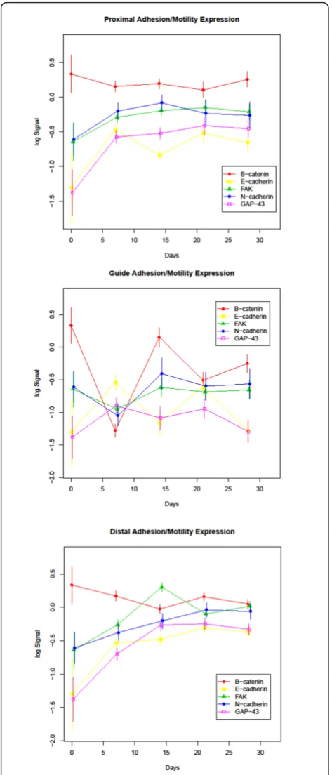
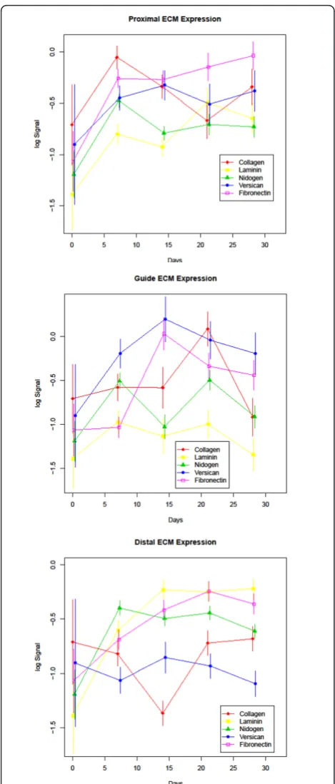
Protein expression profiles throughout the 28 day experimental period are displayed in Figures 3, 4, and 5. The antibody panel selected for this study is divided into three groups. The first group is adhesion and cell motility consisting of E- and N-cadherin, b-catenin, and focal adhesion kinase (FAK) (Figure 3). The growth fac-tors NGF, FGF-1, FGF-2, epidermal growth factor (EGF) and CNTF comprise the second group (Figure 4). ECM proteins laminin, fibronectin, collagen I/III, nidogen and versican make up the third group (Figure 5). While
Figure 1Antibody Validation: Validation of selected antibodies in western blot analysis showing detection of a single band for fibronectin (A), N-cadherin (B),b-catenin (C), E-cadherin (D), laminin
g-1 (E), nidogen (F), versican (G), FGF-2 (H), and CNTF (I).
B
B
Figure 2GAP-43 RPPA: (A) RPPA probed with GAP-43 antibody showing an increase in protein expression over a 28-day
Figure 3Adhesion and Motility Protein Expression: Expression profiles of the adhesion and motility group of study proteins divided into proximal, guide and distal segments. Time (in days) is plotted on the X-axis and log normalized data on the Y axis.
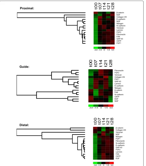
GAP-43 plays direct roles in different aspects of nerve regeneration with functional effects spanning more than one of these groups, it is grouped with the adhesion and motility proteins. A summary of expression of the com-plete panel of proteins under study is shown in Figure 6 presented in a heatmap format.
Adhesion/motility associated proteins
Members of the classic cadherin family, E- and N-cad-herin, displayed an overall increase in expression throughout the course of regeneration. In the proximal and distal segments of the regenerating nerve this increase was significant for E-cadherin when the expres-sion level from day 0 (intact nerve) was compared with day 28 (q-values of 0.022 and 0.002 respectively). The change in N-cadherin expression between day 0 and day 28 was significant in the distal portion only (q-value 0.036). Interestingly, the levels of N-cadherin in the proximal segment appeared to peak at day 14 and the expression level at 14 days was significantly higher than baseline (q-value 0.024). Both E-cadherin and N-cad-herin displayed continued elevated expression up to 21 days in the distal segment followed by a plateau in expression up to 28 days. There was no significant change in E- or N-cadherin levels in the guide contents when comparing the day 0 to the day 28 time point. The guide segment results compare intact nerve (day 0 expression levels) to the regenerate which, at least for the early time points, contains no nerve. Since the guide segment contains only regenerated tissue, the lack of significant difference from day 0 represents a return to the baseline protein levels seen in intact nerve. For N-cadherin this return to baseline occurs by day 14 while fluctuations in E-cadherin expression were recorded throughout the 28 day regenerative period with above baseline peaks at ≤ 7 and 21 days and troughs that approached baseline levels at 14 and 28 days. In con-trast, b-catenin, an integral member of the cadherin complex, showed a significant change from baseline only at day≤ 7 in the proximal (q-value 0.024) and tal (q-value 0.029) segments, but in the guide tissue dis-played peaks and troughs that mirrored N-cadherin, but were opposite to that recorded for E-cadherin in the same site.
FAK has been shown to promote neurite outgrowth and in this model displayed an increase in expression throughout the regenerative process in the proximal and distal segments with an initial, significant drop at day≤ 7 (q-value 0.002) and recovery to higher than baseline levels by day 14 in the guide portion. The overall
t0
0
t0
7
t1
4
t2
1
t2
8
Bct c EGF C13 Ecd NGF NID Ncd VER LG1 FG2 FBN FAK G43 CNT FG1
-3.0 -1.5 0 1.5 3.0
ȕ-catenin EGF Collagen I/III E-cadherin NGF Nidogen N-cadherin Versican Laminin FGF2 Fibronectin FAK GAP-43 CNTF FGF1
Proximal:
t0
0
t0
7
t1
4
t2
1
t2
8
FBN FG2 VER C13 FG1 G43 LG1 Ecd NID Bct FAK Ncd CNT EGF NGF
-3.0 -1.5 0 1.5 3.0
Fibronectin FGF2 Versican Collagen I/III FGF1 GAP-43 Laminin E-cadherin Nidogen
ȕ-catenin FAK N-cadherin CNTF EGF NGF
Guide:
t0
0
t0
7
t1
4
t2
1
t2
8
Bct c C13 VER NGF NID FG1 FBN Ncd Ecd G43 FG2 LG1 FAK CNT EGF
-3.0 -1.5 0 1.5 3.0
Distal:
ȕ-catenin Collagen I/III Versican NGF Nidogen FGF1 Fibronectin N-cadherin E-cadherin GAP-43 FGF2 Laminin FAK CNTF EGF
increase in FAK levels between day 0 and day 28 was significant in the distal segment only (q-value 0.016).
In the distal nerve stump GAP-43 levels increased rapidly up to 14 days followed by a relative plateau for the remainder of the experimental course. A sharp increase in GAP-43 was also seen in the proximal nerve up until 7 days with a slower, but marked increase through 28 days. In contrast, GAP-43 protein level showed no significant change from baseline throughout the course of the experiment in the guide contents indi-cating an early up-regulation of GAP-43 in the regener-ating tissue. The overall increase in GAP-43 between day 0 and day 28 was significant in the proximal and distal segments only (q-values 0.002 and 0.001 respectively).
Growth factors
Amongst the molecules included in the growth factor group, a significant increase in the expression level of FGF-1, FGF-2 and EGF was recorded between 0-14 days in the regenerating distal nerve followed by a plateau in expression between 14 and 28 days (the q-value for the day 0 to day 14 comparisons were 0.007, 0.004, and 0.009 respectively). In the proximal nerve segment, expression levels of each of the aforementioned growth factors con-tinued to rise past 14 days although none of the changes after day 14 reached significance. Comparing day 0 to day 28, both FGF-1 and FGF-2 demonstrated a significant increase in protein levels in the proximal nerve segment, but EGF did not (q-values for FGF-1 and FGF-2 were 0.006 and < 0.001 respectively). Interestingly, significantly reduced EGF expression was recorded in the guide tissue over the 28 day regeneration period (q-value < 0.001). In contrast, a comparison of day 0 to day 28 protein levels for FGF-1 and FGF-2 did not reveal a significant change in the guide contents indicating a return to the baseline levels of intact nerve and both proteins displayed a temporary increase in expression that peaked at 14 days and reached significance for FGF-2 (q-value 0.037).
CNTF displayed only a limited, insignificant increase in expression in the proximal and distal segments relative to the other growth factors in the study and, like EGF, sig-nificantly reduced expression in the guide tissue between day 0 and day 28 (q-value < 0.001). However, unlike EGF, CNTF did not decrease throughout the entire regenera-tive period, instead displaying an increase in expression at 21 days followed by a further decline at 28 days.
NGF displayed a biphasic expression profile in the regenerating distal nerve with significant peaks occur-ring at 14 and 28 days post-axotomy (q values < 0.001 and 0.006 respectively when comparing day 14 and day 28 to baseline). No significant change in NGF expres-sion was recorded in the proximal segment or guide compartment of the regenerating nerve between baseline
and day 28. In the guide compartment the level of NGF was not significantly different than baseline by day≤ 7 indicating an early recovery of NGF protein in this segment.
Extracellular matrix proteins
The ECM proteins have been shown to play a role in growth promotion and cell migration involving direc-tional guidance, both important processes in nerve regeneration. Fibronectin displayed a significant increase in protein levels over baseline at 21 and 28 days in the proximal segment (q-values 0.014 and 0.024 respec-tively), 21 days in the distal segment (q-value 0.036), and even at the earliest time point of≤7 days no signif-icant change from intact nerve in the guide segment. A significant increase from baseline levels was observed in the guide segment at day 14 and day 21 (q-values < 0.001 and 0.023 respectively). Laminin displayed a sig-nificant increase in both the proximal and distal seg-ments between 0 and 28 days (q values 0.009 and < 0.001 respectively), whereas the guide contents did not show a significant change when comparing the same time points indicating a return to the levels of intact nerve. Like fibronectin, day≤ 7 laminin expression was also not significantly different than baseline in the guide segment. Collagen I/III had a distinct temporal sequence of expression which peaked at days≤ 7 and 21 in the proximal and guide segments, respectively, and was sig-nificantly reduced compared to baseline in the distal segment (q-value 0.011) at 14 days followed by restora-tion to expression levels recorded in the intact nerve for the remainder of the experimental course. At day ≤ 7 the guide segment had recovered to baseline collagen I/ III expression levels. Nidogen and versican protein levels were elevated overall although the change was only sig-nificant in the distal segment (q-values for both compar-isons were 0.014). Nidogen, a potentiator of Schwann cell proliferation, showed biphasic expression with peaks at ≤7 and 21 days post-axotomy most notably in the guide segment indicating an early increase even higher than baseline expression. Versican protein levels were elevated early in the proximal and guide segments with a significant increase at 14 days (q-values 0.011 and ≤ 0.001 respectively) followed by a slow decline with levels increased, but not significantly different than baseline at 28 days. In the distal segment protein levels were signifi-cantly lower than baseline at day ≤7 and day 28 (q-values 0.018 and 0.014 respectively).
Discussion
that have been implicated as promotional in the regen-erative process. Knowledge of the expression pattern of proteins essential to regeneration have helped predict the best candidates for exogenous administration, how-ever this information must be pooled from individual studies with variable models in order to generate a glo-bal expression profile. In this study we used RPPA to facilitate the simultaneous temporal and spatial expres-sion mapping of key proteins in the nerve regeneration process using a rat sciatic nerve transection injury model. The benefit of RPPA analysis is that many sam-ples can be analyzed simultaneously. Since this techni-que requires very little starting material, it also enables the analysis of a large number of proteins. The limited number of proteins reported in this study was not due to a limitation in source material, but instead the limited availability of antibodies that cross-react with rat pro-teins and were suitable for use in RPPA. In this study every antibody was first validated by western blot and any antibody that did not produce a single predominant band by western was excluded. Although an analysis of 15 proteins does not represent the full capability of the RPPA technique, we believe that this paper provides sig-nificant insight into the regulation of these proteins and sets the stage for further proteomic evaluation using this model.
The phases of regeneration in a permeable, non-resorbable nerve guide chamber have been described and include a fluid, matrix, cellular, and axonal phase [62]. Within a day after axotomy and guide insertion the guide fills with fluid that contains neuronotrophic factors. A mainly acellular matrix forms within a week followed by the immigration of cells (including Schwann, fibroblast, and endothelial) from both the proximal and distal nerve stumps after 7-14 days. Finally, after 2 weeks, axonal elongation begins with myelination occurring a few days later [62]. According to this description, the first time point used in the current study (≤7 days) would capture the regenerative process at the matrix phase with the sec-ond time point (14 days) representing the cellular phase. The final time points of 21 and 28 days represent the axonal phase with the later time point likely to have a higher percentage of myelinated axons.
Protein expression analysis in the guide content com-pares intact nerve (day 0 expression levels) to the regen-erate which initially contains no nerve. Based on the described regenerative process, an early increase (mani-fested as a return to baseline levels) in the proteins included in the ECM category would be expected in the guide portion of the regenerate. Indeed, all of the ECM proteins studied demonstrate not only a return to base-line, but an increase in expression at ≤ 7 days over intact nerve. In contrast, several of the cellular proteins such asb-catenin, FAK, and N-cadherin show an initial
decline from the protein level of intact nerve in the guide segment and peak later at day 14 corresponding with the cellular phase of regeneration. This initial decline from baseline is to be expected since the guide segment does not contain intact nerve at the earlier time points. Interestingly, the growth factors represent a mixed group in the guide portion with an early return to baseline or increase over intact nerve for NGF, FGF-1 and FGF-2, and overall decline in the levels of CNTF and EGF in the solid guide contents. Overall, we saw a decrease in the expression of all growth factors in the guide contents from day 21 to day 28, the time at which axonal migration and myelination is underway. This is consistent with a previous study that demonstrated that the neuronotrophic activity of the fluid that fills the guide chamber is highest in the first few days of regen-eration suggesting that the role of at least some of these molecules peaks early in the regenerative process [63]. The general patterns of protein expression seen by RPPA analysis in this study are consistent with estab-lished knowledge and warrant more thorough analysis.
GAP-43 is present in the presynaptic terminal of novel neuromuscular junctions [64], and has been shown to be expressed in regenerating axons following nerve injury [65-68]. GAP-43 reportedly plays a promotional role in nerve sprouting post axotomy [69]. In our model we found an overall increase in GAP-43 levels throughout the experiment. Several groups have reported an early up-regulation of GAP-43 protein or mRNA following nerve injury [68,70,71]. We observed a pattern of early, increased expression in both the proximal and distal nerve segments that appeared to plateau by 14 days. At its peak, the distal nerve segment has a higher expres-sion level of GAP-43 than the proximal nerve and the guide contents show no significant change in GAP-43 compared to intact nerve even at the earliest time point. These results differ slightly than those of Plantinga et al. who reported no increase in proximal GAP-43 mRNA levels, but this difference might be explained by differ-ences in the experimental models, most notably that the nerve ends in the former study were coagulated and ligated while ours were allowed to regenerate. The dis-crepancy could also be explained by poor correlation between mRNA and protein levels [68].
E-cadherin in all segments at the day≤ 7 time point. In the distal and proximal segments, day 28 levels remain significantly higher than baseline while in the guide they return to the baseline level of intact nerve.
As cadherin complexes assemble, there is a distinct hierarchy of catenin association led by E-cadherin/b -catenin binding followed by additional -catenin members. Interestingly,b-catenin that binds to E-cadherin within the cadherin complex, revealed no significant change in expression profile throughout the regenerative process in the proximal and distal segments in our study. b -catenin levels in the guide decreased significantly at≤ 7 days followed by a rebound to baseline levels and a sig-nificant but lower decline from baseline at 21 and 28 days. Our observation is in contrast to a report by Hatoko et al. that showed increased b-catenin expres-sion in the graft portion of a nerve graft model of regen-eration, however this difference might be explained by the fact that the previous study was evaluating expres-sion in intact nerve grafts where our study included just the regenerated contents within a hollow guide [75].
N-cadherin has been shown to be present on axonal growth conesin vitro[76] and is thought to play a role in the stabilization of myelin sheaths based on its distri-bution and low expression levels in normal nerves [77,78]. Several groups have reported increased N-cad-herin levels at 15 days post-axotomy [79,80]. We con-firmed an increase in N-cadherin expression during regeneration that reached statistical significance at 14 days in the proximal segment and 21 days in the distal segment, but did not observe significantly higher expres-sion in the distal segment than the proximal segment as reported by Thorton and colleagues [80]. This discre-pancy may be explained by differences in the models used where in the Thorton study the nerve ends were capped following axotomy and not allowed to regener-ate. We also showed that in the distal stump, N-cad-herin expression peaks at 21 days which is later than the 14 day peak in the proximal segment and suggests a temporal sequence of events. The guide contents show an initial (day≤ 7) decrease in N-cadherin expression followed by a higher than baseline peak at 14 days and slight decline similar to the pattern observed in the proximal segment. Since N-cadherin has been shown to be expressed around regenerated axons [79,81], the drop in N-cadherin expression in the guide contents at day ≤ 7 is likely due to the absence of axon or axon-Schwann cell interactions at this location at such an early stage in the regenerative process.
FAK has been identified as a regulator of Schwann cell proliferation in the developing nervous system [82] and inhibition of FAK activity inhibited neurite growth in an
in vitromodel [83]. In the current study, FAK expres-sion was increased in both the distal and proximal nerve
segments although only the increase in the distal por-tion reached statistical significance. In the guide there was a drop in FAK levels at day ≤ 7, likely due to the absence of intact nerve and corresponding reduction in Schwann cells at this time point, followed by a return to baseline for the remainder of the experiment. Activation of FAK leads to autophosphorylation at Tyr397 and an analysis of activated FAK levels will be needed to further elucidate the role of FAK in regeneration.
Numerous growth factors have been implicated in per-ipheral nerve regeneration with different members of the FGF family playing a prominent role in this process [reviewed in [84] and [85]]. Several groups have reported enhancement of nerve regeneration using FGF-1 and -2 [33,34,86-89] and FGF-2 levels have shown to be up-regu-lated following peripheral nerve injury at both the mRNA and protein levels [[90-92] and others]. Surprisingly, a decrease in FGF-1 protein levels or mitogenic activity after crush or transection has also been reported [93,94]. We observed an increase in FGF-1 and FGF-2 levels in both the proximal and distal ends of the regenerating nerve. Interestingly, in our study, FGF-1, FGF-2, and NGF were the only growth factors to show an early increase in the guide contents suggesting that these factors play a role in the initial phase of the regenerative process.
While EGFR mRNA and protein levels increase fol-lowing nerve injury [95], and an EGF homolog has been shown to promote axonal regeneration in CNS neurons
in vitro [96], Dubuisson et al. found that exogenous EGF did not enhance peripheral nerve regeneration in their model [97]. We found that EGF protein levels were increased significantly in the distal segment. The proximal segment showed an insignificant increase and there was an overall decrease in EGF in the solid con-tents of the guide. This differential in EGF expression could imply a role for EGF in the maturation of periph-eral nerve tissue rather than in the more active state of regeneration.
CNTF increases neuronal survival, neurite outgrowth, and axonal regeneration with exogenous administration [41,42,100,101], but Ito and colleagues reported low CNTF mRNA levels post-axotomy and a similar obser-vation has been made in a crush model [102,103]. In our study CNTF displayed only a limited, insignificant increase in expression in the proximal and distal seg-ments and an overall significant decrease in protein levels within the solid guide contents consistent with these later reports.
ECM components play an essential role in nervous system development and repair. Schwann cells assemble a fibrillar network that consists of fibronectin, laminin, and collagen type IV which is thought to play a role in proliferation [104]. The different ECM proteins likely exert their influence on different facets of nerve regen-eration influencing multiple cellular processes. Fibronec-tin has previously been shown to be a potent chemoattractant for Schwann cell migrationin vitroand
in vivo [13,14,105,106] and fibronectin protein and mRNA levels were found to be elevated in several mod-els of nerve injury [107-109]. We found an increase in fibronectin levels that reached significance at day 21 proximally and distally. Within the guide there was an early return to baseline levels by day≤7 and significant increase over baseline by day 14. Fibronectin levels increased more rapidly in the proximal segment than the distal segment and were significantly higher at ≤ 7 days in the proximal segment consistent with the find-ings of Lefcort et al. who demonstrated highest fibro-nectin expression in the vicinity of the injury at the same time point [107].
Laminins are key components of the basal lamina and laminin has been shown to play a prominent promo-tional role in nerve regenerationin vivo by regulating axonal growth [110,111]. The laminin gamma-1 (B2) chain is one of the most abundant of the 12 known chains that compose the laminin heterotrimer and is present in laminin isoforms 1-4 and 6-11 [112]. There is conflicting evidence regarding gamma-1 mRNA expres-sion with Wallquist and colleagues showing an up-regu-lation after sciatic transection (most notably in the proximal stump) and Doyu et al. reporting a decrease when normalized to total RNA levels [113,114]. In our experiment, gamma-1 protein expression increased sig-nificantly between day 0 and day 28 both proximally and distally, and there was an early return to baseline gamma-1 levels in the guide itself. Our results are most consistent with those of Wallquist [113]. In both our model and the Wallquist model the nerve ends were allowed to regenerate, while in the Doyu studies they were not [113,114]. This difference in experimental design may explain the conflicting results.
Collagen is the major component of the ECM and several groups have shown an increase in collagen I and III mRNA after nerve injury as well as functional
improvement with exogenous administration
[108,115-118]. In the current study, we used an antibody that detected both collagens I and III and found no sig-nificant change in levels between day 0 and day 28 in the proximal and distal segments. In the guide there was an early return to baseline protein levels by day≤ 7 followed by a significantly higher than baseline expres-sion level at day 21 and a significantly lower level than baseline level by day 28 suggesting that, at least at the protein level, the role of collagens I/III is predominantly in the regenerating portion of the nerve.
Nidogen is a component of the basement membrane. Nidogen protein and mRNA levels have been shown to be up-regulated post transection injury [15] and nidogen appears to be required for neurite outgrowth after axot-omy [119]. Our results support these findings as we found an increase in nidogen levels early in the regen-erative process with a significant increase by≤7 days in all segments. The guide contents displayed a biphasic increase in the guide with significant peaks at≤ 7 and 21 days and baseline levels at the 14 and 28 days. Clearly nidogen plays a role in many aspects of the regenerative process.
The chondroitin sulphate proteoglycan, versican, iso-form V2 has been shown to be an inhibitor of axonal growth while V1 has been shown to promote neurite outgrowth [120-122]. We looked at the versican V0/V1 isoforms and found a significant increase in the proxi-mal and guide segments that peaked at 14 days followed by a slight decline to the end of the study period consis-tent with these reports. Interestingly, versican levels remained low throughout the experiment in the distal segment suggesting that versican plays more of a role in the proximal region of the regenerating nerve during the timeframe we analyzed.
Analysis of expression levels in the guide content is particularly interesting as this compares intact nerve (day 0 expression levels) to the regenerate which, at least for the early time points, contains no nerve. It is not surprising that a greater number of proteins show an initial decline in this segment, but interestingly many are up-regulated even at the earliest time point. In the guide segment groupings more closely follow functional roles. For example, all of the ECM proteins are repre-sented in a single branch (early up-regulated expression) and the majority of the adhesion and motility proteins in the other (early down-regulated expression). The pat-tern of expression correlates to the temporal role of these proteins groups in the regenerative process where ECM proteins play an early role in the matrix phase and adhesion and motility proteins a later role in the cellular phase of regeneration. GAP-43 is clustered in the up-regulated branch and grouped closely with laminin. A functional relationship between GAP-43 and laminin has been implicated in several other studies and this expression pattern may reflect the complex role of GAP-43 in the regenerative process [124,125].
Conclusions
The expression of multiple cellular and extracellular molecules is finely orchestrated and integrated leading to the regeneration of a new nerve. Past work using this or similar models of peripheral nerve regeneration have taken a reductionist approach evaluating the contribu-tion of individual or small numbers of proteins in the regenerative process. The RPPA approach taken in this study allows expression profiling of multiple proteins from a large number of samples simultaneously. In this study we focused on proteins known to be involved in the regenerative process and with known expression profiles. The results both validated previous observations and provided new detail and complexity, but the appli-cation can be easily expanded to include any protein with a suitable, validated antibody including posttransla-tional modifications such as phosphorylation. In this study we have demonstrated that RPPA is a reliable, high-throughput approach for the analysis of overall protein expression profiles with the potential to identify the temporal sequence of events underlying complex, multifaceted, biological processes.
Methods
Surgical procedure
All animal experiments were performed with the approval of the Institutional Animal Care and Use Com-mittee at Lahey Clinic. Forty male Sprague Dawley rats weighing between 225-250 g served as the model for nerve guide repair. All animals were anesthetized via an intraperitoneal injection of 45 mg kg-1 sodium
pentobarbital. All underwent an initial surgery during which the right sciatic nerve was exposed via a three centimeter incision made just posterior to the femoral head. The dissection was carried down through the glu-teal muscle at which point the sciatic nerve was identi-fied. The remainder of the dissection was performed under an operating microscope at 30 × magnification. This same approach was carried out during a second harvesting procedure.
A length of at least 20 mm of nerve was exposed. A 10 mm section was removed via sharp transection with straight micro-scissors leaving at least a 5 mm proximal and distal sciatic nerve stump. A 14 mm long polyethy-lene nerve guide (1.67 mm ID, Becton Dickinson) was sutured in place with 10-0 nylon suture, using a hori-zontal mattress stitch. The suture was placed 2 mm from the end of the guide and through the epineurium on each respective nerve stump. The full circumference of each stump was therefore drawn entirely inside the guide with 2 mm of length lying inside each end of the guide, resulting in the proximal and distal stumps posi-tioned 10 mm apart. The 10 mm resected section of nerve was divided into two 5 mm sections (5 mm to act as a per-animal control, and 5 mm to be used for anti-body validation) and the two sections were placed in individual tubes, and frozen immediately in liquid nitro-gen. The gluteal muscle was then closed with three bur-ied interrupted 3-0 chromic sutures and the skin was closed with a running 4-0 braided polyglycolic acid suture.
Antibody validation
Nerve lysate for antibody validation was prepared by crushing a pooled sample of the excised normal 5 mm nerve segments under liquid nitrogen, using a mortar and pestle. The finely ground tissue was lysed in hot sample buffer (2 × ESB - 0.08 M Tris, pH 6.8; 0.07 M SDS, 10% glycerol, 0.001% bromophenol blue and 1 mM CaCl2) and sheared through a 26-gauge needle.b-mercaptoethanol (1%) was added to each sample which was boiled for 5 min. The resulting lysate was loaded across the top of a 7.5% polyacrylamide resolving gel, with one lane contain-ing a marker. Proteins were transferred overnight onto nitrocellulose. Membranes were blocked in 10% milk in TBS with 0.05% Tween-20, cut into strips and placed on primary antibody overnight at 4°C. Blots were washed in TBS with 0.05% Tween-20, three times for 15 min each, and secondary antibody linked to horseradish peroxidase was incubated with the blots for 60 min at room tempera-ture. Blots were then washed as described above and developed with an ECL kit (Amersham, Arlington Heights, IL). Only antibodies that produced a single band by wes-tern blot analysis were used for RPPA.
Antibodies
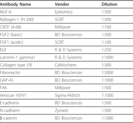
Validated antibodies used were: NGF (Epitomics, Burlin-game, CA); Nidogen and FGF1 (Santa Cruz Biotechnol-ogy, Santa Cruz, CA); CNTF and FAK (Millipore, Billerica, MA); FGF2, Fibronectin, GAP-43, E-cadherin andb-catenin (BD Biosciences, San Jose, CA) EGF and laminin-1 gamma-1 (R&D Systems, Minneapolis, MN); Collagen Type I/III (Calbiochem, San Diego, CA); Versi-can (Sigma-Aldrich, St. Louis, MO); and N-cadherin (Zymed, San Francisco, CA). The manufacturer of the fifteen antibodies as well as the concentrations used in RPPA and western blot are shown in Table 2.
Preparation of tissue lysates
Each nerve sample was crushed under liquid nitrogen with a clean mortar and pestle and suspended in 1.0 ml ice-cold PBS and centrifuged at 4000 rpm for 2 min.
After removing the PBS, a volume of lysis buffer, com-posed of 9 M Urea, 4% Chaps, 2% Pharmalyte pH 8.0-10.5 and 65 mM Dithiothreitol, equal to the size of the pellet, was added and aspirated up and down 3 times. Samples were centrifuged at 14,000 rpm for 30 min. The supernatant was collected and stored at -80°C and the pellet was discarded. All steps following the initial PBS wash were performed at 4°C. Ten, 2-fold serial dilu-tions were made from each lysate with a buffer contain-ing 6 M urea, 2.7% CHAPS, 1.3% Pharmalyte (pH 8.0-10.5), and 43.6 mM Dithiothreitol [126,127].
Reverse-phase protein arrays
Samples were arrayed using the Aushon 2470 solid pin, contact microarrayer equipped with 185 μm pins (Aushon BioSystems, Billerica, MA). The print matrix was a nitrocellulose-cast glass slide (Grace BioLabs, Bend, OR). Each slide contained 10, 2-fold serial Table 1 Experimental Overview
Number of Animals Included in Day 0 (Baseline) Measurements
Number of Rats Harvested at End Point
Number of Rats with Solid Guide Contents at End Point
Day≤
7
40 10 5
Day 14
10 2
Day 21
10 6
Day 28
10 4
At each time point proximal, distal and solid guide contents were harvested from each animal. Only tissue from animals with solid guide content was included in the analysis
Table 2 Antibody Name, Vendor, and Dilutions Used for Western Blotting and RPPA Applications
Antibody Name Vendor Dilution
NGF-b Epitomics 1:500
Nidogen-1 (H-200) SCBT 1:500
CNTF (4-68) Millipore 1:100
FGF2 (basic) BD Biosciences 1:500
FGF1 (acidic) SCBT 1:100
EGF R & D Systems 1:250
Laminin-1 gamma1 R & D Systems 1:1000
Collagen type I/III Calbiochem 1:300
Fibronectin BD Biosciences 1:5000
GAP-43 BD Biosciences 1:1000
FAK Millipore 1:500
Versican V0/V1 Sigma-Aldrich 1:1000
E-cadherin BD Biosciences 1:500
N-cadherin Zymed 1:500
dilutions of each sample and included a technical repli-cate totaling 3,360 lysate spots per slide. All printing was carried out at 80% relative humidity.
Developing
Printed nitrocellulose-coated slides were washed for two 15-min periods in deionized water. Slides were blocked for 1 h in I-Block (Life Technololgies-0.2% I-Block, 0.01% Tween-20 in phosphate buffered saline) with con-tinuous shaking at room temperature. Immunostaining of microarrays was performed on an automated stainer (Autostainer Plus, Dako, Carpinteria, CA) using a tyra-mide-based catalyzed signal amplification system according to the manufacturer’s protocol (K1500, Dako, Carpinteria, CA). A negative control was included using a non-specific antibody solution (Dako, Carpinteria, CA) substituting for the primary antibody.
Image processing & quantification
The slides were scanned at 2400 dpi resolution in an uncompressed, 16-bit TIFF image format using an opti-cal flatbed scanner. Images were processed by the P-SCAN and ProteinScan (version 0.21) programs written using Matlab software (MathWorks, Natick, MA) by the National Institutes of Health (NIH campus, Bethesda, MD). Outliers traceable to defects in the arrays were eliminated and the data were analyzed. The numerical output for each sample was generated by a modified Dose Interpolation algorithm, DI25[128]. ProteinScan is a program package that automates the modified DI algo-rithm. The DI25value of each nerve sample was normal-ized for total protein concentration determined by colloidal gold staining.
Statistical analysis
The protein intensity data was log-transformed to ensure data normality. To investigate the spatial and temporal differences in the protein expression levels, a linear mixed effects model was applied to each protein separately. The models contained fixed effects for differ-ent times and sample types and are adjusted for baseline protein expression levels. To account for correlated data structure random rat effect was used in the models. To determine which particular times and samples were sig-nificantly different, Scheffe’s test [129] was used. The significance results were presented as q-values, false dis-covery rate computed based on p-values to adjust for multiple testing [[130,131], see Additional file 1-q values]. All statistical analyses were performed using R version 2.9.1 [132] and R packages nlme [133] and q-value [134]. The clustering and heatmaps were produced using dChip software [135-137]. Heatmap data was row-standardized to have a mean of 0.
Additional material
Additional file 1: q-values; The statistical significance of expression data was determined and results are presented as q values, false
discovery rate adjusted p-values.
Abbreviations
ECM: Extracellular matrix; NGF: Nerve growth factor; FGF: Fibroblast growth factor; CNTF: Ciliary neurotrophic factor; RPPA: Reverse phase protein array; GAP-43: Growth associated protein 43; FAK: Focal adhesion kinase; EGF: Epidermal growth factor
Acknowledgements
This work was supported by a grant from the Leisa V. Clayton Foundation and the Robert E. Wise, MD Research and Education Foundation.
Author details
1Tissue Engineering Laboratory, Lahey Clinic Medical Center, Burlington,
Massachusetts, USA.2Ian C. Summerhayes Cell and Molecular Biology Laboratory, Lahey Clinic Medical Center, Burlington, Massachusetts, USA.
3
Institute for Clinical Research and Health Policy Studies, Tufts Medical Center, Boston, Massachusetts, USA.4Aushon BioSystems Inc., Billerica,
Massachusetts, USA.5Department Surgery, Section of General Surgery, Saint Joseph Mercy Hospital, Ann Arbor, Michigan, USA.6Department of Plastic
and Reconstructive Surgery, Lahey Clinic Medical Center, Burlington, Massachusetts, USA.
Authors’contributions
CRL and JVM performed animal surgeries. CRL performed antibody validation experiments, reverse phase microarray processing and assisted with manuscript preparation. DJB contributed to study design, data interpretation, and critical manuscript review. TL performed statistical analysis. AHH contributed to study design and data interpretation, performed reverse phase microarray printing and drafted the manuscript. JA-performed reverse phase microarray printing and critical review of the manuscript. ICS contributed to study design and data interpretation. KMR-C contributed to study design, data interpretation, and critical review of the manuscript. All authors read and approved the final manuscript.
Competing interests
AHH and JA were employees of Aushon BioSystems (manufacturer of the 2470 microarrayer) at the time that this study took place. AHH’s affiliation at the time of publication is with Lahey Clinic.
Received: 19 September 2011 Accepted: 10 February 2012 Published: 10 February 2012
References
1. Noble J, Munro CA, Prasad VS, Midha R:Analysis of upper and lower extremity peripheral nerve injuries in a population of patients with multiple injuries.J Trauma1998,45(1):116-122.
2. Terenghi G:Peripheral nerve regeneration and neurotrophic factors.J Anat1999,194(Pt 1):1-14.
3. Abe N, Cavalli V:Nerve injury signaling.Curr Opin Neurobiol2008,
18(3):276-283.
4. Lundborg G, Dahlin LB, Danielsen N, Gelberman RH, Longo FM, Powell HC, Varon S:Nerve regeneration in silicone chambers: influence of gap length and of distal stump components.Exp Neurol1982,76:361-375. 5. Danielsen N, Dahlin LB, Lee YF, Lundborg F:Axonal growth in mesothelial
chambers. The role of the distal nerve segment.Scand J Plast Reconstr Surg1983,17(2):119-125.
6. Fu SY, Gordon T:The cellular and molecular basis of peripheral nerve regeneration.Mol Neurobiol1997,14:(1-2):67-116.
7. Ide C:Peripheral nerve regeneration.Neurosci Res1996,25(2):101-121. 8. Parrinello S, Napoli I, Ribeiro S, Digby PW, Fedorova M, Parkinson DB,
9. Gallo G, Lefcort FB, Letourneau PC:The trkA receptor mediates growth cone turning toward a localized source of nerve growth factor.J Neurosci1997,17(14):5445-5454.
10. Dodla MC, Bellamkonda RV:Differences between the effect of anisotropic and isotropic laminin and nerve growth factor presenting scaffolds on nerve regeneration across long peripheral nerve gaps.Biomaterials2008,
29(1):33-46.
11. Brushart TM:Motor axons preferentially reinnervate motor pathways.J Neurosci1993,13(6):2730-2738.
12. Abernethy DA, Rud A, Thomas PK:Neurotropic influence of the distal stump of transected peripheral nerve on axonal regeneration: absence of topographic specificity in adult nerve.J Anat1992,180(Pt 3):395-400. 13. Bailey SB, Eichler ME, Villadiego A, Rich KM:The influence of fibronectin
and laminin during Schwann cell migration and peripheral nerve regeneration through silicon chambers.J Neurocytol1993,22(3):176-184. 14. Bryan DJ, Tang JB, Holway AH, Rieger-Christ KM, Trantolo DJ, Wise DL,
Summerhayes IC:Enhanced peripheral nerve regeneration elicited by cell-mediated events delivered via a bioresorbable PLGA guide.J Reconstr Microsurg2003,19(2):125-134.
15. Lee HK, Seo IA, Park HK, Park YM, Ahn KJ, Yoo YH, Park HT:Nidogen is a prosurvival and promigratory factor for adult Schwann cells.J Neurochem
2007,102(3):686-698.
16. Vanlair C:De la régénération des nerfs périphériques par la procédé de la suture tubulaire.Arch Biol1882,3:379-496.
17. Gu X, Ding F, Yang Y, Liu J:Construction of tissue engineered nerve grafts and their application in peripheral nerve regeneration.Progress Neurobio2010,93(2):204-230.
18. Siemionow M, Bozkurt M, Zor F:Regeneration and repair of peripheral nerves with different biomaterials: review.Microsurgery2010,
30(7):574-588.
19. Wang KK, Costas PD, Bryan DJ, Eby PL, Seckel BR:Inside-out vein graft repair compared with nerve grafting for nerve regeneration in rats.
Microsurgery1995,16(2):65-70.
20. Yang Y, Ding F, Wu J, Hu W, Liu W, Liu J, Gu X:Development and evaluation of silk fibrin-based nerve grafts used for peripheral nerve regeneration.Biomaterials2007,28(36):5526-5535.
21. Kim YT, Haftel VK, Kumar S, Bellamkonda RV:The role of aligned polymer fiber-based constructs in the bridging of long peripheral nerve gaps.
Biomaterials2008,29(21):3117-3127.
22. Oh SH, Kim JH, Song KS, Jeon BH, Yoon JH, Seo TB, Namgung U, Lee IW, Lee JH:Peripheral nerve regeneration within an asymmetrically porous PLGA/Pluronic F127 nerve guide conduit.Biomaterials2008,
29(11):1601-1609.
23. Bian YZ, Wang Y, Aibaidoula G, Chen GQ, Wu Q:Evaluation of poly(3-hydroxybutyrate-co-3-hydroxyhexanoate) conduits for peripheral nerve regeneration.Biomaterials2009,30(2):217-225.
24. Bini TB, Gao S, Xu X, Wang S, Ramakrishna S, Leong KW:Peripheral nerve regeneration by microbraided poly(L-lactide-co-glycolide) biodegradable polymer fibers.J Biomed Mater Res A2004,68(2):286-295.
25. Matsuyama T, Mackay M, Midha R:Peripheral nerve repair and grafting techniques: a review.Neurol Med Chir (Tokyo)2000,40(4):187-199. 26. Dvali L, Mackinnon S:Nerve repair, grafting, and nerve transfers.Clin Plast
Surg2003,30(2):203-221.
27. Nichols CM, Brenner MJ, Fox IK, Tung TH, Hunter DA, Rickman SR, Mackinnon SE:Effects of motor versus sensory nerve grafts on peripheral nerve regeneration.Exp Neurol2004,190(2):347-355.
28. Hoke A, Redett R, Hameed H, Jari R, Zhou C, Li ZB, Griffin JW, Brushart TM:
Schwann cells express motor and sensory phenotypes that regulate axon regeneration.J Neurosci2006,26(38):9646-9655.
29. Cohen S, Levi-Montalcini R, Hamburger V:A Nerve Growth-Stimulating Factor Isolated from Sarcomas 37 and 180.Proc Natl Acad Sci USA1954,
40(10):1014-1018.
30. Rich KM, Alexander TD, Pryor JC, Hollowell JP:Nerve growth factor enhances regeneration through silicone chambers.Exp Neurol1989,
105(2):162-170.
31. Boyd JG, Gordon T:Neurotrophic factors and their receptors in axonal regeneration and functional recovery after peripheral nerve injury.Mol Neurobiol2003,27(3):277-324.
32. Huang EJ, Reichardt LF:Neurotrophins: roles in neuronal development and function.Annu Rev Neurosci2001,24:677-736.
33. Cordeiro PG, Seckel BR, Lipton SA, D’Amore PA, Wagner J, Madison R:
Acidic fibroblast growth factor enhances peripheral nerve regeneration in vivo.Plast Reconstr Surg1989,83(6):1013-1021.
34. Walter MA, Kurouglu R, Caulfield JB, Vasconez LO, Thompson JA:Enhanced peripheral nerve regeneration by acidic fibroblast growth factor.
Lymphokine Cytokine Res1993,12(3):135-141.
35. Midha R, Munro CA, Dalton PD, Tator CH, Shoichet MS:Growth factor enhancement of peripheral nerve regeneration through a novel synthetic hydrogel tube.J Neurosurg2003,99(3):555-565.
36. Ohta M, Suzuki Y, Chou H, Ishikawa N, Suzuki S, Tanihara M, Mizushima Y, Dezawa M, Ide C:Novel heparin/alginate gel combined with basic fibroblast growth factor promotes nerve regeneration in rat sciatic nerve.J Biomed Mater Res A2004,71(4):661-668.
37. Wang S, Cai Q, Hou J, Bei J, Zhang T, Yang J, Wan Y:Acceleration effect of basic fibroblast growth factor on the regeneration of peripheral nerve through a 15-mm gap.J Biomed Mater Res A2003,66(3):522-531. 38. Bryan DJ, Holway AH, Wang KK, Silva AE, Trantolo DJ, Wise D,
Summerhayes IC:Influence of glial growth factor and Schwann cells in a bioresorbable guidance channel on peripheral nerve regeneration.Tissue Eng2000,6(2):129-138.
39. Wells MR, Kraus K, Batter DK, Blunt DG, Weremowitz J, Lynch SE, Antoniades HN, Hansson HA:Gel matrix vehicles for growth factor application in nerve gap injuries repaired with tubes: a comparison of biomatrix, collagen and methylcellulose.Exp Neurol1997,146(2):395-402. 40. Sendtner M, Stöckli KA, Thoenen H:Synthesis and localization of ciliary
neurotrophic factor in the sciatic nerve of the adult rat after lesion and during regeneration.J Cell Biol1992,118(1):139-148.
41. Sahenk Z, Seharaseyon J, Mendell JR:CNTF potentiates peripheral nerve regeneration.Brain Res1994,655:(1-2):246-250.
42. Newman JP, Verity AN, Hawatmeh S, Fee WE Jr, Terris DJ:Ciliary neurotrophic factors enhances peripheral nerve regeneration.Arch Otolaryngol Head Neck Surg1996,122(4):399-403.
43. Lewin SL, Utley DS, Cheng ET, Verity AN, Terris DJ:Simultaneous treatment with BDNF and CNTF after peripheral nerve transection and repair enhances rate of functional recovery compared with BDNF treatment alone.Laryngoscope1997,107(7):992-999.
44. Archibald SJ, Krarup C, Shefner J, Li ST, Madison RD:A collagen-based nerve guide conduit for peripheral nerve repair: an electrophysiological study of nerve regeneration in rodents and nonhuman primates.J Comp Neurol1991,306(4):685-696.
45. Li ST, Archibald SJ, Krarup C, Madison RD:Peripheral nerve repair with collagen conduits.Clin Mater1992,9:(3-4):195-200.
46. Kauppila T, Jyväsjärvi E, Huopaniemi T, Hujanen E, Liesi P:A laminin graft replaces neurorrhaphy in the restorative surgery of the rat sciatic nerve.
Exp Neurol1993,123(2):181-191.
47. Whitworth IH, Brown RA, Doré C, Green CJ, Terenghi G:Oriented mats of fibronectin as a conduit material for use in peripheral nerve repair.J Hand Surg Br1995,20(4):429-436.
48. Bryan DJ, Miller RA, Costas PD, Wang KK, Seckel BR:Immunocytochemistry of skeletal muscle basal lamina grafts in nerve regeneration.Plast Reconstr Surg1993,92(5):927-940.
49. Yoshii S, Oka M, Shima M, Taniguchi A, Akagi M:30 mm regeneration of rat sciatic nerve along collagen filaments.Brain Res2002,949 :(1-2):202-208.
50. Perlson E, Medzihradszky KF, Darula Z, Munno DW, Syed NI, Burlingame AL, Fainzilber M:Differential proteomics reveals multiple components in retrogradely transported axoplasm after nerve injury.Mol Cell Proteomics
2004,3(5):510-520.
51. Jiménez CR, Stam FJ, Li KW, Gouwenberg Y, Hornshaw MP, De Winter F, Verhaagen J, Smit AB:Proteomics of the injured rat sciatic nerve reveals protein expression dynamics during regeneration.Mol Cell Proteomics
2005,4(2):120-132.
52. Melle C, Ernst G, Grosheva M, Angelov DN, Irintchev A, Guntinas-Lichius O, von Eggeling F:Proteomic analysis of microdissected facial nuclei of the rate following facial nerve injury.J Neurosci Methods2009,185(1):23-28. 53. Michaelevski I, Segal-Ruder Y, Rozenbaum M, Medzihradszky KF, Shalem O,
Coppola G, Horn-Saban S, Ben-Yaakov K, Dagan SY, Rishal I, Geschwind DH, Pilpel Y, Burlingame AL, Fainzilber M:Signaling to transcription networks in the neuronal retrograde injury response.Sci signal2010,3(130):ra53. 54. Paweletz CP, Charboneau L, Bichsel VE, Simone NL, Chen T, Gillespie JW,
microarrays which capture disease progression show activation of pro-survival pathways at the cancer invasion front.Oncogene2001,
20(16):1981-1989.
55. Nishizuka S, Charboneau L, Young L, Major S, Reinhold WC, Waltham M, Kouros-Mehr H, Bussey KJ, Lee JK, Espina V, Munson PJ, Petricoin E, Liotta LA, Weinstein JN:Proteomic profiling of the NCI-60 cancer cell lines using new high-density reverse-phase lysate microarrays.Proc Natl Acad Sci USA2003,100(24):14229-14234.
56. Espina V, Mehta AI, Winters ME, Calvert V, Wulfkuhle J, Petricoin EF, Liotta L:
Protein microarrays: molecular profiling technologies for clinical specimens.Proteomics2003,3(11):2091-2100.
57. Grubb RL, Calvert VS, Wulfkuhle JD, Paweletz CP, Linehan WM, Phillips JL, Chuaqui R, Valasco A, Gillespie J, Emmert-Buck M, Liotta LA, Petricoin EF:
Signal pathway profiling of prostate cancer using reverse phase protein arrays.Proteomics2003,3(11):2142-2146.
58. Wulfkuhle JD, Aquino JA, Calvert VS, Fishman DA, Coukos G, Liotta LA, Petricoin EF:Signal pathway profiling of ovarian cancer from human tissue specimens using reverse-phase microarrays.Proteomics2003,
3(11):2085-2090.
59. Hermann PC, Gillespie JW, Charboneau L, Bichsel VE, Paweletz CP, Calvert VS, Kohn EC, Emmert-Buck MR, Liotta LA, Petricoin EF:
Mitochondrial proteome: Altered Cytochrome oxidase subunit levels in prostate.Proteomics2003,3(9):1801-1810.
60. Liotta LA, Espina V, Mehta AI, Calvert V, Rosenblatt K, Geho D, Munson PJ, Young L, Wulfkuhle J, Petricoin EF:Protein microarrays: meeting analytical challenges for clinical applications.Cancer Cell2003,3(4):317-325. 61. Wang K-K, Nemeth IR, Seckel BR, Chakalis-Haley DP, Swann DA, Kuo JW,
Bryan DJ, Cetrulo CL Jr:Hyaluronic acid enhances peripheral nerve regeneration in vivo.Microsurgery1998,18:270-275.
62. Williams LR, Longo FM, Powell HC, Lundborg G, Varon S:Spatial-temporal progress of peripheral nerve regeneration with a silicone chamber: parameters for a bioassay.J Comp Neurol1983,218:460-470. 63. Longo FM, Skaper SD, Manthorpe M, Willimas LR, Lundborg G, Varon S:
Temporal changes of neuronotrophic activities accumulatingin vivo within nerve regeneration chambers.Exp Neurol1983,81:756-769. 64. Verhaagen J, Oesteicher AB, Edwards PM, Veldman H, Jennekens FG,
Gispen WH:Light and electron-microscopical study of phosphoprotein B-50 following denervation and reinnervation of the rat soleus muscle.J Neurosci1988,8(5):1759-1766.
65. Verhaagen J, van Hooff CO, Edwards PM, De Graan PN, Oestreicher AB, Schotman P, Jennekens FG, Gispen WH:The kinase C substrate protein B-50 and axonal regeneration.Brain Res Bull1986,17(6):737-741.
66. Verkade P, Oestreicher AB, Verkleig AJ, Gispen WH:The increase in B-50/ GAP-43 in regenerating rat sciatic nerve occurs predominantly in unmyelinated axon shafts: a quantitative ultrastructural study.J Comp Neurol1995,356(3):433-443.
67. Verkade P, Schrama LH, Verkleij AJ, Gispen WH, Oestreicher AB:
Ultrastructural co-localization of calmodulin and B-50/growth-associated protein-43 at the plasma membrane of proximal unmyelinated axon shafts studied in the model of the regenerating rat sciatic nerve.
Neuroscience1997,79(4):1207-1218.
68. Plantinga LC, Verhaagen J, Edwards PM, Hol EM, Bar PR, Gispen WH:The expression of B-50/GAP-43 in Schwann cells is up-regulated in degenerating peripheral nerve stumps following nerve injury.Brain Res
1993,602(1):69-76.
69. Buffo A, Holtmaat AJ, Savio T, Verbeek JS, Oberdick J, Oestreicher AB, Gispen WH, Verhaagen J, Rossi F, Strata P:Targeted overexpression of the neurite growth-associated protein B-50/GAP-43 in cerebellar Purkinje cells induces sprouting after axotomy but not axon regeneration into growth permissive transplants.J Neurosci1997,17(22):8778-8791. 70. Van der Zee CEEM, Nielander HB, Vos JP, da Silva SL, Verhaagen J, Oestreicher AB, Schrama LH, Schotman P, Gispen WH:Expression of growth-associated protein B-50 (GAP-43) in dorsal root ganglia and sciatic nerve during regenerative sprouting.J Neurosci1989,
9(10):3505-3512.
71. Tetzlaff W, Zwiers H, Lederis K, Cassar L, Bisby MA:Axonal transport and localization of B-50/GAP-43-like immunoreactivity in regenerating sciatic and facial nerves of the rat.J Neurosci1989,9(4):1303-1313.
72. Tricaud N, Perrin-Tricaud C, Brusés JL, Rutishauser U:Adherens junctions in myelinating Schwann cells stabilize Schmidt-Lanterman incisures via
recruitment of p120 catenin to E-cadherin.J Neurosci2005,
25(13):3259-3269.
73. Hasegawa M, Seto A, Uchiyama N, Kida S, Yamashima T, Yamashita J:
Localization of E-cadherin in peripheral glia after nerve injury and repair.
J Neuropathol Exp Neurol1996,55(4):424-434.
74. Tada H, Hatoko M, Tanaka A, Kuwahara M, Mashiba K, Yurugi S:The difference in E-cadherin expression between nonvascularized and vascularized nerve grafts: study in the rat sciatic nerve model.J Surg Res
2001,100(1):57-62.
75. Hatoko M, Tanaka A, Kuwahara M, Yurugi S, Iioka H, Niitsuma K:Expression of alpha, beta, and gamma catenins in vascularized and nonvascularized nerve grafts during the regeneration process.J Reconstr Microsurg2003,
19(4):271-278.
76. Bixby JL, Lilien J, Reichardt LF:Identification of the major proteins that promote neuronal process outgrowth on Schwann cells in vitro.J Cell Biol1988,107(1):353-361.
77. Matsunaga M, Hatta K, Nagafuchi A, Takeichi M:Guidance of optic nerve fibers by N-cadherin adhesion molecules.Nature1988,334(6177):62-64. 78. Cifuentes-Diaz C, Nicolet M, Gondou D, Rieger F, Mege RM:N-cadherin
expression in developing adult and denervated chicken neuromansular system: accumulations at both the neuromuscular junction and the node of Ranvier.Development1994,120(1):1-11.
79. Hatoko M, Tada H, Tanaka A, Kuwahara M, Yurugi S:The differential expression of N-cadherin in vascularized and nonvascularized nerve grafts: a study in a rat sciatic nerve model.Ann Plast Surg2001,
47(3):322-327.
80. Thornton MR, Mantovani C, Birchall MA, Terenghi G:Quantification of N-CAM and N-cadherin expression in axotomized and crushed rat sciatic nerve.J Anat2005,206(1):69-78.
81. Shibuya Y, Mizoguchi A, Takeichi M, Shimada K, Ide C:Localization of N-cadherin in the normal and regenerating nerve fibers of the chicken peripheral nervous system.Neuroscience1995,67(1):253-261.
82. Grove M, Komiyama NH, Nave KA, Grant SG, Sherman DL, Brophy PJ:FAK is required for axonal sorting by Schwann cells.J Cell Biol2007,
176(3):277-282.
83. Tucker BA, Rahimtula M, Mearow KM:Src and FAK are key early signaling intermediates required for neurite growth in NGF-responsive adult DRG neurons.Cell Signal2008,20(1):241-257.
84. Grothe C, Wewetzer K:Fibroblast growth factor and its implications for developing and regenerating neurons.Int J Dev Biol1996,40(1):403-410. 85. Ebadi M, Bashir RM, Heidrick ML, Hamada FM, Refaey HE, Hamed A, Helal G,
Baxi MD, Cerutis DR, Lassi NK:Neurotrophins and their receptors in nerve injury and repair.Neurochem Int1997,30:(4-5):347-374.
86. Danielsen N, Pettmann B, Vahlsing HL, Manthorpe M, Varon S:Fibroblast growth factor effects on peripheral nerve regeneration in a silicone chamber model.J Neurosci Res1998,20(3):320-330.
87. Laird JM, Mason GS, Thomas KA, Hargreaves RJ, Hill RG:Acidic fibroblast growth factor stimulates motor and sensory axon regeneration after sciatic nerve crush in the rat.Neuroscience1995,65(1):209-216. 88. Trigg DJ, O’Grady KM, Bhattacharyya T, Reinke M, Toriumi DM:Peripheral
nerve regeneration: comparison of laminin and acidic fibroblast growth factor.Am J Otolaryngol1998,19(1):29-32.
89. Jungnickel J, Haase K, Konitzer J, Timmer M, Grothe C:Faster nerve regeneration after sciatic nerve injury in mice over-expressing basic fibroblast growth factor.J Neurobiol2006,66(9):940-948.
90. Meisinger C, Grothe C:Differential regulation of fibroblast growth factor (FGF)-2 and FGF receptor 1 mRNAs and FGF-2 isoforms in spinal ganglia and sciatic nerve after peripheral nerve lesion.J Neurochem1997,
68(3):1150-1158.
91. Grothe C, Meisinger C, Claus P:In vivo expression and localization of the fibroblast growth factor system in the intact and lesioned rat peripheral nerve and spinal ganglia.J Comp Neurol2001,434(3):342-357.
92. Grothe C, Meisinger C, Hertenstein A, Kurz H, Wewetzer K:Expression of fibroblast growth factor-2 and fibroblast growth factor receptor 1 messenger RNAs in spinal ganglia and sciatic nerve: regulation after peripheral nerve lesion.Neuroscience1997,76(1):123-135.
93. Eckenstein FP, Shipley GD, Nishi R:Acidic and basic fibroblast growth factors in the nervous system: Distribution and differential alteration of levels after injury of central versus peripheral nerve.J Neurosci1991,