Laser capture microdissection as a tool
to evaluate human papillomavirus
genotyping and methylation as
biomarkers of persistence and
progression of anal lesions
Alyssa M Cornall,1,2Jennifer M Roberts,3Monica Molano,1Dorothy A Machalek,1,2 Samuel Phillips,1,2Richard J Hillman,4Andrew E Grulich,5Fengyi Jin,5
I Mary Poynten,5David J Templeton,5,6Suzanne M Garland,1,7Sepehr N Tabrizi,1,7 on behalf of the SPANC Study Team
To cite:Cornall AM, Roberts JM, Molano M,et al. Laser capture
microdissection as a tool to evaluate human
papillomavirus genotyping and methylation as biomarkers of persistence and progression of anal lesions.BMJ Open2015;5: e008439. doi:10.1136/ bmjopen-2015-008439
▸ Prepublication history for this paper is available online. To view these files please visit the journal online (http://dx.doi.org/10.1136/ bmjopen-2015-008439).
SMG and SNT are joint last authors.
Received 8 April 2015 Revised 18 June 2015 Accepted 19 July 2015
For numbered affiliations see end of article.
Correspondence to Dr Alyssa M Cornall; [email protected]
ABSTRACT
Introduction:Anal squamous cell carcinoma is
preceded by persistent infection with high-risk human papillomavirus (HPV) and the cancer precursor, high-grade squamous intraepithelial lesion (HSIL). Detection of specific HPV genotypes and HPV-related biomarkers may be an option for primary anal screening. However, more data on the natural history of HPV-related anal lesions are required. The outcomes from this study will enhance our understanding of the clinical and biological behaviour of HPV-related anal lesions and inform the development of future HPV genotype and/or biomarker screening tests.
Methods and analysis:negative and
HIV-positive men who have sex with men, aged 35 years and over, recruited from community-based settings in Sydney, Australia, attend 6 clinic visits over 3 years. At the first 5 visits, participants undergo a digital anorectal examination, an anal swab for HPV genotyping and anal cytology, and high-resolution anoscopy with directed biopsy of any visible abnormalities that are suggestive of any abnormality suspicious of SIL. Tissue sections from participants diagnosed with histologically confirmed HSIL at the baseline clinic visit will undergo laser capture microdissection, HPV detection and genotyping, and quantitation of CpG methylation in baseline and follow-up biopsies. Histological and cytological findings in combination with HPV genotyping data will be used to identify persistent HSIL. HSIL will be stratified as non-persistent and non-persistent based on their status at 12 months. The performance of HPV genotype and methylation status in predicting disease persistence at 12 months will be assessed, along with associations with HIV status and other covariates such as age.
Ethics and dissemination:The St Vincent’s Hospital
Ethics Committee granted ethics approval for the study. Written informed consent is obtained from all
individuals before any study-specific procedures are performed. Findings from this study will be disseminated to participants and the community
through study newsletters, and through peer-reviewed publications and international conferences.
INTRODUCTION
Human papillomavirus (HPV)-associated anal squamous cell carcinoma (ASCC), like cer-vical cancer, is preceded by persistent infec-tion with high-risk HPV (HR-HPV) and precancerous high-grade squamous intrae-pithelial lesions (HSILs). In developed nations, rates of ASCC in both genders have been increasing for decades.1Subpopulations at greatest risk of ASCC include men who have sex with men (MSM), HIV-positive men and women, solid organ transplant recipients on immunosuppressant drugs, and women with previous HPV-related cervical, vulvar or vaginal disease.2 MSM have a 20-fold
Strengths and limitations of this study
▪ One of only a very small number of cohorts with prospectively collected longitudinal anal cytology and histology samples from HIV-positive and HIV-negative participants.
▪ Participants are not routinely treated for high grade squamous intraepithelial lesion, allowing for longitudinal follow-up of lesions.
▪ Use of laser capture microdissection to isolate lesion tissue.
▪ Challenges relating to sampling and diagnostic uncertainty and establishing whether or not a lesion is persistent from one visit to the next.
▪ Some lesions will be missed during high reso-lution anoscopy; therefore, the study uses both histological diagnoses and cytological findings.
on September 22, 2020 by guest. Protected by copyright.
increased incidence. HIV-positive people have at least a 30-fold increased incidence, and anal cancer has become the most commonly occurring non-AIDS-defining cancer in developed nations. HIV-positive MSM are the group at highest risk, with 50-fold to 150-fold increased inci-dence.3–6 Women with previous HPV-related anogenital disease have a 5-fold to 20-fold increased risk of anal cancer,7 and solid organ transplant recipients have a 5-fold increased incidence.8
Vaccination against HPV infection is likely to reduce anal cancer rates in the long term, but not within the next few decades. The quadrivalent HPV (qHPV) vaccine (targeting HPV 6, 11, 16 and 18) has been shown in phase III clinical trials to greatly reduce the risk of development of anal cancer precursor lesions in MSM.9 10The introduction of prophylactic qHPV vaccin-ation of young Australian females in 2007 led to a 77% reduction in prevalence of vaccine preventable HPV types,11 as well as a reduction in the incidence of cer-vical precancerous lesions in young women.12 13 Vaccination of Australian high school-aged boys with the qHPV vaccine began in 2013.14 A new nonavalent HPV vaccine, targeting an extended range of genotypes (HPV 6, 11, 16, 18, 31, 33, 45, 52 and 58) has recently com-pleted clinical trials.15 This vaccine has been approved for use by the US Food and Drug Administration (FDA),16and decisions about the use of this vaccine are yet to be made for general and at-risk populations, and ongoing studies are generating data to inform these decisions.
Currently available HPV vaccines are not therapeutic and do not protect against disease development in indi-viduals already HPV DNA-positive for vaccine geno-types,17 18although individuals who are seropositive and DNA-negative are protected from future disease.19 Because vaccination does not protect against existing ongoing infections, it may not be as effective in adults, and consequently widespread vaccination of adults over the age of 26 years has not been adopted by any country to date. To reduce ASCC morbidity in the unvaccinated populations, other approaches are necessary. Organised population-based cytology screening programmes have led to a continuing decline in incidence and mortality from cervical cancer in Australia and other industria-lised nations, where high standards of quality assurance and quality control are in place and there is high cover-age of the target population.20 Epidemiological, aetio-logical and pathoaetio-logical similarities between squamous cancers of the cervix and anus have led to suggestions that similar screening programmes in high-risk groups could also reduce the incidence of ASCC;21 22however, it has been established that anal cytology has not been shown to be as effective as cervical cytology in terms of screening to prevent cancer.21 It has also been deter-mined that genotyping of anal swabs is not predictive for lesion-specific genotypes.23 Existing data indicate that approximately 20–40% of MSM have anal HSIL at any one time,4with only a very small proportion progressing
to cancer even in the absence of treatment.24Therefore, the specificity of anal cytological screening is likely to be low. More specific screening targets, such as DNA or RNA from the most high-risk genotypes, and/or other biomarkers of cancer risk, may be a necessary part of any screening programme.
Between 70% and 90% of ASCC are associated with HPV type 16 in single or mixed infections; the contribu-tions of other HPV genotypes are not well established, but may predominantly be restricted to a small subset of causative genotypes.25 26In line with ongoing changes to cervical screening practices in Australia and the USA,27 28 detection of specific HPV genotypes and HPV-related biomarkers, such as methylation, HPV mRNA transcript patterns and protein markers, may be options for primary anal screening. Host-specific biomar-kers such as increased methylation in regulatory regions of tumour suppressor genes have also shown promise as a potential triage marker for cervical high-grade disease,29 and there are some data to support this
finding in the anal canal.30
HPV DNA-based anal screening algorithms and vaccin-ation policy for anal cancer prevention in high-risk populations will rely on more accurate HPV genotype data than are currently available. Genotype data for anal cancers are limited; however, they suggest that the distri-bution of HPV genotypes associated with anal cancers is different to that observed in cervical cancers, with a higher proportion of HPV16-positive ASCC and a higher, but still low, proportion of low-risk HPV
(LR-HPV) and possible/probable HR-HPV
geno-types.25 31 32 Reliable genotype data for anal HSIL are even more limited than for cancers. In addition to the limited quantity of data,33–36 the interpretation of these data are confounded by the common occurrence of multiple HPV genotypes and multiple squamous lesions in the anal canals of MSM37 and, to a lesser extent, women.38 While each individual lesion or cancer is caused by a single genotype,23 39up to a quarter of anal cancer specimens from men and women25 40 41 and up to half of anal HSIL biopsies contain multiple detectable genotypes,23 most of which will be incidental to the lesion of interest. Consequently, attributions of genotype to lesions, based on swab or biopsy samples, are indirect and often rely on the use of mathematical algorithms to determine the most likely causative genotype.35 42
A more direct method, laser capture microdissection (LCM) combined with sensitive genotype testing, is a highly effective approach to assign HPV genotypes to individual lesions.23 39 LCM utilises lasers to precisely isolate and capture specifically targeted tissue, from a single cell to a selected segment of an intraepithelial lesion, from which nucleic acid can be extracted and tested. We describe here the use of LCM to study the natural history of longitudinally collected HPV-associated anal lesions. The data generated using the methods described below will enable us to accurately determine which HPV genotypes present the highest risk of anal
on September 22, 2020 by guest. Protected by copyright.
HSIL persistence and/or progression to cancer, investi-gate patterns of HPV-related biomarkers related to per-sistence of HSIL, and inform the development of screening and vaccine strategies for the prevention of anal cancer in at-risk populations.
METHODS AND ANALYSIS Participant recruitment
Approximately 600 MSM, 35 years and older, recruited from community-based settings in Sydney, Australia, are being enrolled into this 3-year cohort study. Participant recruitment and sample collection have been previously described in detail.43 Once recruited, participants attend a baseline clinic visit and four follow-up visits at 6, 12, 24 and 36 months after baseline. LCM is per-formed on lesions collected at baseline and at follow-up visits from consenting participants with biopsy-confirmed HSIL diagnosed at baseline.
Sampling of anal lesions
At each clinic visit, participants undergo a digital anorec-tal examination, intra-anal swabbing for HPV DNA geno-typing as well as anal cytology, which is followed by high-resolution anoscopy (HRA) and biopsy of any lesions suspicious of SIL as described elsewhere.43 Clinical impressions of intra-anal abnormalities and their anatomical positions observed during HRA are recorded on a preformatted form by the physician and digital photographs of the area are taken. All clinical reporting by the anoscopist is carried out with the knowledge of the participants’ HIV status, cytology and HRA results from previous visits. Biopsies are placed into a 10% formalin solution and sent to Douglass Hanly Moir Pathology (Sydney, Australia) where they are routinely processed
and embedded in paraffin. Formalin-fixed
paraffin-embedded (FFPE) biopsies are sectioned and stained with H&E, and an initial diagnosis obtained, as described.43 Reporting of the biopsies is performed in accordance with criteria, terminology and recommenda-tions of the Lower Anogenital Squamous Terminology (LAST) Project.44 45 In addition to the LAST criteria, anal low-grade squamous intraepithelial lesions (LSILs) are routinely subdivided into exophytic (eLSIL) andflat (fLSIL), based on architectural criteria (eLSIL corre-sponding to condyloma acuminatum).43 46 47
HPV genotyping of anal cytology samples
A 1 ml aliquot of intra-anal swab material in ThinPrep PreservCyt (Hologic, Inc, Marlborough, Massachusetts, USA) is centrifuged at 13 000×g for 20 min and the cell pellet resuspended in 200 µL of sterile phosphate-buffered saline. DNA is extracted using the MagNAPure LC (Roche Diagnostics GmbH, Penzberg, Germany) using the DNA-I kit (blood cells high-performance protocol) or on the MagNAPure 96 using the DNA and Viral Nucleic Acid Small Volume Kit (Pathogen Universal 200 protocol). Extracted DNA is eluted into a final volume of 100 µL.
HPV genotyping is performed on all samples using the Linear Array HPV Genotyping Test (Roche Diagnostics), which detects and identifies 37 of the most common mucosal HPV genotypes. Samples which return an unas-sessable result on Linear Array are retested using DNA diluted 1 in 2 in nuclease-free water, to dilute any sub-stances which may be inhibiting PCR amplification. Samples which test positive for HPV52/33/35/58 and one or more of HPV33, 35 and/or 58 are further tested on a quantitative PCR (qPCR) assay to determine whether HPV52 is also present, as previously described.48 Samples that are negative for HPV16 or 18 will be tested on a type-specific qPCR assay for these genotypes, as previously described.49
Selection of biopsies
Participants diagnosed with biopsy-confirmed HSIL at the baseline visit and a further 50 participants diagnosed with LSIL are included for comparison. Participants are excluded if no tissue remains in the biopsy block, the biopsy is negative for any SIL on review, or if the partici-pant did not give consent for their tissue to be stored and used for procedures other than histological diagno-sis. Participants who have had ablative treatment are also excluded. More than one biopsy may be selected from a single participant.
Follow-up of HSILs
By reference to clinical notes and photographs taken during HRA (figure 1A), biopsies from follow-up visits where available are selected for sectioning. A HSIL is con-sidered persistent if it is (1) detected at least 12 months after the baseline visit within the same octant or up to one octant either side of the baseline lesion (figure 1B); (2) where possible confirmed by reference to photographs taken during HRA and annotated by a clinician; and (3) if it contains the same HPV genotype following LCM and genotyping. An anal intraepithelial neoplasia grade 2 (AIN2) could be considered the same lesion as an AIN3 diagnosed at a subsequent visit, and vice versa, as both are HSIL. An LSIL diagnosed on follow-up is not necessarily considered to be the same lesion as a HSIL diagnosed at a previous visit; however, exceptions may include low-grade lesions that are p16-positive and positive for the same HPV genotype, if an eligible HSIL is detected at the next visit. In cases where it is not clear whether a HSIL is still present at a follow-up visit, additional data including cytology results and reference to HRA photographs will be used in accordance with the algorithm in figure 2. HSIL will be defined as persistent for <6 or <12 (not persistent), or ≥12 months ( persistent).
Sandwich sectioning and diagnosis of FFPE samples for LCM
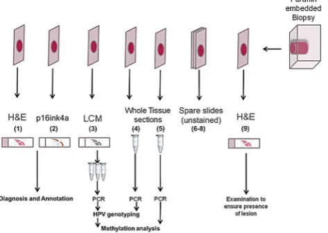
For each biopsy, a series of nine sections are cut and processed for histological analysis using a sandwich
sec-tioning method modified from refs 39 and 49
(figure 3). To minimise cross-contamination, the
on September 22, 2020 by guest. Protected by copyright.
microtome blade is changed and the microtome surface is cleaned with xylene after sectioning of each sample. Where possible without resetting the blocks, biopsies are cut from the basal side towards the epithelial/superficial
side to minimise carryover of unrelated viral DNA to lesional tissue. The two outer sections (sections 1 and 9) are cut to 3 µm thickness and stained with H&E. The
first inner section (section 2) is also cut to 3 µm Figure 1 (A) Photograph of the anal transformation zone taken through a speculum during high-resolution anoscopy. A digital photograph at 10× magnification of an area treated with 3% acetic acid. An area suspicious for high-grade disease is outlined; this was confirmed by biopsy to be anal intraepithelial neoplasia grade 3 (AIN3). (B) Clock face diagram of anal transformation zone segmented into eight quadrants. A high-grade squamous intraepithelial lesion is considered to be potentially persistent if it was biopsied from within the same quadrant or up to one quadrant either side of the baseline lesion at follow-up clinic visits.
Figure 2 Flow chart for defining probable persistence of high-grade squamous intraepithelial lesion (HSIL) detected at baseline. A HSIL biopsy detected at consecutive visits which tests positive for the same human papillomavirus (HPV) genotype following laser capture microdissection (LCM) and is located in the same octant or an octant either side of the baseline biopsy is considered persistent. In the event that not all of these criteria are met, additional information including the cytology result and swab HPV genotype result and extent of disease on high-resolution anoscopy (HRA) will be considered. A HSIL lesion is considered to be not persistent if there were no follow-up biopsy taken at 6 months meeting the criteria for persistence, and if the cytology and swab results also do not support persistence. If there is doubt, 12-month samples will be taken into consideration and if not found to support the existence of a persistent HSIL, the result will be‘not persistent’.
on September 22, 2020 by guest. Protected by copyright.
thickness and automatically stained with CINtec anti-p16 (Roche, Tucson, Arizona, USA) and counterstained with Gills No. 2 haematoxylin (Australian Biostain, Traralgon, Australia) on the Ventana Benchmark Ultra instrument (Ventana Medical Systems, Inc, Oro Valley, Arizona, USA). Sections 1, 2 and 9 are reviewed by a histopath-ologist experienced in reading anal biopsy specimens ( JMR), to assess p16 positivity and lesion grade. Biopsies are classified as negative for intraepithelial neoplasia, eLSIL, fLSIL, HSIL-AIN2, HSIL-AIN3 or ASCC, and assessment of p16 staining is performed, both in accord-ance with accepted international criteria.45 Hence, p16 staining is reported as positive if there is continuous strong nuclear or nuclear plus cytoplasmic staining of the basal cell layer with extension upwards involving at least one-third of the epithelial thickness. Other patterns are regarded as negative. The second inner section (section 3) is cut to 9 µm thickness and placed onto an Arcturus polyethylene naphthalate (PEN) membrane glass slide (Applied Biosystems, Foster City, California, USA) for LCM and allowed to dry. The next two sections (sections 4 and 5, designated whole tissue sections (WTS)) are cut to 9 µm thickness, placed into sterile 1.5 mL microfuge tubes as scrolls and stored away from light. An additional three sections are cut to 3 µm thick-ness, mounted and stored for future immunohistochem-ical staining.
Digestion and DNA extraction of WTS
Section 5 is archived at 4°C and section 4 is digested with a mixture of histolene (800 µL) and ethanol (400 µL). The tissue is pelleted at 14 000×g for 10 min, and the resultant pellet washed twice with 500 µL of
70% ethanol. The tissue is air dried to ensure that no residual ethanol is present and is subsequently incu-bated with 400 µg of proteinase K in 200 µL of tissue lysis buffer (Roche Molecular Systems) on a 55°C heat block for 1 h followed by 37°C overnight. Samples which are not fully digested are vortexed briefly and incubated for a further hour following the addition of a further 10 µL of proteinase K (10 mg/mL in tissue lysis buffer). The digested tissue is then transferred onto an auto-mated MagnaPure 96 (Roche) isolation and purification system, using the DNA and Viral NA small volume kit, Universal Pathogen 200 protocol. The final volume of eluted DNA in solution is 100 µL.
Dewaxing, LCM and DNA extraction of lesions
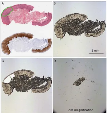
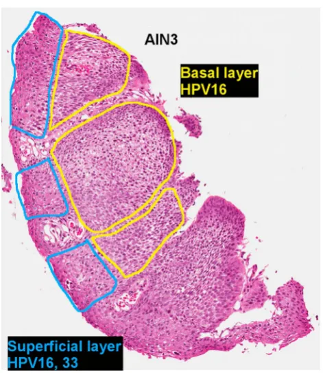
Immediately prior to tissue capture by LCM, PEN mem-brane slides are de-paraffinised via sequential 5 min incubations in 100% xylene (twice) and 100% ethanol (twice) and then allowed to dry thoroughly prior to laser capture. An annotated photograph of the H&E-stained section is used to guide selection of abnor-mal tissue to be captured. Tissue sections are visualised and dissected with an ultraviolet cutting laser on a Veritas 704 Laser Capture Microdissection System (Arcturus Bioscience, Mountain View, California, USA) and a single lesion is captured onto each thermoplastic CapSure Macro LCM Cap (Applied Biosystems) with an infrared capture laser (figure 4). Care is taken to avoid capturing the superficial layers of the epithelium
(figure 5). For any lesions which test positive for more
than one HPV genotype (genotype testing described below), additional sections will be captured by LCM from the basal layer at either end of the originally dissec-tion area of tissue to test for multiple colliding lesions or contamination (figure 6). DNA is extracted from each cap with the Arcturus PicoPure DNA Extraction Kit (Applied Biosystems) as per protocol F of the PicoPure DNA Extraction Kit User guide.
Genotyping of DNA extracted from WTS and LCM samples A 110 bp region of the humanβ-globin gene is detected by quantitative real-time PCR to confirm successful DNA extraction, as previously described.49 DNA from WTS-extracted and LCM-extracted tissue sections are both tested for HPV genotype on the RHA kit HPV SPF10-LiPA25, V.1 (Labo Bio-medical Products BV, Rijswijk, The Netherlands) according to the manufac-turer’s instructions. Because of an inability to discrimin-ate between HPV68 and 73 by the SPF10-LiPA25 assay, samples which are positive for HPV68/73 are tested on HPV68 and 73 type-specific qPCR assays targeting the E6 region as previously described,49 using the following primers and probes: HPV68 (forward primer 50-CTG TGCAGGACATTGGACAC-30; reverse primer 50-TTGGC ATGCAGCAAATGGTA-30; probe 50-LC610-TGCAGAAGG
CAACTACAACGGACA-BHQ2–30); HPV73 (forward
primer 50-CCAATTCAGAAGAACGACCA-30; reverse primer 50-CGTTGGCAAAACACACAGTC-30; probe 50 -Figure 3 Sandwich sectioning of formalin-fixed,
paraffin-embedded anal biopsy tissue samples and
downstream testing of each section. (1) First H&E slide, 3 µm section; (2) p16-ink4a slide, 3 µm section; (3) membrane slide to be used for LCM, 9 µm section; (4 and 5) whole tissue sections, unmounted, 9 µm sections; (6–8) unstained tissue sections mounted on slides, 3 µm sections; (9) last H&E slide, 3 µm section. HPV, human papillomavirus; LCM, laser capture microdissection.
on September 22, 2020 by guest. Protected by copyright.
FAM-CTTCGTCACATAACGCTTGTAGCTTG-BHQ1-30). Confirmed lesions that are positive forβ-globin, but not detectable on the SPF10-LiPA25, are tested for HPV DNA on the DNA ELISA kit HPV SPF10, V.1 (Labo Bio-medical Products BV) which is validated for
detec-tion of 44 HPV genotypes. Samples which are
ELISA-positive are genotyped on the RHA Kit HPV SPF+ (Labo Bio-medical Products BV) which is able to identify HPV26, 30, 55, 61, 62, c64, 67, 69, 71, 71sub, 82, 83, 84, 85, 87, 89, 90 and 91. ELISA-negative samples are tested on a qPCR assay targeting the E6 region of HPV16 and 18 as previously described,49 in addition to HPV45
(forward primer 50-CTACAAGACGTATCTATTGCC
TGTG-30; reverse primer 50-CCAGTGTCTCTCCATATA CAGAGTTT-30; probe 50-Cy5-CCTCTGTGCGTTCCAAT GTTGCTT-BHQ3-30). Confirmed lesions which are still negative on ELISA and/or all previous HPV tests are tested for the presence of β-papillomavirus (cutaneous or skin) genotypes as follows. Samples are PCR
ampli-fied using degenerate PCR primers CP65 (50 -CARGGTCAYAAYAATGGYAT-30) and CP70 (50-AAYT
TTCGTCCYARAGRAWATTGRTC-30)50 as follows: 5 µL of DNA template is included in a final reaction volume of 50 µL (2.5U AmpliTaq Gold and 1× Buffer II (Applied Biosystems, Foster City, California, USA); 3.6 mM MgCl2;
0.2 mM of each dNTP; 1 µM of each primer).
Amplicons are visualised on a 2% agarose gel. These samples are also genotyped using the RHA Kit Skin (β) HPV (Diassay B.V., Rijswijk, The Netherlands) which is able to identify HPV5, 8(I), 8(II), 9, 12, 14, 15, 17, 19, 20, 21, 22, 23, 24, 25 36, 37, 38, 47, 49, 75, 80, 92, 93 and 96, or by Sanger sequencing of the CP65/CP70 amplicon and comparison with the (National Center for Biotechnology Information) NCBI Blast nucleotide data-base (http://www.ncbi.nlm.nih.gov/Blast.cgi) and/or HPV-QUEST database (http://www.ijbcb.org/HPV/)51 to identify the closest matching known sequence. Samples that are negative for both β-globin and HPV, after re-extraction by LCM if possible, are denoted‘ unas-sessable’. Samples that are positive forβ-globin but nega-tive for all HPV tests are denoted ‘HPV not detected’. Samples that are positive for HPV DNA ELISA but not Figure 4 Laser capture microdissection of high-grade squamous intraepithelial lesion (HSIL). (A) Adjacent H&E stained and p16 stained biopsy section with HSIL annotated by a histopathologist in green. (B) Dewaxed, unstained biopsy section on polyethylene naphthalate membrane slide ready for laser capture microdissection. (C) Lesion excised with ultraviolet laser, thermoplastic cap heated with infrared laser and fragments lifted off. (D) Excised fragment ready for DNA extraction.
on September 22, 2020 by guest. Protected by copyright.
able to be genotyped are denoted ‘HPV X’. Samples that are negative for mucosal HPV types (negative on SPF10 LiPA, SPF+ LiPA and E6 qPCR) and positive for
β-papillomavirus DNA gel electrophoresis assay but not able to be genotyped are denoted‘HPVβ’. Additionally, all WTS samples that are negative for HPV16 or 18 will be tested on a type-specific qPCR assay for these genotypes.
Viral genome methylation sequence analysis
DNA from HPV16-positive HSIL is subjected to methyla-tion sequencing of viral genomic regions associated with regulation of oncogene expression using modifications of published protocols52 53 on anal swab, WTS and LCM-extracted tissue. DNA from the second WTS sample (section 5) is extracted using the QIAmp DNA FFPE Tissue Kit (Qiagen GmbH, Hilden, Germany). The same DNA sample used for HPV genotyping of swab and LCM samples is also used for methylation ana-lysis. Known controls for low (SiHa cell line, ATCC HTB35; American Type Culture Collection (ATCC), Manassas, Virginia, USA) and high (CaSki cell line, ATCC CRL1550; ATCC) methylation at the target sites are included in each experiment as well as no-template controls. For each HPV16-positive HSIL, 24 µL of
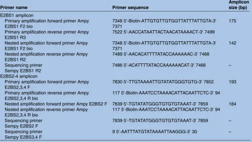
extracted DNA is subjected to bisulfite conversion and DNA cleanup using the Methylamp DNA Modification Kit (Epigentek, Farmingdale, New York, USA), accord-ing to the manufacturer’s instructions. This procedure converts unmethylated cytosine (C) residues into tyro-sine (T) residues. Following conversion, two targeted sections of the HPV16 genome are amplified by nested PCR; samples are amplified in a first round of PCR using the primary pair of sequences, and a 1 µL aliquot is then amplified in a second round of PCR using the secondary (nested) PCR primers, as listed in table 1. The amplified sections are the E2BS1 region from nucleotide position 7348 to 7489 (final amplicon size 142 bp), and E2BS2-4 region from nucleotide position 7839 to 117 (final amplicon size 184 bp). Primary PCR amplifications are performed in reaction mixes with a
final volume of 25μL (12.5μL of Hot Start Taq Master mix (Qiagen); 0.25μmol of each primer; 5μL of
bisul-fite modified template DNA; nuclease-free water tofinal volume). Thermocycler parameters are as follows: 15 min at 95°C, followed by 50 cycles of (40 s at 95°C, 30 s at 50°C and 40 s at 72°C), followed by 6 min at 72°C. Amplicons are visualised on a 2% agarose gel. The secondary (nested) PCR amplification for the E2BS1 region is performed in reaction mixes with a final volume of 25μL (12.5μL of Hot Start Taq Master mix (Qiagen); 0.25μmol of each nested primer; 1μL of amplicon from the primary PCR; nuclease-free water to
final volume). Thermocycler parameters are as follows: 15 min at 95°C, followed by 50 cycles of (40 s at 95°C, 30 s at 56°C and 40 s at 72°C), followed by 6 min at 72°C. The secondary PCR amplification for the E2BS2-4 Figure 5 Laser capture microdissection (LCM) of the basal
layer to avoid superficial contamination with incidental human papillomavirus (HPV) genotypes. H&E section of an anal canal biopsy containing an anal intraepithelial neoplasia grade 3 (AIN3). The basal and superficial layers of the AIN3 were dissected separately by LCM. The basal layers of the lesion were positive for HPV16 and the biopsy also had superficial contamination with HPV33 DNA.
Figure 6 Example of a biopsy containing colliding lesions each positive for different HPV genotypes. H&E section of an anal canal biopsy with three lesions positive for HPV16, 53 and 56, respectively. Shaded areas indicate tissue dissected by LCM and genotyped. The bottom two lesions collide, and the original LCM section tested positive for both HPV53 and 56. Subsequent sections each tested positive singly for either HPV53 or 56. HPV, human papillomavirus; LCM, laser capture microdissection; LSIL, low-grade squamous intraepithelial lesion.
on September 22, 2020 by guest. Protected by copyright.
region is performed in reaction mixes with a final volume of 25μL (12.5μL of Hot Start Taq Master mix (Qiagen); 0.25μmol of each nested primer; 1μL of amplicon from the primary PCR; nuclease-free water to
final volume). Thermocycler parameters are as follows: 15 min at 95°C, followed by 50 cycles of (40 s at 95°C, 30 s at 50°C and 40 s at 72°C), followed by 6 min at 72°C. CpG sequence analysis and quantitation of methy-lation at nucleotide positions 7426, 7432, 7453, 7459, 7860, 31, 37, 43, 52 and 68 on the HPV16 viral genome52 is performed using pyrosequencing technol-ogy on the PyroMark Q24 system (Qiagen) at the Australian Genome Research Facility (AGRF). Assay setup, pyrosequence run and analysis are performed by PyroMark Q24 Software. Sequencing primers for E2BS1, E2BS2 and E2BS3,4 regions are listed intable 1.
Other biomarkers
Additional biomarker analyses may include host genome marker methylation sequencing, viral genome integra-tion frequency and relative transcripintegra-tion levels of HPV mRNA species, contingent on sample availability and assay performance.
Statistical considerations
In total, about 600 participants (30–40% of whom will be HIV-positive) will be recruited by June 2015. As of the end of 2014, 480 participants have been recruited with 125 (26%, 95% CI 22% to 30%) men diagnosed with biopsy-confirmed LSIL and 143 (30%, 95% CI 26%
to 34%) men with biopsy-confirmed anal HSIL. Therefore, we anticipate between 130 and 180 of the total 600 participants will be diagnosed with LSIL, and between 150 and 200 will be diagnosed with biopsy-confirmed HSIL at baseline.
HPV prevalence in LCM-isolated lesions, including low-risk (including β-papillomavirus or HPV X), high-risk and individual genotypes will be calculated overall and stratified by HIV status as the proportion of lesions that tested positive for a specific HPV type or risk group. The exact binomial method will be used to calculate the
corresponding 95% CIs for prevalence values.
Biomarker positivity associated with lesion type will be calculated using the same method. Univariate and multi-variate logistic regression models will be carried out to measure the strength of association between HPV status and each biomarker in lesions at baseline.
To determine the performance of biomarkers in pre-dicting disease persistence at 12 months, univariate and multivariate Cox regressions will be performed to assess the strength of the association between biomarker posi-tivity and anal HSIL persistence at 12 months in those who are diagnosed with anal HSIL at baseline. The sen-sitivity and specificity of each biomarker detected on anal swab to predict persistent HSIL at 12 months will be calculated alone and in combination with HPV geno-typing results. In addition, univariate logistic regression models will be carried out to measure the strength of each test in relation to a diagnosis of HSIL. A multivari-ate logistic regression model will be constructed to Table 1 Primer sets for determination of human papillomavirus 16 CpG methylation using pyrosequencing
Primer name Primer sequence
Amplicon size (bp) E2BS1 amplicon
Primary amplification forward primer Ampy E2BS1 F2 bio
7348 50-Biotin-ATTGTGTTGTGGTTATTTATTGTA-30 7371
175
Primary amplification reverse primer Ampy E2BS1 R3
7522 50-AACCATAATTACTAACATAAAACT-307499
Nested amplification forward primer Ampy E2BS1 F2 bio
7348 50-Biotin-ATTGTGTTGTGGTTATTTATTGTA-30 7371
142
Nested amplification reverse primer Ampy E2BS1 R2
7489 50-AACACATTTTATACCAAAAAAC-307468
Sequencing primer Sempy E2BS1 R2
7486 50-ACATTTTATACCAAAAAACAT-307466 –
E2BS2-4 amplicon
Primary amplification forward primer Ampy E2BS2,3,4 F
7830 50-TTGTAAAATTGTATATGGGTGTG-307852 193
Primary amplification reverse primer Ampy E2BS2,3,4 R bio
117 50-Biotin-AAATCCTAAAACATTACAATTCTC-3094
Nested amplification forward primer Ampy E2BS2 F 7839 50-TGTATATGGGTGTGTGTAAAT-307859 184 Nested amplification reverse primer Ampy
E2BS2,3,4 R bio
117 50-Biotin-AAATCCTAAAACATTACAATTCTC-3094
Sequencing primer Sempy E2BS2 F
7839 50-TGTATATGGGTGTGTGTAAAT-307859 –
Sequencing primer Sempy E2BS3,4 F
9 50-AATTTATGTATAAAATTAAGGG-3030 –
on September 22, 2020 by guest. Protected by copyright.
determine a combination of these biomarkers that best predict persistent HSIL and to assess the association with HIV status and other covariates such as age.
Percentages (%) agreement andκstatistic will be used to determine HPV concordance in anal swab, WTS and LCM sample ( poor ifκ≤0.20; fair if 0.21≤κ≤0.40; mod-erate if 0.41≤κ≤0.60; substantial if 0.61≤κ≤0.80; good if
κ>0.80). Differences in HPV genotypes prevalence between samples will be assessed by the McNemar test.
Currently available algorithms to determine the most likely lesion-associated genotype based on swab samples42will be compared with actual lesion-associated genotypes determined using LCM. Percentage detection and 95% CIs will be used to calculate the performance of hierarchical and proportional attribution methods against LCM-determined lesion-associated genotypes.
Ethics and dissemination
Written informed consent is obtained from all indivi-duals before any study-specific procedures are per-formed. As clinical guidelines do not indicate treatment and there is currently no scientific evidence to support routine treatment of anal HSIL,54 55 participants diag-nosed with HSIL are not routinely treated, except where deemed clinically necessary; this may include severely abnormal blood vessels, persistent large lesions or other risk factors for ASCC, such as prolonged immunosup-pression, low CD4 nadir and history of smoking.
Findings from this study will be disseminated to parti-cipants and the community through study newsletters, and through peer-reviewed publications and inter-national conferences.
DISCUSSION
Importance of this study
With continuing increases in ASCC incidence,3 56–58 future prevention of this disease will rely on effective vac-cination and screening measures and these in turn will depend on accurate data pertaining to the association of individual HPV genotypes with ASCC and with anal HSIL. Persistent anal HSIL is widely accepted to be the precursor to ASCC, and is the anal counterpart to cer-vical HSIL, the main disease end point in cercer-vical screening programmes. The end point for anal screen-ing will likely differ from cervical screenscreen-ing as rates of progression from anal HPV infection and/or HSIL to anal cancer are very low,4 24 and the populations that are likely to be most frequently screened contain a high
proportion of individuals with HR-HPV and/or
HSIL.4 59 Effective anal screening programmes will therefore require very high specificity for an appropriate end point, to limit unnecessary procedures on a large number of low-risk individuals, as well as high sensitivity to identify the small population of high-risk individuals. This research will contribute to the determination of appropriate end points for anal screening.
Approximately 70–90% of cases of ASCC are attribu-ted to infection with HPV16.25 26 33 58 60 However, the exact contribution of HPV16, and perhaps more import-antly other HPV genotypes, to the load of ASCC cases is not known, as up to a quarter of anal cancer specimens test positive for multiple HPV genotypes.25 40 41 Where HPV16 is present, it is usually assumed to be the causa-tive genotype; given that HPV16 is a commonly found genotype in any population of sexually active adults, it is highly likely to be incidental to the cancer in a number of cases and therefore its contribution to cancer devel-opment overestimated. Conversely, an increasing quan-tity of evidence suggests that so-called LR-HPV genotypes—particularly HPV6 and 11—contribute to a larger proportion than expected based on cervical cancer data sets25 31 49 60 61and present data collection methods probably lead to under-estimation of their con-tribution to anal cancer rates.
In addition to HPV genotype detection, methylation patterns also have potential utility as biomarkers in cancer screening,52 62particularly with rapid advances in sequencing technology and the increasing recognition in other medicalfields of the diagnostic potential of epi-genetic markers.63 Currently available data on methyla-tion of viral DNA targets are insufficient to inform development of methylation as a screening marker for HPV-related cancers, and are derived predominantly from cervical tissue and cervical cancer cell lines with only a single study presenting any methylation sequence data for the HPV genome in anal lesions.64
Because it is thepersistence of HR-HPV and HSIL that lead to increased risk of anal cancer development, longi-tudinal studies provide more useful information than cross-sectional data, particularly for rare events such as anal cancer incidence. Longitudinal or natural history studies provide data to allow more accurate risk categor-ies to be assigned to single screening results, so that indi-vidual patients can be triaged appropriately. This prospective, longitudinal study is designed to provide data on risk factors associated with persistent HSIL— and by extension increased risk of anal cancer develop-ment—to inform future decisions on anal cancer prevention in at-risk populations.
Strengths of this study
This participant cohort is a unique resource, as one of only a very small number of cohorts with prospectively collected longitudinal cytology and histology samples from HIV-positive and HIV-negative participants diag-nosed with the full spectrum of HPV-associated anal lesions. A team of intensively trained anoscopists have their diagnostic skills monitored regularly with extensive quality control metrics, ensuring high-quality clinical data. Participants in this cohort are not routinely treated when HSIL is detected unless it is determined to be clin-ically necessary, for example, severely abnormal blood vessels, persistent large lesions or other risk factors for ASCC, such as prolonged immunosuppression, low CD4
on September 22, 2020 by guest. Protected by copyright.
nadir and history of smoking. This approach allows for longitudinal follow-up of lesions, and therefore allows assessment of when treatment is likely to be most beneficial.
A major strength of the present study is the use of LCM to distinguish lesions from adjacent normal tissue, which allows for precise genotype and biomarker testing
from which to infer the natural history of
HPV-associated anal lesions. Methylation patterns and the behaviour of other biomarkers are also often tissue-specific or lesion-specific, and can be heterogeneous within even a small tissue sample.53 The use of LCM generates extremely low-concentration DNA samples, and the assays being used in this study are very sensitive for low copies of DNA targets, and have a small ampli-con size for the detection of fragmented DNA, which is partially degraded in the fixation process. These methods have been proven sensitive enough for HPV genotyping and methylation sequence analysis in pub-lished studies23 39 53and in our own laboratory.
In routine diagnostic work, p16 immunostaining is uti-lised, as per LAST criteria, for confirming a diagnosis of HSIL-AIN2 and for distinguishing true HSIL-AIN3 from atypical immature metaplasia. This latter differential diagnosis is more challenging in the anal canal that in the cervix (T Darragh, personal communication, 2013). Routine p16 staining is not indicated for diagnostic samples due to the uncertain significance of p16-positive LSIL,45but it is a useful research tool for further explor-ing the natural history of p16-positive LSIL in conjunc-tion with HPV genotype and/or biomarker analysis and potentially clarifying the significance of these lesions.
Anticipated challenges
The major challenges of this study relate to diagnostic uncertainty and establishing whether or not a lesion is persistent from one visit to the next. It is inevitable that some lesions will be missed during HRA, and therefore multiple sources of data will be used to define the per-sistence of individual lesions, as indicated in figure 2. The study uses both cytology and histology (based on HRA-guided biopsy collection) to establish disease grade in participants at each visit. It is known that anal cytology and HRA/histology each have their limitations,21 65 and therefore both methods will be used in a complemen-tary fashion to derive a more accurate diagnosis. Longitudinal sampling and identification of the same individual lesion has not been attempted before in a sys-tematic manner. This adds an extra dimension of com-plexity to the project. Defining persistent lesions will not be straightforward due to the difficulties inherent in sampling the anal canal, the possibility of missed lesions, the probability of multiple lesions and the uncertainties in locating and identifying the same lesion at consecu-tive visits.23 The presence of multiple distinct lesions caused by the same genotype cannot be ruled out. All anoscopists are aware of each participant’s previous clinic results and have access to HRA photographs; this
improves the likelihood of sampling the same lesion during subsequent HRA visits. It is known that the anal canal may rotate up to 90° between clinic visits ( per-sonal communication R J Hillman, 2012), making precise location of anatomical features difficult; this has been acknowledged and follow-up biopsies will be con-sidered if they are taken up to one octant either side of the baseline biopsy to reduce the chance of missing a persistent lesion. The use of features in HRA photo-graphs will also aid in determining lesion location, and in conjunction with anoscopists’ notes will be used to indicate the extent of disease (ie, across multiple octants). Each original biopsy diagnosis is made by one of a team of three experienced anogenital pathologists, and this is the basis on which tissues are selected for inclusion. The review diagnoses, however, are all made by one of these three pathologists ( JMR) based on the new H&E and p16 slides as described in the Methods section. The main issue resulting from this will be
con-firming persistence of HSIL when the review diagnosis at follow-up has been downgraded to LSIL or negative for SIL. In these cases, the study investigators will make a decision based on the original diagnosis, the HPV genotype detected by LCM, and the histological diagno-sis at the next clinic visit.
This study will use precise molecular biology techni-ques and international expertise to characterise the natural history and biological markers of individual anal lesions in a population at high risk of anal cancer. This is an ambitious and unique study which will enhance our understanding of the clinical and biological behav-iour of HPV-related anal lesions and the potential prog-nostic implications of future HPV genotype or biomarker screening tests.
Author affiliations
1Regional HPV Labnet Reference Laboratory, Department of Microbiology and Infectious Diseases, The Royal Women’s Hospital, Parkville, Victoria, Australia 2Murdoch Children’s Research Institute, Parkville, Victoria, Australia 3Douglass Hanly Moir Pathology, Sydney, New South Wales, Australia 4Western Sydney Sexual Health Centre, University of Sydney, Sydney, New South Wales, Australia
5The Kirby Institute, UNSW Australia, Kensington, New South Wales, Australia 6RPA Sexual Health, Sydney Local Health District, Camperdown, New South Wales, Australia
7Department of Obstetrics and Gynaecology, University of Melbourne
Collaborators The SPANC study team includes: AEG, IMP, FJ, Brian Acraman, Garrett Prestage, Leonie Crampton, Patrick McGrath, Kathy Petoumenos, Matthew O’Dwyer, Robert Mellor, Piero Pezzopane, Matthew Law (Kirby Institute, University of New South Wales); Carmella Law, Winnie Tong, Daniel Seeds, Eddie Fraissard and Andrew Carr (St Vincent’s Hospital, Sydney); DJT and Rick Varma (RPA Sexual Health, Sydney); Kit Fairley (Melbourne Sexual Health Centre); RJH, Kirsten McCaffery and Kirsten Howard (University of Sydney); Annabelle Farnsworth, JMR, Adele Richards and Julia Thurloe (Douglass Hanly Moir Pathology, Sydney); SMG, SNT, AMC and SP (The Royal Women’s Hospital, Melbourne); and Lance Feeney and Russ Gluyas (community representatives).
Contributors All authors contributed to study design. AMC drafted the manuscript. JMR provided histopathological expertise. SP and MM provided molecular biology expertise. DAM provided statistical expertise. RJH provided clinical expertise. AEG, FJ, IMP, DJT, SMG and SNT collectively conceived of
on September 22, 2020 by guest. Protected by copyright.
the study and provided epidemiological and molecular biology expertise. All authors read and approved of the final manuscript.
Funding The SPANC study is funded by Australian Commonwealth Government National Health and Medical Research Council Program grants (#568971 and #1071269) and a Cancer Council New South Wales Strategic Research Partnership Program grant (#13-11).
Competing interests None declared.
Ethics approval The St Vincent’s Hospital Ethics Committee.
Provenance and peer review Not commissioned; externally peer reviewed.
Open Access This is an Open Access article distributed in accordance with the Creative Commons Attribution Non Commercial (CC BY-NC 4.0) license, which permits others to distribute, remix, adapt, build upon this work non-commercially, and license their derivative works on different terms, provided the original work is properly cited and the use is non-commercial. See: http:// creativecommons.org/licenses/by-nc/4.0/
REFERENCES
1. Grulich AE, Poynten IM, Machalek DA,et al. The epidemiology of anal cancer.Sex Health2012;9:504–8.
2. Grulich AE, Jin F, Conway L,et al. Cancers attributable to human papillomavirus infection.Sex Health2010;7:244–52.
3. Jin F, Stein AN, Conway EL,et al. Trends in anal cancer in Australia, 1982–2005.Vaccine2011;29:2322–7.
4. Machalek DA, Poynten M, Jin F,et al. Anal human papillomavirus infection and associated neoplastic lesions in men who have sex with men: a systematic review and meta-analysis.Lancet
2012;13:487–500.
5. van Leeuwen MT, Vajdic CM, Middleton MG,et al. Continuing declines in some but not all HIV-associated cancers in Australia after widespread use of antiretroviral therapy.AIDS2009;23:2183–90. 6. Daling JR, Weiss NS, Hislop TG,et al. Sexual practices, sexually
transmitted diseases, and the incidence of anal cancer.N Engl J Med1987;317:973–7.
7. van der Zee RP, Richel O, de Vries HJC,et al. The increasing incidence of anal cancer: can it be explained by trends in risk groups?Neth J Med2013;71:401–11.
8. Grulich AE, van Leeuwen MT, Falster MO,et al. Incidence of cancers in people with HIV/AIDS compared with immunosuppressed transplant recipients: a meta-analysis.Lancet2007;370:59–67. 9. Giuliano AR, Palefsky JM, Goldstone S,et al. Efficacy of
quadrivalent HPV vaccine against HPV infection and disease in males.N Engl J Med2011;364:401–11.
10. Palefsky JM, Giuliano AR, Goldstone S,et al. HPV vaccine against anal HPV infection and anal intraepithelial neoplasia.N Engl J Med
2011;365:1576–85.
11. Tabrizi SN, Brotherton JML, Kaldor JM,et al. Fall in human papillomavirus prevalence following a national vaccination program.
J Infect Dis2012;206:1645–51.
12. Brotherton JML, Fridman M, May CL,et al. Early effect of the HPV vaccination programme on cervical abnormalities in Victoria, Australia: an ecological study.Lancet2011;377:2085–92. 13. Gertig DM, Brotherton JM, Budd AC,et al. Impact of a
population-based HPV vaccination program on cervical abnormalities: a data linkage study.BMC Med2013;11:227. 14. Wilkinson E. Australia leads way on HPV vaccination in boys.Lancet
Infect Dis2012;12:831–2.
15. Luxembourg A; On behalf of the V503 Program Team.An overview of the 9-valent HPV L1 virus-like particle vaccine clinical
development program. Florence, Italy: EUROGIN International Congress, 2013.
16. U.S. Food and Drug Administration. News & Events. http://www.fda. gov/NewsEvents/Newsroom/PressAnnouncements/ucm426485.htm (accessed 18 Feb 2015).
17. Ault KA., The Future II Study Group. Effect of prophylactic human papillomavirus L1 virus-like-particle vaccine on risk of cervical intraepithelial neoplasia grade 2, grade 3, and adenocarcinoma in situ: a combined analysis of four randomised clinical trials.Lancet
2007;369:1861–8.
18. Haupt RM, Wheeler CM, Brown DR,et al. Impact of an HPV6/11/16/ 18 L1 virus-like particle vaccine on progression to cervical
intraepithelial neoplasia in seropositive women with HPV16/18 infection.Int J Cancer2011;129:2632–42.
19. Joura EA, Garland SM, Paavonen J,et al. Effect of the human papillomavirus (HPV) quadrivalent vaccine in a subgroup of women
with cervical and vulvar disease: retrospective pooled analysis of trial data.BMJ2012;344:e1401.
20. Farnsworth A. Screening for the prevention of cervical cancer in the era of human papillomavirus vaccination: an Australian perspective.
Acta Cytol2011;55:307–12.
21. Darragh TM, Winkler B. Anal cancer and cervical cancer screening: key differences.Cancer Cytopathol2011;119:5–19.
22. Palefsky JM, Holly EA, Hogeboom CJ,et al. Anal cytology as a screening tool for anal squamous intraepithelial lesions.J Acquir
Immune Defic Syndr1997;14:415–22.
23. Richel O, Quint KD, Lindeman J,et al. One lesion, one virus: individual components of high grade anal intraepithelial neoplasia in HIV+ men contain a single HPV type.J Infect Dis
2014;210:111–20.
24. Cachay E, Agmas W, Mathews C. Five-year cumulative incidence of invasive anal cancer among HIV-infected patients according to baseline anal cytology results: an inception cohort analysis.
HIV Med2014;16:191–5.
25. Hillman RJ, Garland SM, Gunathilake MPW,et al. Human papillomavirus (HPV) genotypes in an Australian sample of anal cancers.Int J Cancer2014;135:996–1001.
26. Ouhoummane N, Steben M, Coutlee F,et al. Squamous anal cancer: patient characteristics and HPV type distribution.Cancer
Epidemiol2013;37:807–12.
27. Australian Government Department of Health. National Cervical Screening Program: Overview of the Renewal. http://www. cancerscreening.gov.au/internet/screening/publishing.nsf/Content/ overview-of-the-renewal (accessed 18 Feb 2015).
28. Huh WK, Ault KA, Chelmow D,et al. Use of primary high-risk human papillomavirus testing for cervical cancer screening: interim clinical guidance.J Low Genit Tract Dis2015;19:91–6.
29. Bierkens M, Hesselink AT, Meijer CJLM,et al. CADM1 and MAL promoter methylation levels in hrHPV-positive cervical scrapes increase proportional to degree and duration of underlying cervical disease.Int J Cancer2013;133:1293–300.
30. Zhang J, Martins CR, Fansler ZB,et al. DNA methylation in anal Intraepithelial lesions and anal squamous cell carcinoma.
Clin Cancer Res2005;11:6544–9.
31. Alemany L, Saunier M, Alvarado-Cabrero I,et al. HPV DNA prevalence and type distribution in anal carcinomas worldwide.Int J
Cancer2015;136:98–107.
32. Walboomers JMM, Jacobs MV, Manos MM,et al. Human papillomavirus is a necessary cause of invasive cervical cancer worldwide.J Pathol1999;189:12–19.
33. De Vuyst H, Clifford GM, Nascimento MC,et al. Prevalence and type distribution of human papillomavirus in carcinoma and intraepithelial neoplasia of the vulva, vagina and anus: a meta-analysis.Int J Cancer2009;124:1626–39.
34. Gohy L, Gorska I, Rouleau D,et al. Genotyping of human papillomavirus DNA in anal biopsies and anal swabs collected from HIV-seropositive men with anal dysplasia.J Acquir Immune Defic
Syndr2008;49:32–9.
35. Sahasrabuddhe VV, Castle PE, Follansbee S,et al. Human papillomavirus genotype attribution and estimation of preventable fraction of anal intraepithelial neoplasia cases among HIV-infected men who have sex with men.J Infect Dis2013;207:392–401. 36. Wong AK, Chan RC, Aggarwal N,et al. Human papillomavirus
genotypes in anal intraepithelial neoplasia and anal carcinoma as detected in tissue biopsies.Mod Pathol2010;23:144–50.
37. Machalek DA, Grulich AE, Jin F,et al. The epidemiology and natural history of anal human papillomavirus infection in men who have sex with men.Sex Health2013;9:527–37.
38. Nyitray AG. The epidemiology of anal human papillomavirus infection among women and men having sex with women.
Sex Health2012;9:538–46.
39. Quint W, Jenkins D, Molijn A,et al. One virus, one lesion—individual components of CIN lesions contain a specific HPV type.J Pathol
2012;227:62–71.
40. Abramowitz L, Jacquard A-C, Jaroud F,et al. Human papillomavirus genotype distribution in anal cancer in France: the EDiTH V study.
Int J Cancer2010;129:433–9.
41. Steinau M, Unger ER, Hernandez BY,et al. Human papillomavirus prevalence in invasive anal cancers in the United States prior to vaccine introduction.J Low Genit Tract Dis2013;17:397–403. 42. Wentzensen N, Schiffman M, Dunn ST,et al. Multiple HPV genotype
infections in cervical cancer progression in the Study to Understand Cervical Cancer Early Endpoints and Determinants (SUCCEED).
Int J Cancer2009;125:2151–8.
43. Machalek DA, Grulich AE, Hillman RJ,et al. The Study of the Prevention of Anal Cancer (SPANC) design and methods of a three-year prospective cohort study.BMC Public Health2013;13:946.
on September 22, 2020 by guest. Protected by copyright.
44. Darragh TM, Colgan TJ, Cox JT,et al. The Lower Anogenital Squamous Terminology Standardization Project for HPV-associated lesions: background and consensus recommendations from the College of American Pathologists and the American Society for Colposcopy and Cervical Pathology.J Low Genit Tract Dis
2012;16:205–42.
45. Darragh TM, Colgan TJ, Cox JT,et al. The Lower Anogenital Squamous Terminology Standardization Project for HPV-associated lesions: background and consensus recommendations from the College of American Pathologists and the American Society for Colposcopy and Cervical Pathology.Arch Pathol Lab Med
2012;136:1266–97.
46. Pirog EC, Quint KD, Yantiss RK. P16/CDKN2A and Ki-67 enhance the detection of anal intraepithelial neoplasia and condyloma and correlate with human papillomavirus detection by polymerase chain reaction.Am J Surg Pathol2010;34:1449–55.
47. Oh K-T, Palefsky J. Diseases of the anus. In: CP C, KR L, eds. Diagnostic gynecologic and obstetric pathology. Philadelphia: Elsevier Saunders, 2006:199–227.
48. Stevens MP, Garland SM, Tabrizi SN. Development and validation of a real-time PCR assay specifically detecting human
papillomavirus 52 using the Roche LightCycler® 480 system.J Virol
Methods2008;147:290–6.
49. Cornall AM, Roberts J, Garland SM,et al. Anal and perianal cancers exclusively associated with low-risk HPV genotypes 6 and 11.Int J
Cancer2013;133:2253–8.
50. Berkhout RJM, Tieben LM, Smits HL,et al. Nested PCR approach for detection and typing of epidermodysplasia
verruciformis-associated human papillomavirus types in cutaneous cancers from renal transplant recipients.J Clin Microbiol
1995;33:690–5.
51. Yin L, Yao J, Gardner BP,et al. HPV-QUEST: a highly customized system for automated HPV sequence analysis capable of processing next generation sequencing data set.Bioinformation
2012;8:388–90.
52. Chaiwongkot A, Vinokurova S, Pientong C,et al. Differential methylation of E2 binding sites in episomal and integrated HPV 16 genomes in preinvasive and invasive cervical lesions.Int J Cancer
2013;132:2087–94.
53. Vinokurova S, von Knebel Doeberitz M. Differential methylation of the HPV 16 upstream regulatory region during epithelial differentiation and neoplastic transformation.PLoS ONE2011;6: e24451.
54. Macaya A, Munoz-Santos C, Balaguer A,et al. Interventions for anal canal intraepithelial neoplasia.Cochrane Database Syst Rev2012 (10):CD009244.
55. Richel O, de Vries HJC, van Noesel CJM,et al. Comparison of imiquimod, topical fluorouracil, and electrocautery for the treatment of anal intraepithelial neoplasia in HIV-positive men who have sex with men: an open-label, randomised controlled trial.Lancet Oncol
2013;14:346–53.
56. Joseph DA, Miller JW, Wu X,et al. Understanding the burden of human papillomavirus-associated anal cancers in the US.Cancer
2008;113(S10):2892–900.
57. Brewster DH, Bhatti LA. Increasing incidence of squamous cell carcinoma of the anus in Scotland, 1975–2002.Br J Cancer
2006;95:87–90.
58. Frisch M, Glimelius B, van den Brule AJC,et al. Sexually transmitted infection as a cause of anal cancer.N Engl J Med
1997;337:1350–8.
59. Coutlee F, de Pokomandy A, Franco EL. Epidemiology, natural history and risk factors for anal intraepithelial neoplasia.Sex Health
2012;9:547–55.
60. Daling JR, Madeleine MM, Johnson LG,et al. Human
papillomavirus, smoking, and sexual practices in the etiology of anal cancer.Cancer2004;101:270–80.
61. Guimera N, Lloveras B, Lindeman J,et al. The occasional role of low-risk human papillomaviruses 6, 11, 42, 44, and 70 in anogenital carcinoma defined by laser capture microdissection/PCR
methodology: results from a global study.Am J Surg Pathol
2013;37:1299–310.
62. Kalantari M, Calleja-Macias IE, Tewari D,et al. Conserved methylation patterns of human papillomavirus type 16 DNA in asymptomatic infection and cervical neoplasia.J Virol
2004;78:12762–72.
63. Partin AW, Van Neste L, Klein EA,et al. Clinical validation of an epigenetic assay to predict negative histopathological results in repeat prostate biopsies.J Urol2014;192:1081–7.
64. Wiley DJ, Huh J, Rao JY,et al. Methylation of human papillomavirus genomes in cells of anal epithelia of HIV-infected men.J Acquir Immune Defic Syndr2005;39:143–51.
65. Berry JM, Palefsky JM, Jay N,et al. Performance characteristics of anal cytology and human papillomavirus testing in patients with high-resolution anoscopy-guided biopsy of high-grade anal intraepithelial neoplasia.Dis Colon Rectum2009;52: 239–47.
on September 22, 2020 by guest. Protected by copyright.