TECHNIQUES AND INSTRUMENTATION
Paul PG&Khan Shabnam&Sheetal Avinash Bhosale& Harneet Kaur&Prathap Talwar&Tony Thomas
Received: 25 February 2013 / Accepted: 6 August 2013 / Published online: 3 September 2013
#Springer-Verlag Berlin Heidelberg 2013
Abstract Hysterectomy in frozen pelvis is a challenging surgical condition whether done by laparotomy or laparosco-py. We describe an alternative technique of total laparoscopic hysterectomy with retrograde adhesiolysis in patients with frozen pelvis. Total laparoscopic hysterectomy with retro-grade adhesiolysis was done in 25 patients with frozen pelvis between October 2003 and May 2012. The mean (standard deviation; 95 % confidence interval) age of patients was 42.6 (6.00; 40.1–45.07). Body mass index was 27.48 (5.06; 25.3– 29.57). Twenty (80 %) patients had previous abdominal sur-gery, and three (15 %) patients had previous failed surgeries for attempted hysterectomy. Twenty-three patients had frozen pelvis due to severe endometriosis, and two patients had severe abdominopelvic adhesions due to multiple previous surgeries. One patient had intraoperative injury to the sigmoid colon and bladder during adhesiolysis, and laparotomy con-version was performed. The median (range) operating time was 210 (120–300) min, and estimated blood loss was 400 (300–600) ml. Length of post-operative stay was 1 (1–6) days, and the post-operative period was uneventful except in two patients who had paralytic ileus. The median (range) follow-up at 1 month and 6 months was 100 and 68 % (17 of 25), respectively. Our technique of laparoscopic hysterectomy with retrograde adhesiolysis and subsequent removal of ad-nexa is an alternative technique for hysterectomy in frozen pelvis and limits the potential hazards of injury to vital organs; it is associated with fewer complications. We emphasize that adequate surgical experience and expertise still remain the prerequisites for performing hysterectomy in frozen pelvis.
Keywords Retrograde adhesiolysis . Frozen pelvis . Laparoscopic surgery . Severe endometriosis
Background
Hysterectomy in frozen pelvis is a challenging surgical con-dition whether done by laparotomy or laparoscopy. Frozen pelvis refers to a surgical condition where reproductive organs and adjacent structures are distorted by extensive adhesive disease and fibrosis, which obscure the normal anatomical landmarks and surgical planes, making dissection extremely difficult and increasing the risk of injury to vital organs [1]. Common causes are infections, endometriosis and multiple previous surgeries, and other causes are ovarian carcinoma and radiotherapy. Surgery in frozen pelvis can result in serious complications like bowel and urinary tract injury. Most of these cases are referred to oncosurgeons who usually perform the surgery by an open retroperitoneal approach [2, 3]. We have been performing a total laparoscopic hysterectomy with retrograde adhesiolysis technique in frozen pelvis for 10 years in selected cases with gross pelvic distortion where the con-ventional method of laparoscopic hysterectomy is impractical. The concept of hysterectomy in frozen pelvis entails two steps, to perform extensive adhesiolysis first followed by hysterectomy. The highlight of our technique is to perform adhesiolysis sufficient enough to visualize the uterine fundus and cornua and then to proceed with hysterectomy. Difficult posterior bowel adhesions to the uterus and pouch of Douglas are dealt in a retrograde fashion after hysterectomy. Our technique of retrograde adhesiolysis is based on an alternative technique and can be practiced by most gynaecologists.
Material and methods
This is a retrospective case series. We included all patients who had undergone total laparoscopic hysterectomy in frozen pelvis at Paul’s Hospital between October 2003 and May 2012. The aim of this study is to find the laparotomy conversion rate, duration of surgery, blood loss, intra- and post-operative complications with this alternate technique. The institutional P. PG (*)
:
K. Shabnam:
S. A. Bhosale:
H. Kaur:
P. TalwarPaul’s Hospital, Kochi, Kerala, India e-mail: drpaulpg@gmail.com
T. Thomas
Walsall Manor Hospital, Walsall, West Midlands, England
ethical committee of Paul’s Hospital approved the data collec-tion, aggregacollec-tion, deidentification and analysis for this study. Data regarding patient characteristics like age, body mass in-dex, parity and previous surgeries, and intraoperative details like duration of surgery, complications, estimated blood loss, duration of hospital stay and post-operative events are evaluat-ed. Informed consent is obtained from all patients, and risk of conversion to laparotomy and injury to vital structures is explained. Patients are admitted to hospital on the day of surgery and kept nil per os for 6 h prior to surgery. Bowel preparation is performed using sodium phosphate solution en-ema. Antibiotic prophylaxis is given at the time of induction of anaesthesia. Procedures are performed under general anaesthe-sia. Intermittent pneumatic compression device is used for prevention of deep venous thrombosis in all patients. Low molecular weight heparin is administered in high-risk patients.
Patient evaluation
Detailed history is taken regarding severity of abdominal pain, dysmenorrhoea, dyspareunia, dyschezia, haematochezia, diar-rhoea, constipation, previous abdominopelvic infections and surgeries. Clinical examination and transvaginal ultrasonog-raphy are done by the operating surgeon prior to surgery. MRI is not routinely done.
Surgical technique
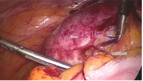
Pneumoperitoneum is created using a Veress needle at Palmer’s point. Peritoneal entry is done by visual technique using a Ternamian Endotip (Karl Storz, Tuttlingen) at the umbilicus or supraumbilicus in patients with large abdominal masses. Abdominopelvic inspection is done for omental and bowel adhesions and to confirm the preoperative diagnosis (Fig.1). The three-accessory-port technique is used, two ports in the lower quadrants lateral to the inferior epigastric artery and the third port in the suprapubic area. First, a secondary trocar is inserted under vision in an adhesion-free area, and other ports
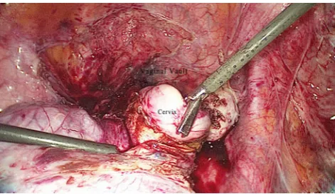
are inserted after adhesiolysis. The port position is modified according to the size of the pelvic mass. Omental adhesions on the abdominal wall are released using a monopolar hook, scis-sors or Harmonic Ace (Ethicon Endosurgery, Cincinnati, OH). Sharp dissection close to the parietal peritoneum is employed to achieve lysis of bowel adherent to the anterior abdominal wall. A Clermont Ferrand uterine manipulator (Karl Storz, Tuttlingen, Germany) with a modified bulger is used. Small-bowel or rectosigmoid adhesions to the uterus and adnexa (Fig. 2) are released by sharp dissection as far as possible. Injection of dilute vasopressin into the myometrium close to bowel adhesions is used to achieve reduction in vascularity. This also facilitates ease of dissection by raising the serosa from the myometrium at the site of adhesion. In dense bowel adhesions, dissection onto the serosa of the uterus is performed. In the majority of cases, complete rectosigmoid adhesiolysis is not possible because of the severity of adhesions and poor accessibility due to the enlarged size of the uterus or adnexa. Enlarged endometriomas, cysts and encysted fluid collections are decompressed for better visibility. Identification of the round ligament and division of the same close to the lateral pelvic wall on both sides are performed. Adnexal adhesions to the uterus are released, and the cornual structures are divided with Enseal or Harmonic Ace. Division of the infundibulopelvic ligament is done at a later stage owing to possible proximity to the ureters. The uterovesical fold is opened and bladder dissection carried out subsequently. The uterine arteries are identified and adhesiolysis done to achieve its safe division. After the uterine arteries were secured, the anterior fornix is opened over the Clermont Ferrand fornix bulger. Once division of the anterior half of the vagina is achieved, the manipulator is removed and the cervix held with a tenaculum and pulled up (Fig.3). A vaginal blocker is employed to prevent loss of pneumoperitoneum. Circumferential dissection of the vagina is continued to completely separate the cervix from the vagina. Rectal adhesions to the posterior vaginal wall are re-leased by application of traction to the anterior and subsequently posterior lip of the cervix. This helps in safe completion of posterior vaginal wall incision. The posterior lip of the cervix is pulled away from posterior rectal adhesions, and by maintaining traction on the cervix, sharp adhesiolysis is done
in retrograde fashion to release the rectum and sigmoid (Figs.4
and5). Once the rectum is completely separated from the uterus, retrieval of the specimen is done vaginally with or without morcellation depending on the size of the uterus. A vaginal blocker is used to achieve reinsufflation of the peritoneal cavity. Removal of the uterus creates space to identify the adnexa and helps with their dissection from the lateral pelvic wall. Adnexectomy is done after identifying the ureter, and the specimen is removed vaginally. Endometriotic implants and nodules over the rectal wall are excised. Vault closure is done laparoscopically (Fig. 6). Cystoscopy is done to confirm the integrity of the bladder and urine reflux from both the ureters. The rectum is inflated with air using a no. 16 F disposable suction catheter per anus to check for any inadver-tent rectosigmoid injury. A drain is kept in the pelvis through one of the secondary ports and the 10-mm port closed with sutures.
Findings
Total laparoscopic hysterectomy with retrograde adhesiolysis was done in 25 patients with frozen pelvis between October 2003 and May 2012. Patient data are shown in Table1. The mean (standard deviation (SD); 95 % confidence interval (CI)) age of patients was 42.6 (6.00; 40.1–45.07), and body mass
index was 27.48 (5.06; 25.3–29.57). Twenty (80 %) patients had previous abdominal surgery (laparotomy, laparoscopy), and three (15 %) patients had previous failed surgeries for hysterectomy. Fifteen patients had previous laparotomies, of which eight had one laparotomy, three had two laparotomies, three had three laparotomies and one had five laparotomies. Eleven patients had previous laparoscopic surgeries, of which six had both laparotomy as well as laparoscopy. Fourteen (56 %) patients were nulliparous, and 11 (44 %) were parous, of which five (20 %) had previous caesarean sections. The presenting complaints of patients were dysmenorrhoea in 17 (68 %) and abdominal pain in 8 (32 %). Twenty-three (92 %) patients had frozen pelvis due to severe endometriosis [4]. Out of these 23 patients, seven had fibroid uterus with severe endometriosis, seven had adenomyosis of the uterus with severe endometriosis and nine had severe endometriosis. Two patients had severe abdominopelvic adhesions due to multiple surgeries in the past.
Laparoscopic hysterectomy with retrograde adhesiolysis was successful in 24 (96 %) patients, and one patient had laparotomy conversion. Operative details are mentioned in Table2. In all the cases, the uterine artery was ligated after complete anterior wall adhesiolysis and before adhesiolysis of the posterior aspect of the uterus. Because of dense adhesions, adnexectomy was done at the end of surgery in all cases. One patient had intraoperative injury to the sigmoid colon and bladder during adhesiolysis, and laparotomy conversion was
Fig. 3 Pulling up the anterior lip of the cervix to visualize the posterior vaginal wall
Fig. 4 Retrograde separation of the uterus from the rectosigmoid
Fig. 5 Final separation of the rectum from the uterus
done with the assistance of a general surgeon; the bladder was repaired in two layers, and since the sigmoid perforation was 2 cm in size with >60 % stricture of a 4-cm segment of the sigmoid colon due to an endometriotic nodule, intestinal
segmental resection and sigmoid colostomy were performed. Two units of blood were transfused. There were no complica-tions related to entry procedures. Post-operative morbidity in the form of paralytic ileus was noted in two (8 %) patients who were managed conservatively. Intraoperative blood transfu-sion was needed only in one patient. The median (range) operating time was 210 (120–300) min, and estimated blood loss was 400 (300–600) ml. Length of post-operative stay was 1 (1–6) days. The median (range) follow-up visit at 1 month and 6 months was 100 and 68 % (17 of 25), respectively, and was uneventful in all except for one patient who had a local-ized haemorrhagic collection in the pelvis measuring 4.5× 5 cm; an ultrasonography-guided aspiration was done at 1 month follow-up. A telephonic inquiry at 1 year was done to confirm the well-being of patients and recurrence of symp-toms. None of our patients had recurrence of symptoms or occurrence of new symptoms pertaining to endometriosis.
Discussion
Hysterectomy is the commonest major gynaecological proce-dure [5]. Most hysterectomies can be performed laparo-scopically including those with large myomas and severe en-dometriosis [6–8]. Various techniques have been described previously to tackle difficult hysterectomies, but the level of expertise needed limits the general use of these techniques [9,
10]. Even experienced gynaecologic laparoscopic surgeons find it very difficult to operate in such condition, and the surgery may be abandoned in some cases. In our series, three patients had failed surgeries with attempted hysterectomy and were abandoned due to the operative difficulties. At our centre, total laparoscopic hysterectomy with retrograde adhesiolysis is cho-sen in all cases of frozen pelvis.
Laparoscopic hysterectomy in frozen pelvis facilitates nexa removal compared to vaginal hysterectomy as the ad-nexa may be adherent to surrounding structures that may be extremely difficult to reach transvaginally [9]. The laparotomy conversion rate for laparoscopic hysterectomy in severe en-dometriosis and large fibroids as described in literature is 4.3 and 7.9 % [8,11]. In our series, one patient had conversion to laparotomy. This was done to facilitate repair of incidental bladder perforation in a densely adherent bladder and to achieve bowel resection for severe bowel endometriosis with bowel stenosis of greater than 60 % [12]. A study involving 115 patients with severe endometriosis undergoing laparo-scopic hysterectomy reported bladder injury in 1 (0.9 %) of 115 cases without any bowel or ureteral injury [8]. In our series, 23 patients had frozen pelvis due to severe endometri-osis. Our surgical approach in these patients is focused pri-marily on symptom-guided approach rather than mandatory oncologic resection [13]. All visible endometriotic lesions
Table 1 Patient data
Variable Valuea Age 42.6 years BMI 27.48 Parity median (range) 0 (0–2) Previous abdominal (laparoscopy + laparotomy)
surgery
20 of 25 (80 %) Previous failed surgeries 3 of 20 (15 %) Previous laparoscopies only 5 of 20 (30 %) Previous laparotomy and laparoscopy 6 of 20 (20 %) Previous laparotomies 15 of 20 (70 %) Previous 1 laparotomy 8 of 20 (40 %) Previous 2 laparotomies 2 of 20 (10 %) Previous 3 laparotomies 3 of 20 (15 %) Previous 5 laparotomies 1 of 20 (5 %) Severe endometriosis with fibroid uterus 7 (28 %) Severe endometriosis with adenomyosis of uterus 7 (28 %) Severe endometriosis 8 (32 %) Severe endometriosis with bowel endometriosis 1 (4 %) Severe abdominopelvic adhesion 2 (8 %) Follow-up 1 month 25 (100 %) Follow-up 6 months 17 (68 %)
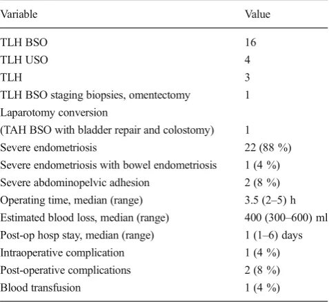
Table 2 Operative data
Variable Value TLH BSO 16 TLH USO 4
TLH 3
TLH BSO staging biopsies, omentectomy 1 Laparotomy conversion
(TAH BSO with bladder repair and colostomy) 1 Severe endometriosis 22 (88 %) Severe endometriosis with bowel endometriosis 1 (4 %) Severe abdominopelvic adhesion 2 (8 %) Operating time, median (range) 3.5 (2–5) h Estimated blood loss, median (range) 400 (300–600) ml Post-op hosp stay, median (range) 1 (1–6) days Intraoperative complication 1 (4 %) Post-operative complications 2 (8 %) Blood transfusion 1 (4 %)
were excised and the cul de sac cleared of endometriotic lesions. Considering the rare progressive nature of deep en-dometriosis, segmental bowel resection was not performed for superficial bowel lesions [14,15]. We routinely identify the ureter during and after dissection and do cystoscopy at the end of the procedure to confirm the integrity of the bladder and patency of the ureters. Even the most experienced surgeon may encounter bowel injury. The golden rule is early recog-nition of injury as time of diagnosis is the most important independent factor determining the outcome. A difficult sur-gical condition can be well anticipated, and the surgeon can arrange for preoperative and, if required, intraoperative surgi-cal or urologisurgi-cal assistance. Surgery should be performed in a setting where such facilities are readily available. Paralytic ileus may occur probably due to extensive bowel adhesiolysis and handling; out of 25 patients, two patients developed paralytic ileus in the post-operative period whereas James et al. reported seven cases with paralytic ileus following major gynaecological procedure in 707 patients [16]. The risk of complications depends upon the extent of bowel involvement, adhesions and extent of endometriosis infiltration, surgeon experience and bowel resection [16,17]. The operating time was 210 min in our study compared to 185±48.7 min men-tioned by Chalermchockchareonkit et al., while it was 131 and 147 min in two case reports by Walid et al. [6] in laparoscopic hysterectomy for severe endometriosis [8]. This can be explained by the severity of the disease process and grossly distorted anatomical planes, necessitating meticulous dis-section and slow progression. Mean (SD; 95 % CI) weight of the uterus was 390.2 (441.59; 207.91–572.49) g. The mean estimated blood loss in our series was 384 ml whereas Chalermchockchareonkit reported a mean blood loss of 302.6 ml [8], while that reported by Walid et al. was 150 ml in laparoscopic hysterectomy for severe endometri-osis [6]. The length of hospital stay was 1.3±1.07 days in our series which is shorter when compared to 3.5±1.1 days re-ported in the literature [8]. One patient (4 %) required blood transfusion in our series, whereas nine (7.8 %) required blood transfusion out of 115 patients undergoing hysterectomy for severe endometriosis [8].
All procedures have their own limitations, and retrograde adhesiolysis during hysterectomy in frozen pelvis requires adequate surgical experience and expertise to change the course of surgery and manage the complications associated with dif-ficult pelvic surgery. Specialized assistance may be needed to manage the complications. The surgeon should be prepared to modify the surgical technique according to the case. The over-all key to success in such cases depends on thorough knowl-edge of pelvic anatomy and operative experience involving varying degrees of pelvic distortion with or without enlarged uteri. This technique of retrograde adhesiolysis can decrease the complications in difficult hysterectomies with gross pelvic
distortion due to adhesions even in the presence of enlarged uteri. This is a retrospective study, and further evidence is required, preferably by adequately powered well-designed multicentre randomized controlled trials (RCTs) before defini-tive conclusions can be given.
Conclusion
Even though frozen pelvis is not a common surgical condition, it is not rare to come across such cases in clinical practice. Our technique of initial partial adhesiolysis followed by hysterec-tomy and retrograde separation of posterior bowel adhesions and subsequent removal of adnexa is an alternative technique. This technique in the hands of a very experienced surgeon might be more appropriate in decreasing the laparotomy con-version rate and the overall complications. This is a retrospec-tive study, and for this rare and difficult situation, it would be impossible and unethical to have a control group; however, adequately powered well-designed multicentre RCTs are re-quired before definitive conclusions can be given.
Conflict of Interest The authors report no conflicts of interest. The authors alone are responsible for the content and writing of the paper.
References
1. Donald PG, Michael JC (2007) Surgical strategies to untangle a frozen pelvis. OBG Management 19(3)
2. Hudson CN (1981) Victor Bonney lecture, 1980. Ovarian cancer—a gynaecological disorder? Ann R Coll Surg Engl 63:118–25 3. Volpi E, Bernardini L, Ferrero AM (2012) The retrograde and
retro-peritoneal totally laparoscopic hysterectomy for endometrial cancer. Int J Surg Oncol 2012:263850
4. Schenken RS, Guzick DS (1997) Revised ASRM classification for endometriosis: 1996. Fertil Steril 67:820
5. Stovall TG (2007) Hysterectomy. In: Berek JS (ed) Berek & Novak’s gynecology, 14th edn. Lippincott Williams & Wilkins, Philadelphia, pp 805–846
6. Walid SM, Heaton RL (2011) Total laparoscopic extirpation of a fixed uterus from benign gynecological disease. Gynecol Surg 8:157–159
7. Sinha R, Sundaram M, Lakhotia S, Mahajan C, Manaktala G, Shah P (2009) Total laparoscopic hysterectomy for large uterus. J Gynecol Endosc Surg 1:34–39
8. Chalermchockchareonkit A, Tekasakul P, Chaisilwattana P, Sirimai K, Wahab N (2012) Laparoscopic hysterectomy versus abdominal hysterectomy for severe pelvic endometriosis. Int J Gynaecol Obstet 116:109–111
9. Pelosi MA III, Pelosi MA (1997) Vaginal hysterectomy for benign uterine disease in the laparoscopically confirmed frozen pelvis. J Laparoendosc Adv Surg Tech 7:345–351
11. Moon MJ, No JH, Jeon YT, Jee BC, Kim YB (2011) Clinical outcomes of 1,041 total laparoscopic hysterectomies: six years of experience in a single center. Korean J Obstet Gynecol 54:618–622 12. Ferrero S, Camerini G, Maggiore U, Venturini PL, Biscaldi E,
Remorgida V (2011) Bowel endometriosis: recent insights and unsolved problems. World J Gastrointest Surg 3:3–38
13. Roman H, Vassilieff M, Gourcerol G, Savoye G, Leroi AM, Marpeau L et al (2011) Surgical management of deep infiltrating endometriosis of the rectum: pleading for a symptom-guided approach. Hum Reprod 26:274–281
14. Koninckx PR, Ussia A, Leila Adamyan L, Arnaud W, Jacques D (2012) Deep endometriosis: definition, diagnosis, and treatment. Fertil Steril 98:564–571
15. Mereu L, Ruffo G, Landi S, Barbieri F, Zaccoletti R, Fiaccavento A et al (2007) Laparoscopic treatment of deep endometriosis with segmental colorectal resection: short-term morbidity. J Minim Invasive Gynecol 14:463–469
16. Donnez J, Squifflet J (2010) Complications, pregnancy and recur-rence in a prospective series of 500 patients operated on by the shaving technique for deep rectovaginal endometriotic nodules. Hum Reprod 25:1949–1958