Iranian Biomedical Journal 16 (3): 121-126 (July 2012) DOI: 10.6091/ibj.1082.2012
Functional Recombinant Extra Membrane Loop of Human CD20,
an Alternative of the Full Length CD20 Antigen
Mahdi Habibi Anbouhi
1, Aida Feiz Barazandeh
1, Saeid Bouzari
2, Mohsen Abolhassani
3,
Hossein Khanahmad
4, Majid Golkar
5, Mohammad Reza Aghasadeghi
6, Mahdi Behdani
1, Ali
Jahanian-Najafabadi
2and Mohammad Ali Shokrgozar
*11National Cell Bank of Iran, Pasteur Institute of Iran, Tehran; 2Molecular Biology Unit, Pasteur Institute of Iran,
Tehran; 3Hybridoma Laboratory, Dept. of Immunology, Pasteur Institute of Iran, Tehran; 4Production and Research
Complex, Pasteur Institute of Iran, Karaj; 5Molecular Parasitology Laboratory, Dept. of Parasitology, Pasteur Institute
of Iran, Tehran; 6Dept. of Hepatitis and AIDS, Pasteur Institute of Iran, Tehran, Iran
Received 28 March 2012; revised 23 June 2012; accepted 25 June 2012
ABSTRACT
Background:Targeting of CD20 antigen with monoclonal antibodies has become the mainstay in the treatment of non-Hodgkin's lymphomas and immunotherapeutic depletion of malignant B cells.Accessibility of antigen is one of the crucial factors in development of monoclonal antibodies against this antigen. One major problem in expression of full length CD20 is aggregation and misfolding. Therefore, production of an alternative polypeptide is easer and favorable comparing to that of a full length transmembrane protein CD20. Methods:In this study, we
expressed the extra membrane loop of hCD20 (exCD20)consisting of a non-glycosylated 47-amino acids region. The exCD20 coding sequence was amplified by PCR and cloned in pET32a(+) expression vector. The desired protein was expressed in fusion with thioredoxin and 6× His tag in E. coli Origami strain. ELISA and Western-blotting data were performed to indicate the functionality of this protein.Results: We have obtained the exCD20
recombinant protein which can be detected in ELISA and Western-blot experiments. This recombinant fusion protein was soluble and stable without aggregation and misfolding problems.Conclusion:The recombinant extra
membrane loop of human CD20 protein in fusion with thioredoxin (exCD20) can be used in function assays and some applications such as ELISA, immuneblotting, affinity purification, immunization, screening, and development of anti-CD20 antibodies.Iran. Biomed. J. 16 (3): 121-126, 2012
Keywords: E. Coli, CD20, Thioredoxin
INTRODUCTION
orld Health Organization has reported that 7.6 million people worldwide died from cancer in 2008. The Non-Hodgkin's lymphoma is expected to be the fifth most common cancer in American men and women [1]. It is a disease in which malignant (cancer) cells are formed in the lymph system. For lymphomas, chemotherapy and radiation therapy have been the mainstay of treatment. On the contrary of immunotherapy, both of these modalities suffer from a lack of specificity [2].
All the currently available anti-CD20 antibodies have been selected and produced against the extra membrane loop of hCD20 via different immunization methods [3]. The human B-lymphocyte-restricted differentiation antigen Bp35 (CD20, MS4A1), a 35-kDa hydrophobic phosphor-protein, is expressed as a
cell surface, non-glycosylated protein during early pre-B-cell development [4] and predicted to span the plasma membrane four times. It is highly expressed on the plasma membrane of almost all plasma B cells, but not on hematological stem cells [5] and plasma cells [6]. This 297 amino acid length protein is consisted of cytoplasmic N- and C-termini and four hydrophobic regions for anchoring the molecule in the membrane. Although the biological function of CD20 remains unclear, some evidence indicated that it might function as a calcium ion channel [7-11]. The importance of CD20 as a target for immunetherapeutic depletion of B cells is irrefutable and anti-CD20 monoclonal antibodies appear to be ideal for the treatment of B-cell malignancies [12].
While for many soluble proteins suitable overexpression systems are used routinely, high-level production of membrane proteins is still challenging.
W
One phenomenon frequently observed in E. coliis that many heterologous proteins become incorrectly folded and accumulate in the cytoplasm as insoluble aggregates, called inclusion bodies [13, 14].Therefore, expression of full length hCD20 protein produces a recombinant protein with incorrect folding and/or probably aggregated forms and using some techniques like liposome technology for hCD20 antigen (Abnova, Taiwan) has not proved yet.
Up to now, a few other studies have focused on expression of full length hCD20 protein in different forms such as purified recombinant protein on the surface of a mammalian cell membrane or transmembrane domain of CD20 in fusion with gIII protein of a phage [15-18]. Misfolding, aggregation and hyper immunogenicity and complexity of fused protein/particle to the recombinant CD20 are the drawbacks that should be avoided.
In this study, we selected the 47 amino acid-extra membrane loops of hCD20 between the third and fourth transmembrane regions [19] to express it in fusion with thioredoxin of pET32a(+) prokaryotic expression vector. After expression, it would be possible to assess the functionality of recombinant exCD20 by ELISA and Western-blotting. The functional exCD20 may be used in some functional assays and applications, such as ELISA, immune-blotting, affinity purification, immunization, screening, and development of anti-CD20 antibodies.
MATERIALS AND METHODS
Cloning and construction of exCD20 expression cassette. Amplification of exCD20 coding sequence
was performed by PCR with 53C of annealing temperature and using specific primers and the pORF9-hCD20a eukaryotic plasmid (InvivoGen, USA) as template. This vector is an expression vector containing the full length of human CD20a (MS4A1) isoform 1 gene.Cloning was facilitated by BamHI and XhoI restriction enzyme sites engineered in the primers
used for amplification (underlined): tCD20F:
TGGATCCAATATTAAAATTTCCCATT and
tCD20R:AATCTCGAGTTATATGCTGTAACAGTA TTGG. The purified PCR product was ligated to pTZ57R/T plasmid (Thermo Scientific, USA) and used to Transform E. coliTOP10 strain competent cells by InsTAclone™ PCR Cloning Kit (Thermo Scientific, USA). Accuracy of PCR amplification and insertion was confirmed by colony PCR using T7 promoter and tCD20R primers and finally by sequencing. The authentic sequence was finally subcloned into the pET32a(+) prokaryotic expression vector (Novagen, USA) for high-level expression of peptide sequence
fused to the 109aa TRX•Tag™ thioredoxin protein and 6× His tag.
Expression and purification of exCD20. Since the
extra membrane loop of hCD20 has a disulfide bond between C167 and C183, the recombinant pET32a(+) plasmid was transformed into E. coliOrigami™ strain
(Novagen, USA) as expression host strain to enhance disulfide bond formation in the cytoplasm. One ml of overnight cultured single colony of the transformed E. coliOrigami™ strain was used to inoculate 300 ml TB medium supplemented with 300 μl 1,000× ampicillin stock (100 μg/ml ampicillin final concentration) in a baffled shaker flask. The flask was incubated at 37°C at 200 rpm reaching optical density (OD600) of 0.6 to 0.9 in an incubator shaker. Expression was induced by adding 0.5 mM isopropyl-beta-D-thiogalacto-pyranoside (Applichem, Germany). Induction was followed by 5 hours of incubation at 30°C and 200 rpm. E. colicells were harvested by centrifugation at 5,000 ×g at 4°C for 8 min. The cell pellet was resuspended in 10 ml lysis buffer (50 mM Tris-HCl, pH 8.0), 25% w/v sucrose (Merck, Germany), 1 mM EDTA (Sigma, Germany), 100 µg/ml lysozyme (Sigma, Germany), and 1 ml complete protease inhibitor cocktail (Roche, Germany) in a 50 ml falcon tube and incubated on ice for 30 min. Then, it was frozen at -20°C and thawed by immersing the tube in 37°C water. Ten ml of the lysis buffer was added to the lysate and centrifuged at 8,000 ×g at 4°C for 8 min. Supernatant was collected and the expressed protein was purified by HisPur™ Ni-NTA Resin (Thermo Scientific, USA) via its 6× His tag. Imidazole removal and complementary purification of exCD20 protein was performed by size exclusion chromatography (SEC) by Ä KTA Explorer FPLC system (GE Healthcare Life Sciences, UK) and two separated peaks were fractionated.
Characterization of exCD20:
ELISA tests. ELISA plates (Nunc, Germany) were
coated with 1 µg per well of exCD20 in PBS at 4°C overnight. Then, they were blocked with 2.5% BSA at 37C for 2 h. After 3 washes, 100 µl of anti-CD20 peptide antibody in PBS (10 µg/ml) [20] was added to the wells and the plates were incubated at 37C for 1.5 h. The wells were washed, and 100 µl of 1:4,000 diluted horseradish peroxidase-conjugated rabbit anti-mouse Ig (Sigma, Germany) was added, then incubated for 1.5 h at 37°C. After 5 washes, 100 µl of tetramethylbenzidine solution (Sigma, Germany) was added, the plates were incubated at room temperature in dark, then the reaction was stopped after 1 min. P5 peptide (the complete extra membrane loop of hCD20 with a disulfide bond between C167 and C183) was
Iran. Biomed. J., July 2012 Recombinant Extra Membrane Loop of Human CD20 123
Table 1. Nucleotide sequence of a gene coding for extra-membrane loop of human CD20 and its amino acid sequence. A 225-amino acid sequence was produced by expression of exCD20 pET32a recombinant vector.
Name Size Sequence
extra-membrane loop of hCD20
Nucleotide 141 bp AATATTAAAATTTCCCATTTTTTAAAAATGGAGAGTCTGAATTTTATTAGAGCTCACACACCATATATTAACATATACAACTGTGAACCAGCTAATC CCTCTGAGAAAAACTCCCCATCTACCCAATACTGTTACAGCATA
Amino acid 47 aa NIKISHFLKMESLNFIRAHTPYINIYNCEPANPSEKNSPSTQYCYSI
Recombinant
exCD20 Amino acid 225 aa
EGDIHMSDKIIHLTDDSFDTDVLKADGAILVDFWAEWCGPCKMIAPILDEI ADEYQGKLTVAKLNIDQNPGTAPKYGIRGIPTLLLFKNGEVAATKVGALS KGQLKEFLDANLAGSGSGHMHHHHHHSSGLVPRGSGMKETAAAKFERQ HMDSPDLGTDDDDKAMADIGSNIKISHFLKMESLNFIRAHTPYINIYNCEP ANPSEKNSPSTQYCYSILEHHHHHH
synthesized (Biomatik, Canada) and used as positive control. Lysate of E. coli Origami strain transformed by empty vector pET32a(+) (TRX), as well as PBS were used as negative controls. OD was measured at 450 nm by ELISA reader (State Fax 2100, USA). All procedures were performed in triplicate.
SDS-PAGE and Western-blot analysis. One
microgram of each two peaks of SEC experiment (Fig. 2) was run on 10% NuPAGE®Novex Bis-Tris Mini Gels (Invitrogen, USA). The separated proteins were transferred to Hybond-C nitrocellulose membrane (GE Healthcare, UK) by using a Bio-Rad protein transfer apparatus (100 v, 250 mA, 90 min). PageRuler™ Prestained Protein Ladder (Thermo Scientific, USA) was used in both experiments. Following protein transfer, the nitrocellulose membrane was blocked by blocking buffer containing 5% skimmed milk plus 0.25% Tween.The exCD20 band was detected by the anti-CD20 peptide antibody (10 µg/ml in the blocking buffer, 1 h, 25C), and alkaline phosphatase-conjugated anti-mouse antibody (Sigma, Germany) (1:2,000 dilution in the blocking buffer, 25C, 1 h) as primary and secondary antibodies, respectively. Alkaline phosphatase enzyme conjugate was detected by nitro-blue tetrazolium chloride and 5-bromo-4-chloro-3'-indolyphosphate p-toluidine salt substrates (Thermo Scientific Pierce, USA).
RESULTS
Cloning and construction of exCD20 expression cassette. In order to construct the exCD20 expression cassette, the 141-nucleotide sequence (47 amino acids) of the extra membrane loop of hCD20 (Table 1) was amplified by PCR and using tCD20F and tCD20R amplification primers. Amplified region gave a band of the expected size (around 150 bp) on a 1.5% agarose gel (Fig. 1A). The PCR product was cleaned up and after digestion with restriction enzymes, cloned into
the pTZ57R/T and then subcloned into the pET32a(+) expression vector. The band of around 720 bp was corresponding to the PCR product of colony PCR with T7 Promoter and tCD20R primers (Fig. 1B). The positive clone was sequenced (Genfanavaran, Iran) using tCD20F and tCD20R primers. The sequencing data was compared with sequence of hCD20 gene in the NCBI website (Gene ID: 931) and the sequence of inserted exCD20 was confirmed.
Expression, purification and characterization of exCD20. After Transformation of recombinant vector
into E. coli Origami™, it was cultivated and induced with isopropyl-beta-D-thiogalactopyranoside. The expressed protein was a 225-amino acid sequence
including the extra membrane loop of hCD20 peptide
Fig. 1. Gel electrophoresis of PCR product and colony PCR. The 141-bp exCD20 coding sequence was amplified by PCR and 2 µl sample of PCR product was run on a 1.5% agarose gel (A). This sequence was cloned into pET32a(+) expression vector and the cloning was confirmed by colony PCR. The amplified product was visualized around 720 bp on a 1% agarose gel (582 bp, from T7 promoter binding site to BamHI cut site + 141 bp, the size of insert)(B).
Fig. 2.Chromatogram of size exclusion chromatography protein peaks in addition to some small protein peaks
imidazole was obtained at the end of size exclusion chromatography
(bolded and underlined), a 109aa thioredoxin protein and two 6× His tags expressed protein was purified by
Ni-according to the manufacture's instruction and exCD20 protein was eluted and collected
1 ml. The Ni-NTA purified protein was loaded on column and fractionated and two main peaks
were obtained. Also, there were some small protein peaks before the main peaks. A conductivity peak corresponding to the imidazole was obtained at the end of SEC experiment (Fig. 2). The ELISA results for exCD20 and P5 peptide were strongly positive comparing to the negative controls. In comparison with the negative control [TRX, the lysate of
Origami which was transfected with empty pET plasmid], the OD450nm values for exCD peptide were significantly higher (Table main peaks were analyzed by SDS-PAGE
Silver staining showed the expected molecular mass of near 30 kDa fusion recombinant protein in both peaks (Fig. 3A). Western-blot analysis showed the reactivity of mouse anti-CD20 peptide
these two bands (Fig. 3B).
DISCUSSION
In the present study, we obtained a
binant extra membrane loop of hCD20 in fusion with thioredoxin and its native disulfide bridge
Since 1988, a few studies have attempted to expres recombinant hCD20 antigen for different purposes 18]. In one study, the full length of hCD
and expressed [18]. However, aggregation,
and inclusion body formation are common phenomena
chromatography. The sample was loaded onto a Superdex 75 column
some small protein peaks (peaks 1 and 2) were separated. A conductivity peak corresponding to the size exclusion chromatography experiment.
aa-TRX•Tag™ (Table 1). The -NTA column instruction and the protein was eluted and collected in volume of loaded on SEC peaks of protein Also, there were some small protein peaks before the main peaks. A conductivity peak corresponding to the imidazole was obtained at the end The ELISA results for peptide were strongly positive In comparison with TRX, the lysate of E. coli Origami which was transfected with empty pET32a(+)
exCD20 and P5 able 2). The two PAGE (10% gel). the expected molecular mass of kDa fusion recombinant protein in both main blot analysis showed the peptide antibody with
novel in fusion with disulfide bridge (exCD20). studies have attempted to express
different purposes [15-the full length of hCD20 was cloned
aggregation, misfolding and inclusion body formation are common phenomena
in expression of full length transmembrane proteins because of hydrophobic regions (membrane
domains) within these types of proteins. study [17], a recombinant vector with
hCD20 was transfected into a mammalian cell line and CD20 was expressed on the surface of the cells in order to study the structure and function of
study [16], the extra-membrane loop of been displayed on the surface of M
this recombinant phage was used to develop immune response in animal. In this approach
antibodies were developed against the phage
the fused peptide, because the phage is a complex and strong immunogenic carrier. In addition, the native disulfide bond of hCD20 does not construct in the normal cytosolic condition in the
engineered strain like E. coliOrigami
Since all the currently available anti antibodies have been selected and produced against the extra membrane loop of hCD20
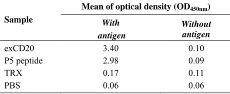
Table 2.ELISA results. One µg of the
lysate of E. coli Origami strain which transformed by empty vector pET32a(+) (TRX), were coated
CD20 peptide antibody and anti-mouse
conjugated antibody were used as primary and secondary antibodies, respectively. ELISA test was performed in triplicate.
Sample
Mean of optical density (
With antigen
exCD20 3.40
P5 peptide 2.98
TRX 0.17
PBS 0.06
column. Two overlapped main A conductivity peak corresponding to the
in expression of full length transmembrane proteins f hydrophobic regions (membrane-spanning domains) within these types of proteins. In another a recombinant vector with full length of was transfected into a mammalian cell line and
expressed on the surface of the cells in order function of CD20. Also, in a membrane loop of hCD20 has ace of M13K07 phage and this recombinant phage was used to develop immune . In this approach, most of the against the phage but not because the phage is a complex and carrier. In addition, the native does not construct in the the bacteria as good as Origami host strain [21]. currently available anti-CD20 been selected and produced against the 20 [3], in our study, we
esults. One µg of the exCD20, P5 peptide or Origami strain which transformed by empty were coated in each well. The
anti-mouse alkaline phosphatase-conjugated antibody were used as primary and secondary antibodies, respectively. ELISA test was performed in triplicate.
ptical density (OD450nm)
Without antigen 0.10 0.09 0.11 0.06
Iran. Biomed. J., July 2012 Recombinant Extra Membrane Loop of Human CD20 125
Fig. 3. SDS-PAGE and Western-blot analyses of size
exclusion chromatography (SEC) columnpurified samples.One µg from peaks 1 and 2 of chromatography-purified SEC fraction was subjected to SDS-PAGE (10% acrylamide gel) under reducing conditions (A). Separated proteins were then transferred to the nitrocellulose membrane and stained by a mouse anti-CD20 peptide antibody and anti-mouse alkaline phosphatase-conjugated antibody as primary and secondary antibodies, respectively (B).
used the extra membrane loop of hCD20 instead of its full length. This region was expressed in a Thioredoxin fusion system in the E. coliOrigami strain to force the formation of disulfide ridge [21] between its two cysteines in the cytosol as native form on the surface of B-cells [22]. This strategy resulted in a stable, soluble and functional protein [23, 24].
As Table 2 shows, in ELISA a high OD450nm value for exCD20 as well as P5 peptide were obtained. This result confirmed that exCD20 had an epitope with appropriate conformation to interact with the anti-CD20 peptide antibody. Western-blotting also confirmed this conclusion (Fig. 3).
Two bands of peak 1 and one band of peak 2 were detected in Western-blot experiment. The bands of about 30 kDa are corresponding to the exCD20 fused with the thioredoxin and His tags and the band of about 60 kDa seems to correspond to its dimer form (Fig. 3).
Purification steps show the necessity of optimization of conditions for purification of exCD20 with Ni-NTA column. Some changes in concentration of imidazole in the washing step, the volume of washing buffer and the time of binding incubation may reduce the contaminations and increase the xCD20purity.
The anti-CD20 peptide antibody which was developed against P5 peptide [20], detected the 30 band exCD20 in the Western-blot. The 60 kDa-band seems to be the exCD20 dimer (Fig. 3).
In conclusion, the functional exCD20 protein can be expressed in E. coli Origami host strain. This recombinant extra membrane loop of hCD20 can be used instead of its full length protein without aggregation and misfolding problems in development of poly- and/or monoclonal anti-hCD20 antibodies. Also, it may be used in ELISA, immunoblotting, affinity purification, immunization and screening.
ACKNOWLEDGEMENTS
We are very grateful to Prof. Dr. Serge Muyldermans (Laboratory of Cellular and Molecular Immunology, Vrije Universiteit Brussel), Dr. Amir Amanzadeh and Mr. Hassan Sanati for their outstanding technical assistances. Financial support of this study has been provided by Pasteur Institute of Iran, Postgraduate Office.
REFERENCES
1. Ferlay J, Shin HR, Bray F, Forman D, Mathers C,
Parkin DM. Estimates of worldwide burden of cancer in
2008: GLOBOCAN 2008. Int J Cancer.2010 Dec;127
(12):2893-917.
2. Furman RR, Coleman M, Muss D, Leonard JP.
Monoclonal antibodies in the treatment of
non-Hodgkin's lymphoma. Cancer Treat
Res.2006;131:221-50.
3. Teeling JL, Mackus WJ, Wiegman LJ, van den Brakel
JH, Beers SA, French RR, et al. The biological activity of human CD20 monoclonal antibodies is linked to unique epitopes on CD20. J Immunol.2006 Jul;177(1):
362-71.
4. Reff ME, Carner K, Chambers KS, Chinn PC, Leonard
JE, Raab R, et al. Depletion of B cells in vivo by a
chimeric mouse human monoclonal antibody to CD20.
Blood.1994 Jan;83(2):435-45.
5. Nishida M, Usuda S, Okabe M, Miyakoda H, Komatsu
M, Hanaoka H, et al. Characterization of novel murine anti-CD20 monoclonal antibodies and their comparison to 2B8 and c2B8 (rituximab). Int J Oncol. 2007 Jul;
31(1): 29-40.
6. Perosa F, Favoino E, Caragnano MA, Prete M,
Dammacco F. CD20: a target antigen for
immunotherapy of autoimmune diseases. Autoimmun
Rev.2005 Nov;4(8):526-31.
7. Robak T, Robak E. New anti-CD20 monoclonal
antibodies for the treatment of B-cell lymphoid
malignancies. BioDrugs.2011 Feb;25(1):13-25.
8. Cragg MS, Walshe CA, Ivanov AO, Glennie MJ. The
biology of CD20 and its potential as a target for mAb
therapy. Curr Dir Autoimmun.2005;8:140-74.
9. Einfeld DA, Brown JP, Valentine MA, Clark EA,
Ledbetter JA. Molecular cloning of the human B cell CD20 receptor predicts a hydrophobic protein with
multiple transmembrane domains. EMBO J. 1988
Mar;7(3):711-7.
10. Stashenko P, Nadler LM, Hardy R, Schlossman SF. Expression of cell surface markers after human B
lymphocyte activation. Proc Natl Acad Sci USA.1981
Jun;78(6):3848-52.
11. Beers SA, Chan CH, French RR, Cragg MS, Glennie MJ. CD20 as a target for therapeutic type I and II
monoclonal antibodies. Semin Hematol. 2010
Apr;47(2):107-14..
12. Deans JP, Li H, Polyak MJ. CD20-mediated apoptosis: signalling through lipid rafts. Immunology.2002 Oct;
107(2):176-82.
13. Hunte EPC. Production and Purification of
Recombinant Membrane Proteins. In: Hunte C, editor. Membrane Protein Purification and Crystallization; A Practical Guide. 2nded. Academic Press; 2003.p. 55-83.
14. Mus-Veteau I. Heterologous Expression of Membrane Proteins for Structural Analysis. In: Mus-Veteau I, editor. Heterologous Expression of Membrane Proteins; Methods and Protocols. 1sted. Humana Press; 2010. p. 1-16.
15. Hong HY, Sun YX, Guo YX, Wang JN, Lai CN, Qi ZT, et al. Cloning and Expression of Human CD20 Gene on
NIH-3T3 Cell Membrane. Sheng Wu Hua Xue Yu Sheng
Wu Wu Li Xue Bao (Shanghai). 2000;32(4):430-3.
16. Zhang XY, Sun ZW, Yu WY, Cheng JZ. Cloning and expression of fusion gene of transmembrane domain of
human CD20 and g3pN in Escherichia coli. Xi Bao Yu
Fen Zi Mian Yi Xue Za Zhi.2004 Jul;20(4):481-3.
17. Bubien JK, Zhou LJ, Bell PD, Frizzell RA, Tedder TE. Transfection of the CD20 Cell Surface Molecule into Ectopic Cell Types Generates a Ca Conductance Found
Constitutively in B Lymphocytes. Cell Biol.1993;121
(5):1121-32.
18. Ernst JA, Li H, Kim HS, Nakamura GR, Yansura DG, Vandlen RL. Isolation and Characterization of the
B-Cell Marker CD20. Biochemistry.2005 Nov;44(46):
15150-8.
19. Ernst JA, Li H, Kim HS, Nakamura GR, Yansura DG, Vandlen RL. Isolation and characterization of the B-cell
marker CD20. Biochemistry.2005;44(46):15150-8.
20. Habibi Anbouhi M, Abolhassani M, Bouzari S,
Khanahmad S, Aghasadeghi M, Madadkar
S.Immunological evaluation of predicted linear B-cell
epitopes of human CD20 antigen. Biotechnol Appl
Biochem.2012;X:1-6.
21. de Marco A. Strategies for successful recombinant expression of disulfide bond-dependent proteins in
Escherichia coli. Microbial Cell Fact.2009 May;8:26.
22. Oscherwitz J, Gribbin TE, Cease KB. A CD20 tandem-epitope immunogen elicits antibody in mice that binds murine cell surface CD20 and depletes splenic B cells in
vivo. Mol Immunol.2010 Apr;47(7-8):1484-91.
23. Nikitina J, Shutova T, Melnik B, Chernyshov S, Marchenkov V, Semisotnov G, et al. Importance of a single disulfide bond for the PsbO protein of photosystem II: protein structure stability and soluble
overexpression in Escherichia coli. Photosynth
Res.2008 Oct-Dec;98(1-3):391-403.
24. Xiong S, Wang YF, Ren XR, Li B, Zhang MY, Luo Y, et al. Solubility of disulfide-bonded proteins in the cytoplasm of Escherichia coli and its "oxidizing"
mutant. World J Gastroenterol.2005
Feb;11(7):1077-82.