M
ACIEJP
ARUZEL1, J
ERZYJ
ABŁECKI1, J
ERZYZ
WOLIŃSKI2Posttraumatic Cerebrospinal Fluid Rhinorrhea
Accompanied by Pneumocephalus – Case Report
Pourazowy płynotok nosowy z odmą śródczaszkową
– opis przypadku
Department of General Surgery, St Jadwiga’s Hospital, Trzebnica, Poland Department of Neurosurgery, 4thMilitary Clinical Hospital, Wrocław, Poland
Adv Clin Exp Med 2007, 16, 6, 819–823 ISSN 1230−025X
LETTER TO EDITOR
© Copyright by Silesian Piasts University of Medicine in Wrocław
Abstract
Cerebrospinal fluid rhinorrhea accompanies about 2% of blunt head traumas and in half of these cases it results from fracture of the anterior cranial fossa. This life−threatening condition may lead to meningitis, often with lethal outcome. Another complication of head injury is pneumatocele. It is present in ca. 0.5–1% of head injuries, but it occurs in up to 30% of patients suffering from rhinorrhea. The main difficulty in diagnosing both pathologies is due to the delayed onset of their symptoms, as was the case of the described patient. The patient did not consent to surgical management, which is the treatment of choice for the considered conditions. The conservative treatment which was introduced resulted in the resolution of both pathologies (Adv Clin Exp Med 2007, 16, 6, 819–823).
Key words: cerebrospinal rhinorrhea, intracranial pneumocephalus, cranial injury, conservative treatment.
Streszczenie
Płynotok towarzyszy około 2% zamkniętych urazów głowy, a u połowy chorych jest związany ze złamaniem prze− dniego dołu czaszki. Zagrożenie życia chorego związane z powstaniem płynotoku wynika z możliwości rozwinię− cia się zapalenia opon mózgowo−rdzeniowych o bardzo ciężkim przebiegu. Innym typem patologii urazów głowy jest odma śródczaszkowa. Towarzyszy ona około 0,5–1% urazów głowy, ale aż w 30% współistnieje z płynoto− kiem. Trudności związane z rozpoznaniem obu chorób wynikają przede wszystkim z późniejszego ich wystąpie− nia, tak jak to miało miejsce u leczonego przez autorów chorego. Chory nie wyraził zgody na leczenie operacyjne, które jest bezwzględnie wskazane w tego typu przypadkach. Prowadzone z konieczności leczenie zachowawcze doprowadziło do ustąpienia obu patologii (Adv Clin Exp Med 2007, 16, 6, 819–823).
Słowa kluczowe:płynotok, odma śródczaszkowa, urazy głowy, leczenie zachowawcze.
Cerebrospinal fluid leakage through the nose, known as cerebrospinal fluid rhinorrhea (CSFR), is a pathology caused by the formation of a fistula between the cranium and the roof of the nasal cavi− ty, ethmoid air cells, frontal or sphenoid sinus, and tympanic membrane [1]. CSFR may be caused by a trauma, a neoplasm that destroys the bone struc− ture, osteitis, hydrocephalus, and congenital bone structure abnormalities; it may also have no apparent cause [2, 3] (Fig. 1). The fistula forms a potential gate of infection from the nasal cavity and paranasal sinuses directly into the cranium, which may lead to cerebrospinal meningitis and encephalitis.
Pneumocephalus is another life−threatening condition that accompanies 0.5–1% of head injuries; however, it can be present in up to 30% of patients with posttraumatic CSF rhinorrhea [2, 8]. Beside direct trauma, the causes of pneumo− cephalus include inflammation of the facial bones (especially the middle ear) and barotraumas (for instance those related to diving) [9, 10]. The most dangerous is tension pneumocephalus, produced by the formation of a valve at the injury site, because this leads to an increase in intracranial pressure. Pneumocephalus is particularly frequent when the air pressure in the nasal cavity rises rapidly, as occurs during coughing or sneezing. Pneumocephalus comprises a greater risk to the patient because communication between the crani− um and the environment is characterized by the intracranial direction, which obviously increases the risk of meningitis [1].
The relative rarity of the aforementioned pathologies related to head injuries and the serious risk associated with them requires emergency medicine specialists to be prepared for handling both CSF rhinorrhea and pneumocephalus. The aim of this report is to familiarize emergency med− icine professionals with these conditions.
Case Description
A 40−year−old male intoxicated with ethanol, was admitted to the emergency room after experi− encing a transportation accident (Glasgow Coma
Scale 13, BP 130/70 mmHg, HR 108/min, Hb satu− ration 92%, blood alcoholic content 3.62‰). A pre− liminary physical examination revealed ecchymosis on the chest (CXR: fracture of the posterior part of the 7th left rib and frontal part of the 2nd left rib), a minor wound if the left region of the forehead, excoriation of the nose, and minor bleeding from both nasal meatus. No neurological defects of the central nervous system were observed. Similarly, abdominal ultrasonography did not reveal any post− traumatic injuries of the internal organs. The head X−ray made on admission of the patient did not pre− sent any apparent pathology.
In the first days of hospitalization, the patient’s condition did not differ significantly from the norm. On the second day of hospitaliza− tion a neurologist examined the patient and described his state in the following way: “correct praxis; comprehensible and logical speech; absent meningeal reflexes; equal, regular, reactive pupils; extensor plantar response present bilaterally; no focal signs of CNS deterioration” (P. Szada, MD). Based on the results of this examination, the intensive vigilance was interrupted and the patient was being prepared to be discharged from the hos− pital. However, because of the lack of any means of transport, the patient asked to be discharged the following day (i.e. on day 4). On that day, the patient’s state worsened. He reported symptoms of a common cold, suggested by shivers and abun− dant water−clear discharge from the nose; the neu− rological state was correct. An X−ray of the skull, performed again in the typical projections,
rhinorrhoea cerebrospinalis
płynotok nosowy
post traumatic pourazowy
no trauma involvment bez związku z urazem
trauma uraz wypadkowy
iatrogenic trauma uraz jatrogenny
increased intracranial preassure zwiększone ciśnienie wewnątrzczaszkowe
normal intracranial preassure
normalne ciśnienie wewnątrzczaszkowe
tumors guzy
hydrocephalus wodogłowie
congenital defects
suppurative infection of internal ear
osteomyelitis
wady wrodzone zapalenie szpiku kostnego
ropne zapalenie ucha środkowego cloed
zamknięte
communicating komunikujące direct effect
destruction działanie bezpośrednie
destrukcja
indirect effect intracranial high preassure
pośrednie nadciśnienie wewnątrzczaszkowe
Ryc. 1.Przyczyny płynotoku nosowego
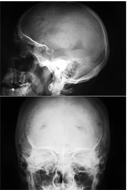
revealed air occupying the cerebral ventricles; the paranasal sinuses were correctly filled with air, with there were no pathologies (Fig. 3).
Since CSF rhinorrhea accompanied by pneu− mocephalus was suspected, antibiotic treatment was instituted and the nasal discharge was exam− ined, and because of its properties (yellowish, tur− bid, protein concentration 1.1 g/100 ml, glucose 63 mg/100 ml, ALP activity 34U/l) it was classified as transudate. Urgent CT of the head confirmed air accumulation in the anterior parts of both lateral ventricles and a few minor accumulations in the paracerebral and subarachnoid spaces. The CT image also revealed “a hypotensive zone at the base of the right frontal lobe – a probable result of the trauma; signs of cortical and subcortical atro− phy with dilatation of the paracerebral CSF space in the region of the left frontal lobe; a basilar skull fracture in the sphenoid plane and in the roof of the sphenoid sinus; abundant liquid occupying the sphenoid sinus, most probably blood. No other pathologies were visible.” (B. Hendrich, MD).
Based on the performed examinations, a com− plex cranio−cerebral injury was diagnosed. The patient was therefore transferred to a neurosurgical ward, where CSF rhinorrhea intensification was observed. The patient complained of nausea, headache, and vertigo since admission to the hospi− tal. On inclination of the patient’s head, the CSFR intensified. CSF diversion was performed twice with subsequent examination of the liquid: color clear yellow, transparency slightly turbid, benzidine reaction positive (++), RBC 10,000/µl, pleocytosis 60/3, lymphocytes 68%, and polymorphonuclear leukocytes 32%. After centrifugation the color was clear yellow, transparency totally transparent, ben− zidine probe negative, Pandy’s reaction ambiguous
(+, –), Nonne−Apelt reaction positive (+), and pro− tein concentration 41.5 mg/100 ml.
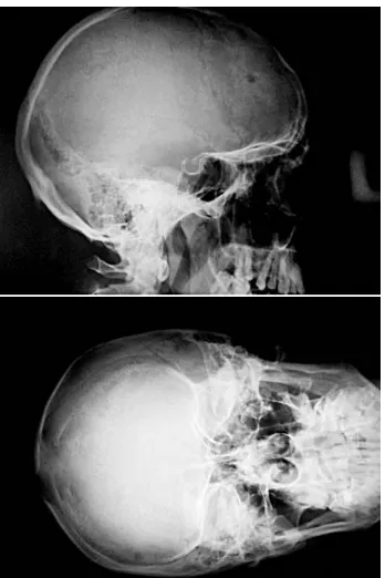
For surgical treatment, the patient was offered a duraplasty and closing of the CSF fistula. However, he did not consent to this therapy. The conservative treatment was thus continued, i.e. obligatory semisupine position and a 10−day−long antibiotic treatment (Oframax, 2 g/day). In this way, the CSF leak resolved successfully. During tilt trials, the patient complained of pains and ver− tigo; however, these symptoms resolved sponta− neously. Moreover, the onset of an insignificant retrograde amnesia of the pre−trauma period was noted. The patient was dismissed from the neuro− surgical ward on day 21 of hospitalization. Later he appeared regularly to the outpatient clinic. The follow−ups revealed no neurological disorders; the cough trial and the Valsalva maneuver were nega− tive. An X−ray of the skull done two weeks after leaving the hospital showed no air presence in the cranial cavities (Fig. 4). The last follow−up, per− formed 11 months after the trauma, showed no deviations from the norm, and the patient did not report any episodes of fever, headaches, sinusitis, or rhinitis.
Fig. 2. Skull X−ray made on admission – no apparent pathology
Ryc 2. RTG czaszki w chwili przyjęcia do oddziału – bez widocznej patologii
Fig. 3. Skull X−ray on the 4thday of hospitalization –
note the pneumocephalus
Discussion
A fistula through which CSF leaks forms most commonly where the bones composing the base of the skull are relatively thin. That is why CSFR fistu− las are mainly located in the lamina ethmoidea or in the roof of the anterior/posterior part of the laby− rinthus ethmoidalis (ca. 40%). The leak may also occur from the posterior wall of the frontal sinus (15%) or from the roof of the sphenoid sinus (15%). Occasionally, cerebrospinal fluid may leak into the nasal cavity from the middle ear through the audito− ry tube [2–4]. A common cause of these leaks is a fracture of the roof of the tympanic cavity, which constitutes a part of the base of the skull. Leakage of CSF into the throat represents the main sign of this type of CSFR. The rhinorrhea may start soon after the trauma or a few days later when the tissular edema resolves and the coagula are resorbed, thus opening new communication between the fossa cranii mediaand the paranasal sinuses [1, 4, 7].
The differential diagnosis of CSF rhinorrhea often presents a challenging problem. Examining the glucose level in the leaking fluid is very helpful,
since a glucose concentration > 30 mg/100 ml con− firms the CSFR diagnosis. Of great value is also an immunological assay that detects β−2 transferritin because this protein is present only in the CSF and the vitreous, so blood presence in the sample does not influence the result [1]. Nevertheless, this test is hardly available in Poland. If a fluid specimen can− not be obtained or the very presence of a fistula is doubtful, scintigraphy might be applied. In this way, after intrathecal administration of a radioactive trac− er (for instance 99mTc), radioactivity of the gastric content may be shown if the fluid leaks onto the posterior wall of the throat [1, 5, 7].
Air presence in the skull is perceived as “flows inside the head”, and in senile patients it may cause awareness disorders [2, 11]. The air usually accumulates in the highest level of the subarach− noid space and/or in the cerebral ventricles, as occurred in our patient. Highly dangerous is the valvular mechanism of pneumocephalus; in this case, air accumulates in the skull with every inspi− ration. This quickly leads to an increase in intracranial pressure and requires an urgent neuro− surgical procedure, such as a trephinopuncture [2, 8]. Most commonly, pneumocephalus forms during the six hours after an acute head trauma [1]. Nonetheless, cases are known in which pneumo− cephalus occurred weeks after the trauma [11].
There are various techniques of managing CSFR [3, 4, 7]. However, it seems that CSFR
Fig. 4. CT scan depicts air presence in the cerebral lat− eral ventricles
Ryc. 4.Badanie TK – obecność powietrza w komorach bocznych mózgu
Fig. 5.Follow−up X−ray made two weeks after dis− charge from the hospital shows no pathologies
should be treated surgically because the scar has an impaired resistance, which may result in recurrence of the CSF leaks even years after the trauma [4, 5, 12–14]. CSFR accompanied by pneumocephalus is a stronger indication for surgical treatment than CSFR alone. In such a case, the operation should take place ca. two weeks after the trauma [2, 8]. Szurmacz suggests that surgical treatment allows for closing the fistula permanently in 95% of patients, while 25% of patients treated conserva− tively died three years after the trauma due to pyo− genic cerebrospinal meningitis [4]. Unfortunately, it was impossible to operate our patient (suspected of alcoholism) since he refused to consent to the oper− ation although we provided him detailed informa− tion on possible complications.
The clinical picture of our patient is relatively uncommon. Its following features need to be stres− sed: no apparent lesions in the preliminary X−ray, onset of CSFR and pneumocephalus a few days after the trauma with no accompanying neurological dis− orders, and resolution of CSFR and pneumo− cephalus in the third week of conservative treatment. A one−year−long observation of the patient is obviously not enough for estimating the risk of CSFR recurrence, but that is not the aim of this report. Awareness of the diagnostic difficulties asso− ciated with CSFR and pneumocephalus as well as their insidious course is of practical importance for traumatologists. With the above data in mind, it is no exaggeration to state that even the apparently most trivial head injuries require scrupulous observation.
References
[1] Czernicki Z, Walecki J:Urazy czaszkowo−mózgowe. W: Podstawy chirurgii, podręcznik dla lekarzy specjalizu− jących się w chirurgii. Red.: Szmidt J. Medycyna Praktyczna, Kraków 2003, s. 627–650.
[2] Czernicki Z.Uraz czaszkowo−mózgowy. W: Chirurgia głowy i szyi. Red.: Kryst L, PZWL, Warszawa 1999, 13–60.
[3] Lindstrom DR, Toohill RJ, Loehrl TA, Smith TL:Management of cerebrospinal fluid rhinorrhea: the Medical College of Wisconsin experience. Laryngoscope 2004, 114, 6, 969–974.
[4] Szurmacz L:Bezpośrednie i odległe wyniki leczenia płynotoków nosowych. Praca doktorska, Akademia Me− dyczna w Gdańsku, Gdańsk 1997.
[5] Gassner HG, Ponikau JU, Sherris DA, Kern EB:CSF rhinorrhea: 95 consecutive surgical cases with long term follow−up at the Mayo Clinic Am J Rhinol 1999, 13, 6, 439–447.
[6] Kowalina I, Kopczyński S:Samoistny płynotok nosowy – opis 2 przypadków. Neur Neurochir Pol 1992, 26, 399–402.
[7] Kowalina I, Kopczyński S:Płynotok nosowy. Leczenie operacyjne. Pol Przegl Lek, 1993, supl. 2, 224–228.
[8] Bayassi S:Odroczona odma śródmózgowa w przebiegu urazu czaszkowo−podstawnego. Opis przypadku i prze− gląd literatury. Neur Neurochir Pol 1997, 31, 5, 1047–1052.
[9] Cowie RA, Harris P:Spontaneous infected pneumatocoele secondary to chronic otitis media. Acta Neurochir 1979, 49, 3–4, 227–234.
[10] Goldmann RW: Pneumocephalus as a consequence of barotraumas JAMA 1986, 255, 22, 3154–3156.
[11] Chan YP, Yau CY, Lewis RR, Kinirons MT: Acute confusion secondary to pneumocephalus in an elderly pa− tient. Age Ageing 2000, 29, 4, 365–367.
[12] Sanan A, Heines SJ: Repairing holes in the head: a history of cranioplasty. Neurosurgery 1997, 41, 999–1004.
[13] Spetzler RF, Wilson CB:Management of recurrent CSF rhinorrhea of the middle and posterior fossa. J Neuro− surgery 1988, 49, 393–397.
[14] Mc Cormac B, Cooper PR, Persky M:Extracranial repair of cerebrospinal fluid fistulas: technique and results in 37 patients. Neurosurgery 1990, 27, 412–417.
Address for correspondence:
Maciej Paruzel
St. Jadwiga’s Hospital in Trzebnica Prusicka 53
55−100 Trzebnica Poland
E−mail: [email protected] Conflict of interest: None declared Received: 24.01.2007