Magdalena Grzonkowska
a, b, d, Mateusz badura
a–c, Jakub Lisiecki
a, b,
Michał Szpinda
d–f, Mariusz baumgart
c, Marcin Wiśniewski
cGrowth Dynamics of the Triceps Brachii Muscle
in the Human Fetus
department of Normal anatomy, Ludwik Rydygier collegium Medicum in bydgoszcz, Nicolaus copernicus University in Toruń, Poland
A – research concept and design; B – collection and/or assembly of data; C – data analysis and interpretation; D – writing the article; E – critical revision of the article; F – final approval of article; G – other
Abstract
Background. The triceps brachii muscle, the strongest extensor of the elbow joint, is characterized by the three heads: long, lateral and medial.
Objectives. In the present study we aimed to examine the linear parameters (length, width) of the fetal triceps brachii muscle and to provide their growth dynamics.
Material and Methods. Using anatomical dissection, digital image analysis (Multiscan v.14.02), and statistics (Student’s t-test, regression analysis) we measured in mm the length and width of the triceps brachii in 30 fetuses of both sexes (12♂,18♀) aged 12–29 weeks.
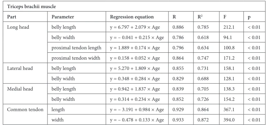
Results. Neither sex nor laterality differences were found. all the parameters studied increased proportionately with age. The linear functions were computed as follows: y = 6.797 + 2.079 x age (r = 0.886) for length of the long head’s belly, y = – 0.041 + 0.215 × age (r = 0.786) for width of the long head’s belly, y = 1.889 + 0.174 × age (r = 0.796) for length of the long head’s proximal tendon, y = 0.158 + 0.052 × age (r = 0.864) for width of the long head’s proximal tendon, y = 5.270 + 1.809 × age (r = 0.855) for length of the lateral head’s belly, y = 0.348 + 0.284 × × age (r = 0.829) for width of the lateral head’s belly, y = 0.942 + 1.837 × age (r = 0.839) for length of the medial head’s belly, y = 0.314 + 0.234 × age (r = 0.852) for width of the medial head’s belly, y = – 3.191 + 0.984 × age (r = 0.929) for lenght of the common tendon, and y = – 0.478 + 0.133 × age (r = 0.933) for width of the common tendon.
Conclusions. Neither male-female nor right-left differences are observed in morphometric parameters of the tri-ceps brachii muscle. The long head’s belly is the thinnest, while the lateral head’s belly is the widest one. The long head is the longest and the medial head is the shortest one. The developmental dynamics of the triceps brachii muscle follow proportionately (Adv Clin Exp Med 2014, 23, 2, 177–184).
Key words: triceps brachii muscle, attachments, length, width, regression analysis.
adv clin Exp Med 2014, 23, 2, 177–184 ISSN 1899–5276
ORIGINaL PaPERS
© copyright by Wroclaw Medical University
The triceps brachii muscle, the primary exten-sor of the elbow and shoulder joints and a help-ing adductor at the shoulder joint (long head), is relevant for the normal functioning of the upper limb. as a natural antagonist to the biceps brachii, it solely occupies the posterior compartment of the arm [1]. The long head normally springs from the infraglenoid tubercle. The lateral head originates from the lateral intermuscular septum and from the humerus, superior to the radial groove, while the medial head arises from the medial intermus-cular septum and from the humerus, inferior to the radial groove [2, 3]. The whole muscle extends distally to be inserted via a common tendon onto
the olecranon. according to Keener et al. [4], in 14 specimens aged 71 years, the mean tendon width at its insertion was found to be approximately 78% of the maximum width of the olecranon.
by chronic posterior elbow pain with active exten-sion, mostly in energetic men, e.g. throwing ath-letes at the age of 30–40 years [10]. Triceps bra-chii tendon ruptures have been described as the least common (2%) of all tendon injuries [11–13], as a result of a fall onto an outstretched hand or a direct blow to the triceps tendon. Injury to the triceps brachii occurs at the distal tendon-olecra-non, myotendinous or intramuscular junctions, and proximally at the origin of the lateral head [14, 15]. These conditions spontaneously tend to accompany systemic diseases (rheumatoid arthri-tis, systemic lupus erythematosus, hyperparathy-roidism, chronic renal failure and hemodialysis, Marfan syndrome) and chronic steroid use [16, 17] that alter the structural integrity of the tendon.
To date, little attention has been given to the quantitative anatomy of the growing triceps brachii in the fetus. Therefore, in the present study the fol-lowing three objectives were set in order to exam-ine the following:
1) normal values for the three heads (in terms of their length and width) and two tendons, one proximal and one distal (in terms of their length and width) at varying gestational ages,
2) the growth curves for normal development of the features studied, and
3) the impact of sex and size on the values of the parameters examined.
Material and Methods
This study was performed in accordance with the approval of our University Research Ethics committee (Kb 72/2012). The examination was carried out on 30 human fetuses of both sexes (12 males, 18 females) from spontaneous abortions and stillbirths. The whole fetal material included specimens at the age of 12–29 weeks. all fetuses were preserved in 10% neutral formalin solution for 24–36 months. Every triceps brachii muscle was dissected to visualize its attachments, then recor-dered with a millimeter scale (figs. 1 and 2) using a SONY camera α 330 and analyzed by digital image analysis system (Multiscan v. 14.02). for each muscle the following 10 parameters were computed:
1) length of the long head’s belly, measured from its origin to its termination,
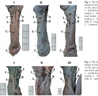
Fig. 1. The triceps brachii muscle in fetuses aged 14 (I), 19 (II), and 29 weeks (III) 1 – lateral head’s proximal tendon, 2 – long head’s prox-imal tendon, 3 – common tendon, a – lateral head’s belly, b – long head’s belly, c – common belly
2) length of the lateral head’s belly, measured from its origin to its termination,
3) length of the medial head’s belly, measured from its origin to its termination,
4) length of the proximal tendon of the long head, measured from the infraglenoid tubercle to its end,
5) length of the common tendon, mea-sured from the end of the common belly to the olecranon,
6–8) three widths, one for every head, mea-sured at their widest levels,
9) width of the proximal tendon of the long head, measured at its widest level,
10) width of the common tendon, measured at its widest level.
The statistical analysis was started by assessing the probability of appearance of statistically signifi-cant differences in values with relation to sex (Stu-dent’s t-test for unpaired variables) and laterality (Student’s t-test for paired variables) using Statis-tica 9.0. The particular parameters were correlated to fetal age. Regression analysis was used to derive the curve of best fit for the plot for each morpho-metric parameter vs. gestational age. differences were considered significant at p < 0.05.
Results
The three heads of the triceps brachii mus-cle always extended typically. all individual nu-merical data obtained for the triceps brachii muscle have been presented in Table 1. Nei-ther laterality nor sex differences (p > 0.05) be-tween the parameters studied were found. as a result, we did not attempt to separate fur-ther the numerical data into sex and the side. as presented in Table 2, the statistically significant correlation between each parameter studied and fetal age was found, being expressed by p value and correlation coefficient (r). The statistically signifi-cant correlation (p < 0.01) between every param-eter studied and fetal age was observed. The ex-amined features revealed a proportionate increase in values when related to advancing fetal age. This was presented using regression analysis, including regression formulae of best fit, statistics values (f), and coefficients of determination (R2) – (Table 2).
When compared to its lateral (37.06 mm) and medial (33.24 mm) heads, the mean length (43.36 mm) of the long head was statistically great-er (p = 0.0002; p = 0.0003). When compared to its both medial (4.42 mm) and long (3.74 mm) head’s bellies, the mean width of the lateral head’s belly measured at its widest level (5.34 mm) was found to be statistically greater (p = 0.001; p = 0.008).
Long Head of Triceps Brachii
In the examined age-group the values for length of the long head ranged from 27.43 to 63.15 mm respectively, and averaged 43.36 mm. The val-ues for width of the long head varied from 2.05 to 5.76 mm respectively, with the mean of 3.74 mm. The values for length of the long head’s tendon ranged from 3.26 to 6.11 mm respectively, and av-eraged 4.95 mm. The values for width of the long head’s proximal tendon fluctuated from 0.79 to 1.45 mm respectively, to average 1.07 mm. all the aforementioned parameters revealed a proportion-ate increase when plotted against gestational age (Table 2).
Lateral Head of Triceps Brachii
during the analyzed period the values for length of the lateral head varied from 23.63 to 55.64 mm, respectively, and averaged 37.06 mm. The values of width of the lateral head ranged from 3.21 to 8.14 mm respectively, to average the value of 5.34 mm. The lateral head increased linearly in both length and width (Table 2).
Medial Head of Triceps Brachii
between 12 and 29 weeks, the values for length of the medial head ranged from 19.74 to 51.45 mm respectively, with the mean of 33.24 mm. The val-ues for width of the medial head varied from 2.67 to 6.44 mm respectively, and averaged 4.42 mm. as indicated in Table 2, the growth of the medial head followed proportionately.
Common Tendon
of Triceps Brachii
In the study period, the values for length of the common tendon ranged from 10.21 to 28.30 mm respectively, and averaged 14.11 mm. The width of the common tendon varied from 1.01 to 3.02 mm, respectively, with the mean of 1.86 mm. both pa-rameters were found to rise in accordance with lin-ear functions (Table 2).
Discussion
Table 1. Individual numerical data of the fetal triceps brachii muscle
Fetal number Age (weeks) Sex F –
Female
M
–
Male
Side R –
right L – left Long head (mm) Lateral head (mm) Medial head (mm) Common tendon (mm)
belly length belly width proximal tendon
length
proximal tendon
width belly length belly width belly length belly width
common tendon
length
common tendon
Fetal number Age (weeks) Sex F –
Female
M
–
Male
Side R –
right L – left Long head (mm) Lateral head (mm) Medial head (mm) Common tendon (mm)
belly length belly width proximal tendon
length
proximal tendon
width belly length belly width belly length belly width
common tendon
length
common tendon
the growth dynamics obtained would be more de-tailed. fortunately, the adequate digital program of Multiscan v. 14.02 appears to compensate for that disadvantage by means of objective, precise, and repeatable semi-automatic evaluation of the mus-cle parameters studied in digital pictures.
during the 5th week of embryogenesis, the
tri-ceps brachii musculature develops from the dorsal muscle mass of the upper limb bud [2]. Seldom do we find variations of the triceps brachii muscle in the medical literature. as it turned out, the fourth head of the triceps brachii may have developed from various points of the shoulder joint capsule, the scapula, the humerus, and the coracoid process [2, 18]. autopsy studies by Eiserloh et al. [3] and Handling et al. [5] confirmed a constant capsular contribution to every orgin of the long head of the triceps brachii. This partly remains contradictory to Huber and Putz [19], who noticed the long ten-don of triceps brachii pass to the glenoid labrum in 38% of individuals. Soubhagya et al. [18] described the fourth head of the triceps brachii, being at-tached to the medial aspect of humerus, just be-low the insertion of the latissimus dorsi and teres major tendons. Macalister [20] found the fourth head of the triceps, as a split of the long head, with a long, slender tendon attached to the shoulder cap-sule. furthermore, Tubbs et al. [2] reported an ac-cessory head, being derived from the medial head with its anomalous tendinous attachment into the posterior aspect of the surgical neck of humerus.
as far as entrapment syndromes are con-cerned, the radial nerve was reported to be com-pressed by both the lateral [21, 22] and me-dial [2, 23] heads of the triceps brachii, with a potential radial nerve palsy. Spinner et al. [24]
described the so-called “snapping elbow syndrome” as a consequence of ulnar nerve entrapment from the hypertrophied medial head of the triceps, when flexing the elbow joint. furthermore, in 17% of in-dividuals the medial head of the triceps may bridge over the ulnar groove of humerus to form the an-coneus epitrochlearis, which compresses the ulnar nerve [23]. On the contrary, according to Tubbs et al. [25], deep fibers of the medial head are re-sponsible for the formation of the anconeus muscle, which tends to tether the medial component of the triceps brachii, thus decreasing the likelihood that it would compress the ulnar nerve.
after reviewing the professional literature, we failed to find reference data, Kędzia et al. [26] aside, concerning triceps brachii dimensions. No signif-icant sex and laterality differences between all the triceps brachii parameters were found. Neither sex nor laterality differences remain in agreement with our previous findings concerning the fetal semi-membranosus [27] and semitendinosus [28] mus-cles. Interestingly enough, according to Kędzia et al. [29], the sartorial muscle length was found to be significantly larger both in female fetuses and on the left side. In the material under examination, the growth dynamics of all muscle features studied followed proportionately, thereby being expressed by linear regressions. The strongest correlations re-ferred to the following: the lengths of the common tendon, long, lateral, and medial heads, and long head’s proximal tendon, the widths of the common tendon, medial and lateral heads, and long head’s proximal tendon, and the weakest to the width of the long head. Our findings confirmed a balanced growth of the triceps brachii, being precisely ex-pressed by straight lines.
Table 2. Statistics of the triceps brachii muscle
Triceps brachii muscle
Part Parameter Regression equation R R2 F p
Of note, Kędzia et al. [26] in 70 fetuses aged 15–28 weeks measured the widths of the three heads of triceps brachii muscle. The mean values for widths of the long, medial and lateral heads ranged 6.0 mm, 4.6 mm, and 6.1 mm, respective-ly. In the material under examination, the lateral head’s belly was also the widest, but the long head’s belly was the thinnest.
In the present study, the balanced, proportion-ate growth of the triceps brachii muscle resembles that of different skeletal muscles, reported in the literature [27, 28, 30–32]. This may be exemplified by the morphometric analysis of the semimem-branosus [27] and semitendinosus [28], the bi-ceps femoris [30], deltoid [31], and bibi-ceps brachii
[32] in the human fetus. The lack of information in the medical literature concerning the param-eters studied obviously limits discussion on this subject. We believe that the normative data of the triceps brachii muscle in the fetus obtained in this study will provide the background for future au-topsy studies.
The authors concluded that neither sex dif-ferences nor laterality difdif-ferences are observed in morphometric parameters of the fetal triceps bra-chii muscle. The lateral head’s belly is the widest, while the long head’s belly is the thinnest. The long head is the longest, while the medial head is the shortest. The growth dynamics of the triceps bra-chii muscle follow proportionately.
References
[1] Piquilloud G, Villani F, Casoli V: The medial head of the triceps brachii. anatomy and blood supply of a new muscular free flap: the medial triceps free flap. Surg Radiol anat 2011, 33, 415–420.
[2] Tubbs RS, Salter EG, Oakes WJ: Triceps brachii Muscle demonstrating a fourth Head. clin anat 2006, 19, 657–660. [3] Eiserloh H, Drez D, Guanche CA: The long head of the triceps: a detailed analysis of its capsular origin. J Shoulder
Elbow Surg 2000, 9, 332–335.
[4] Keener JD, Chafik D, Kim HM, Galatz LM, Yamaguchi K: Insertional anatomy of the triceps brachii tendon. J Shoulder Elbow Surg 2010, 19, 399–405.
[5] Handling MA, Curtis AS, Miller SL: The origin of the long head of the triceps: a cadaveric study. J Shoulder Elbow Surg 2010, 19, 69–72.
[6] Landin D, Thompson M: The shoulder extension function of the triceps brachii. J Electromyogr Kinesiol 2011, 21, 161–165.
[7] Bekler H, Wolfe VM, Rossenwasser MP: a cadaveric Study of Ulnar Nerve Innervation of the Medial Head of Triceps brachii. clin Orthop Relat Res 2009, 467, 235–238.
[8] Cheema P, Singla R: four headed triceps brachii muscle: IJaV 2011, 4, 43–44.
[9] Athwal GS, McGill RJ, Rispoli DM: Isolated avulsion of the Medial Head of the Triceps Tendon: an anatomic Study and arthroscopic Repair in 2 cases. arthroscopy 2009, 25, 983–988.
[10] Jafarnia K, Gabel GT, Morrey BF: Triceps Tendinitis. Oper Tech Sports Med 2001, 9, 217–221.
[11] Kibuule LK, Fehringer EV: distal triceps tendon rupture and repair in an otherwise healthy pediatric patient: a case report and review of the literature.J Shoulder Elbow Surg 2007, 16, 1–3.
[12] Sharma SC, Singh R, Goel T, Singh H: Missed diagnosis of triceps tendon rupture: a case report and review of literature. J Orthop Surg (Hong Kong) 2005, 13, 307–309.
[13] Sierra RJ, Weiss NG, Shrader MW, Steinmann SP: acute triceps ruptures: case report and retrospective chart review. J Shoulder Elbow Surg 2006, 15, 130–134.
[14] Blackmore SM, Jander RM, Culp RW: Management of distal biceps and Triceps Ruptures. J Hand Ther 2006, 19, 154–169.
[15] Tatebe M, Horii E, Nakamura R: chronically ruptured triceps tendon with avulsion of the medial collateral liga-ment: a report of 2 cases. J Shoulder Elbow Surg 2007, 16, 5–7.
[16] Petre BM, Grutter PW, Rose DM, Belkoff SM, McFarland EG, Petersen SA: Triceps tendons: a biomechanical comparison of intact and repaired strength. J Shoulder Elbow Surg 2011, 20, 213–218.
[17] Tagliafico A, Gandolfo N, Michaud J, Perez MM, Palmieri F, Martinoli C: Ultrasound demonstration of distal triceps tendon tears. Eur J Radiol 2012, 81, 1207–1210.
[18] Soubhagya RN, Ashwin K, Madhan KSJ, Latha VP, Vasudha S, Merin MT: four-headed biceps and triceps brachii muscles, with neurovascular variation. anat Sci Int 2008, 83, 107–111.
[19] Huber WP, Putz RV: Periarticular fiber system of the shoulder joint. arthroscopy 1997, 13, 680–691.
[20] Macalister A: additional observations on muscular anomalies in human anatomy (third series), with a catalogue of the principal muscular variations hitherto published. Trans Roy Irish acad Sci 1875, 25, 1–134.
[21] Mitsunaga MM, Nakano K: High radial nerve palsy following strenuous muscular activity. a case report. clin Orthop Relat Res 1988, 234, 39–42.
[22] Nakamichi K, Tachibana S: Radial nerve entrapment by the lateral head of the triceps. J Hand Surg (am) 1991, 16, 748–750.
[23] Fabrizio PA, Clemente FR: Variation in the triceps brachii muscle. a fourth muscular head. clin anat 1997, 10, 259–263.
[25] Tubbs RS, Oakes WJ, Salter EG: The subanconeus muscle. folia Morphol 2006, 65, 22–25.
[26] Kędzia A, Woźniak J, Dudek K: Metrological analysis of topography of radial nerve in humeral segment during fetal period. Medical and biological Sciences 2009, 23, 51–57.
[27] Badura M, Wiśniewski M, Szpinda M, Siedlaczek W, Ufnal-Brzozowska S: developmental dynamics of the semimembranosus muscle in human foetuses. Medical and biological Sciences 2011, 25, 13–16.
[28] Badura M, Wiśniewski M, Szpinda M, Siedlaczek W: The growth of the semitendinosus muscle in human foe-tuses. Med biol Sci 2011, 25, 17–21.
[29] Kędzia A, Wałek E, Podleśny K, Dudek K: Musculus sartorius metrology in the fetal period. adv clin Exp Med 2011, 20, 567–574.
[30] Szpinda M, Wiśniewski M, Rolka Ł: The biceps femoris muscle in human foetuses – a morphometric, digital and statistical study. adv clin Exp Med 2011, 20, 575–582.
[31] Szpinda M, Paruszewska-Achtel M, Baumgart M, Sobolewska M, Eliminowska-Wenda G: Quantitative growth of the human deltoid muscle in human foetuses. Med biol Sci 2011, 25, 59–64.
[32] Szpinda M, Paruszewska-Achtel M, Dąbrowska M, Badura M, Eliminowska-Wenda G, Sobolewska A, Szpinda A: The Normal Growth of the biceps brachii Muscle in Human fetuses. adv clin Exp Med 2013, 22, 17–26.
Address for correspondence:
Michał Szpindadepartment of Normal anatomy
Ludwik Rydygier collegium Medicum in bydgoszcz Karłowicza 24
85-092 bydgoszcz Poland
Tel.: + 48 52 58 53 705 E-mail: [email protected]
conflict of interest: None declared