This is an open access journal, and articles are distributed under the terms of the Creative Commons Attribution-Non Commercial-ShareAlike 4.0 License, which allows others to remix, tweak, and build upon the work non-commercially, as long as appropriate credit is given and the new creations are licensed under the identical terms.
© 2019 Journal of Advanced Pharmacy Education & Research | Published by SPER Publication
136
Novel potential therapeutic role of Interleukins 2&12 and
taurine combination against human hepatoma cells
propagated EX-Vivo
Tareq H. Jorob
1, Abdelbary M. Prince
2*, Saad M. EL-gendy
1, Motawa E. EL Houseini
11 Department of Tumor Biology, National Cancer Institute, Cairo University, Cairo, Egypt. 2 Department of Biochemistry, Faculty of Veterinary Medicine, Cairo
University, Giza, Egypt.
Correspondence: Abdelbary M. Prince, Department of Biochemistry, Faculty of Veterinary Medicine, Cairo University, Giza, Egypt. E-mail:bioproteomics @ yahoo.com.
ABSTRACT
Background: Hepatocellular carcinoma (HCC) accounts for the fifth most prevalent cancer and the second leading cause of global cancer mortality. The immunotherapy to enhance systemic, long-lived tumor-specific immunity makes it one of the most promising therapeutic modalities with inspiring clinical outcomes for HCC patients. The tumoricidal influences of interleukin-2 (IL-2), interleukin-12 (IL-12), and taurine in human hepatocellular carcinoma (Hep-G2) cells were investigated in the present study. Results: Hep-G2 cells treated either with IL-2 alone, IL-2 plus IL-12, or IL-2, IL-12 and taurine revealed a significant reduction in the cell viability, a significant enhancement in cell cytotoxicity, a non-significant elevation in INF-γ levels and a significant reduction in VEGF, FOXP3, and relative mRNA levels of FOXP3. However, the best results were obtained from the combination of the three agents. Meanwhile, IL-12 or taurine treatments revealed only a significant reduction in FOXP3 and relative mRNA levels of FOXP3. Conclusion: Our results suggested that IL-2, IL-12, and taurine combined treatment elicited powerful anti-proliferative, cytotoxic, and anticancer impacts on Hep-G2 cells. Therefore, the previously mentioned combination could be a promising therapy in the treatment of patients suffering from HCC after further investigation.
Keywords: HCC, Hep-G2, immunotherapy, IL-2, IL-12, taurine, INF-γ, VEGF, FOXP3, qPCR
Introduction
Hepatocellular carcinoma (HCC), the most prevalent primary liver tumor, considered as the most common cancer worldwide and the second major cause of cancer mortality [1].
Only a few strategies such as liver resections and
transplantation are considered effective for HCC. However, not many patients are liver resection candidates and liver donors are barely accessible. Over the last few decades, immunotherapy has been elicited as a promising technique for suppressing cancer progression, metastasis, and recurrence. IL-2, a common cytokine derived from T cells, was the first to be efficiently utilized for cancer treatment as it plays a crucial
role in the growth, and differentiation of natural killer cells, B cells, T cells, and further cell types [2]. It is important in
anti-tumor immunity generation through inducing proliferation and differentiation of native CD8+ T cells into functional memory cells having a conventional central memory phenotype. UFDA verified IL-2 for the treatment of melanoma and renal cell cancer as a result of great success [3]. IL-12, a vital
immune-regulatory cytokine mainly generated by antigen-presenting cells that play a key role in treating a vast number of illnesses including cancers [4]. IL-12 induces the generation of IFN-γ and
Access this article online
Website: www.japer.in E-ISSN: 2249-3379
How to cite this article: Tareq H. Jorob, Abdelbary M. Prince, Saad M. EL-gendy, Motawa E. EL Houseini. Novel potential therapeutic role of Interleukins 2&12 and taurine combination against human hepatoma cells propagated EX-Vivo. J Adv Pharm Edu Res 2019;9(3):136-144.
excites CD4+ T cells differentiation into Th1 cells determining the adaptive immune response type through infection [5]. IL-12 treatment in mice revealed a rapid
reduction in tumor supportive macrophage activities related to the increase in pro-inflammatory and pro- immunogenic activators. Taurine, which is an amino acid derivative, demonstrates antioxidant activity where it acts as a free radical scavenger in numerous cells and tissues [6]. In addition, taurine
employs hepatoprotective effects through inhibiting the extracellular matrix components accumulation in a liver fibrosis experimental model [7]. Taurine decreases genotoxicity
caused by anticancer drugs in germ and somatic tissues and is thought to have therapeutic potential in relieving the risk of secondary tumors following chemotherapy [8]. Furthermore, it
has been capable of inducing the apoptosis of cancer cells displaying an anticancer activity. However, the underlying molecular mechanism of this impact is still not fully revealed.
Methods:
Main reagents
IL-2, IL-12, and taurine were provided from Sigma Chemical Company (USA). 3-[4,5-dimeth-yl-2-thiazolyl]-2,5-diphenyltetrazoliumbromide (MTT) was obtained from Molecular Probes (USA). Fetal bovine serum, Dulbecco’s Modified Eagle’s Medium (DMEM), and penicillin -streptomycin were provided from Life Technologies (USA).
Cell preparation and experimental design
Hep-G2 cell line was provided by the National Cancer Institute (Egypt). Cells were cultured in DMEM and maintained at 37°C in a humidified atmosphere with 5% CO2.
Cells in the logarithmic phase were separated randomly into control group (group A), IL-2 treatment group with concentration of 20 ng/ml (group B), IL-12 treatment group with concentration of 20 ng/ml (group C), taurine treatment group with concentration of 160 mM (group D), IL-2 & IL-12 treatment group (group E), and IL-2, IL-12 & taurine treatment group (group F).
Evaluation of cell viability utilizing MTT
reduction assay
HepG2 cells (1×10⁶ cells/well) were maintained in treated media in 96-well plates at 37 °C for 72 h. Then, 20 μL of MTT solution (5 mg/ml of PBS) was added into each well and maintained in a 5% CO2 atmosphere at 37 °C for 4h. 200 μL
of acidic isopropanol was added into each well, and the plates were read at 570 nm utilizing Spectra Count TM (Packard Instrument Co., Downers Grove, IL).
Evaluation of cytotoxicity utilizing LDH
release assay
HepG2 cells were plated in 96-well culture plates (1×10⁶ cells/well), treated with different compounds, and incubated for 72 hours. 50 μL of the supernatant was transferred to a new plate. The, 50 μL of 6% Triton X-100 was added to the original plate for the assessment of total LDH. 100 μL of
pyruvic acid solution (4.6 mM in potassium phosphate buffer) and 100 μL of reduced NADH solution (0.4 mg/ml of potassium phosphate buffer) was admixed with the supernatant. The plates were read at 340 nm for 1 min utilizing ELISA microplate reader. To evaluate the total LDH U/well, the same process was repeated with 50 μL of the total cell lysate. 1 U/L of LDH activity is equivalent to a modification of 0.001 absorbance units/min [9]. The LDH
released into the media was divided by the total LDH following cell lysis in the same well to calculate the percentage of LDH release.
Evaluation of VEGF levels utilizing ELISA
technique
Human VEGF was examined utilizing VEGF human ELISA Kit (Invitrogen) based on the manufacturer protocol. 50 µL of the incubation buffer was added to all wells, except the chromogen blank. 100 µL of the standard was added to the appropriate wells (samples and controls). 50 µL of standard diluent buffer was added followed by 50 µL of the sample. The plate was then maintained for 2 hours at room temperature. The solution was aspirated and wells were rinsed 4 times. Then, 100 µL of VEGF Biotin Conjugate solution was added into each well except the chromogen blanks. The plate was covered and maintained for 60 min at room temperature. The solution was aspirated and wells were rinsed 4 times. Then, 100 µL of 1X Streptavidin-HRP solution was added into each well except the chromogen blanks. The plate was covered and kept for half an hour at room temperature. The solution was aspirated and wells were rinsed four times. 100 µL of chromogen was added to each well. Then, the plate was kept for half an hour in the dark at room temperature. 100 µL of stop solution was added to each well. The absorbance was evaluated at 450 nm.
Evaluation of IFN gamma levels utilizing
ELISA technique
IFN gamma human ELISA measured by a Kit (Invitrogen)
based on the manufacturer’s recommendation. 50 µL of
biotinylated antibody reagent was added to each well. 50 µL of reconstituted standard or test samples were added in duplicate to each well. Then, 50 µL of standard diluent was added to all wells except standards or samples. The plate was covered and incubated for 2 hours at room temperature. The solution was aspirated and wells were rinsed 3 times. 100 µL of prepared Streptavidin-HRP solution was added to each well. A new adhesive plate cover was carefully attached and the plate was maintained at room temperature for half an hour. The solution was aspirated and wells were rinsed 3 times. 100 µL of TMB substrate was added into each well. The plate was kept for half an hour at room temperature. The reaction was stopped by adding 100 µL of stop solution to each well. The absorbance was assessed at 450 nm.
FoxP3 was measured utilizing Elabscience® commercial Elisa
kit according to the vendor’s instructions. The standard
working solution was added to the first two columns; each concentration of the solution was added in duplicate to one well each, side by side (100 µL for each well), and 100 µL of samples were added to the other wells. Then, the plate was covered and kept for 90 min at 37℃. Immediately, 100 μL of biotinylated detection Ab working solution was added to each well. Then, the plate was maintained for 1 hour at 37°C. The solution was aspirated, and 350 µL of wash buffer was added to each well. 100 μL of HRP conjugate working solution was added to each well. Then, the plate was kept at 37°C for half an hour. Then, 90 μL of substrate reagent was added to each well, and the plate was kept for about 15 min at 37 °C in dark. 50 μL of stop solution was added to each well. The optical density (OD value) was determined for each well with a micro-plate reader at 450 nm.
Reverse
transcription-polymerase
chain
reaction (RT-PCR)
Total RNA was extracted from HepG2 cells utilizing the SV
Total RNA Isolation System based on the vendor’s instructions
(Promega). RNA was quantified and purity determined by utilizing a spectrophotometer. Total RNA (1 μg) was reverse transcribed into cDNA. Reverse transcription was performed utilizing random priming and Moloney murine leukemia virus (MMLV) reverse transcriptase (RT) (Promega), in a reaction volume of 20 μl. RNase inhibitor (RNAsin, Promega) was utilized to ensure that RNA degradation did not happen in the mixture before reverse transcription. Negative control reactions from which either MMLV-RT enzyme or RNA were omitted, were included to check for genomic DNA and other contaminants in the samples in the RT components. RNA was admixed with 1 μl of 0.5 mg/ml random hexamers and heated for five minutes at 65°C to remove any secondary structure. Then, it was cooled on ice for a further two minutes to ensure that the secondary structure did not reform. The rest of the reaction mixture was then admixed with 0.5 μl of MMLV-RT (200 U/μl), 4 μl of 5X RT buffer, 5 μl of 2mM dNTPs, and 0.25 μl of RNase inhibitor (40 U/μl, Promega). Then, the samples were incubated at 42°C for 2 hours, followed by 10 minutes at 70°C. Reverse transcription products and negative controls were subject to end-point PCR to ensure that the reverse transcription had worked and that no contamination was present.
Quantitative-Polymerase Chain Reaction
(Q-PCR)
The primers were designed utilizing the LUX primer design program (Invitrogen) with the criteria of a product size of around 100 base pairs, primer length of approximately 20 base pairs, a Tm of between 55°C and 60°C. Primer sequences for
FoxP3 were FoxP3 forward (5′
-TCATCCGCTGGGCCATCCTG-3′) and FoxP3 reverse (5′
-GTGGAAACCTCACTTCTTGGTC-3′). As a part of the design program, predicted product sequences were tested for
specificity using the BLAST program
(http://blast.ncbi.nlm.nih.gov/Blast.cgi). Quantitative PCR (qPCR) was performed on a PrimePro 48 utilizing SYBR-Green Master Mix (Intron Biotechnology, Korea). The most stable reference genes were chosen for each cell line (GAPDH for HepG2 cells). The protocol for amplification of genes of interest was as follows: 95°C for 10 minutes, 45 cycles of 95°C (10 seconds), χ°C (20 seconds; χ=annealing temperature) and 72°C (10 seconds), followed by melt curve analysis (cooling to 65°C followed by heating to 97°C during which a continuous fluorescent reading was recorded) and a cooling step. For reference gene the protocol was as follows: 95°C for 10 minutes, 35 cycles of 95°C (15 seconds), 60°C (30 seconds) and 72°C (10 seconds), followed by melt curve analysis and a cooling step. All data were normalized to a geometric mean derived from the reference gene.
Results:
As represented in Figure 1, HepG2 cells treated with taurine demonstrated the highest viability followed by IL-12 treated cells, while the viability of HepG2 cells treated with IL-2, IL-2 & IL-12, or IL-2, IL-12 & Taurine was greatly influenced. Figure 2 demonstrates the cytotoxicity of IL-2, IL-12 & taurine combination on HepG2 cells after 72 h. It is evident that HepG2 cells treated with taurine indicated no significant difference as contrasted with the untreated control, while IL-2, IL-1IL-2, IL-2 & IL-1IL-2, and IL-IL-2, IL-12 & Taurine demonstrated a potential cytotoxic impact. Figure 3 and Table 1 represent the influence of IL-2, IL-12 & Taurine on IFN-γ
Table 1: LDH, IFN-γ, VEGF and FOXP3 protein levels in culture media of Hep-G2 cells treated with IL-2, IL-12 and Taurine and propagated ex-vivo
Groups Criteria
GA (HGC)
GB (IL-2)
GC (IL-12)
GD (Taurine)
GE (IL2+ IL-12)
GF (IL-2+ IL12+ Taurine)
LDH level 22.6 53.2 43.6 24.2 62 45.4
SD 3.974 7.726 19.806 4.549 10.954 13.069
SE 1.77764 3.45543 8.85776 2.0347 4.898 5.844
IFN-γ 76.8 74 68.8 73.4 88.4 91
SD 13.9 17.0 17.5 26.6 15.3 33.8
SE 6.2 7.6 7.8 11.9 6.8 15.1
VEGF level 282.6 158.4 276.2 237.2 123.4 107.8
SD 75.9 46.6 106.2 159.1 28.8 10.4
SE 33.9 20.8 47.5 71.1 12.9 4.6
FoxP3 protein level 237 197.5 208.3 211.7 144.2 150.6
SD 39 51 45 50 19 37
SE 17 16 14 16 8 16
Figure 1: Viable Hep G2 cells were expressed as a percentage
of treated cells. HepG2 cells treated with taurine showed the highest viability followed by IL-12 treated cells, while the viability of HepG2 cells treated with (IL-2), (IL-2 & IL-12) or
(IL-2, IL-12 & Taurine) were greatly affected, * P<0.05 (compared to control)
Figure 2: Cytotoxicity of IL2, IL12, taurine combination
treatment in HepG2 cells after 72h. HepG2 cells treated with taurine showed no significant difference compared to untreated control, while (IL-2), (IL-12), (IL-2 & IL-12) and (IL-2, IL-12 & Taurine) exerted a potential cytotoxic effect, *
Figure 3: the effect of (IL-2, IL-12 & Taurine) on IFN-γ
levels in culture media of HepG2 cells. All treatments showed no significant difference compared to control untreated group,
* P<0.05 (compared to control).
Figure 4: the effect of (IL-2, IL-12 & Taurine) on Levels of
VEGF released in media of HepG2 cells. The latter treated with (IL-12 & taurine) showed no significant difference, while
that treated with (IL-2), (IL-2 & IL-12) or (IL-2, IL-12 & Taurine) were significantly different compared with control
untreated group, * P<0.05 (compared to control).
Figure 5: The effect of (IL-2, IL-12 & Taurine) on Levels of
FOXP3 protein in culture media of HepG2 cells. All treatments showed significant difference compared to control
untreated group, * P<0.05 (compared to control).
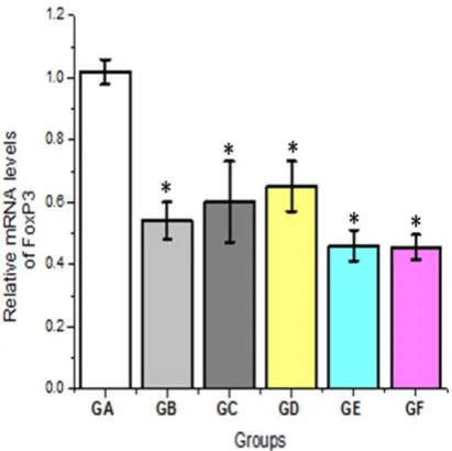
Figure 6: the effect of (IL-2, IL-12 & Taurine) on relative
mRNA levels of FOXP3 in HepG2 cells. All treatments showed significant difference compared to control untreated group, *
Discussion:
HCC has numerous characteristics that make it an immunotherapy attractive target [10]. As approved by their
potential to lyse autologous tumor cells ex vivo, there is active recruitment of tumor-infiltrating lymphocytes capable of perceiving tumor-specific antigens. Mechanisms that tumors follow to avoid immune detection may comprise of secretion of down-regulation of MHC, regulatory T cells, and immuno-inhibitory cytokines [11]. Interleukin-2 (IL-2) is naturally
generated by T cells and is a notable growth and activation factor for cytolytic T lymphocytes (CTLs), macrophages, natural killer (NK) cells, and B lymphocytes. Treatment with IL-2 has revealed definite tumor regression in subjects with advanced cancer. Moreover, there is evidence that immune cells are activated locally and systemically by IL-2 peritumoral injections in cancer patients. Also, IL-2 and IL-12 have potent anti-tumor efficacy [12]. It was revealed in the study that IL-2
treatment (GB) induced cytotoxic impact with no significant difference as contrasted with combination therapy (GF). Meanwhile, the utilization of taurine revealed protective influence against the cytotoxic nature of IL-2 and IL-12 as approved by the non-significant influence of taurine on HepG2 cell viability. The inclusive influence of IL-2 and IL-12 treatment indicated that IL-2 and IL-12 played a crucial role in hepatic carcinoma proliferation, and it might become a potential combination therapy. The influence of Interleukins on IFN-γ and VEGF proteins are well known for immunomodulation, anti-proliferation, and anti-angiogenesis against cancer cells [13].
It has been proved that the three interferon types (IFN-β,
IFN-α, and IFN-γ) are efficient in inhibiting HCC by inducing either tumor cell apoptosis or autophagy [14]. In this
investigation, IFN-γ levels were examined in the culture media of HepG2 cells. The combined IL-2, IL-12 and taurine treatment groups revealed no statistically significant difference as contrasted with the untreated control group. This could be explained in light of the mechanistic action of interleukins. Interleukins were able to selectively recruit natural killer cells to perform the anti-tumor activity in patients with HCC [15].
Interleukins have been utilized to augment anti-tumor responses of the immune system. However, limited investigations concerning these interleukins in humans have been carried out. The efficacy of IL-2 or IL-12 alone or combined together in HCC treatment was assessed in small-scale clinical investigations, but with inconclusive findings [16].
IL-12 anti-tumor mechanism is complex; many investigations have recommended that innate and adaptive immunity are both involved in IL-12 activity [17]. The anti-tumor influences
of IL-12 are mediated by CD4+ or CD8+ T cells or rely on NK and NKT cells [17]. There is persuasive evidence in
pre-clinical researches that antiangiogenic therapy utilizing different approaches inhibits HCC growth. The first one is to
block the angiogenic factors’ influence involved in HCC
angiogenesis, of which VEGF has been targeted repeatedly.
The growth of HCC xenon transplants in a nude mice model has been successfully inhibited by a group of investigators where they utilized VEGF-targeted gene therapy by gene transfer of antisense VEGF. In the present investigation, treatment of HepG2 cells with a combination of IL-2, IL-12, and taurine resulted in a significant reduction in VEGF production by HepG2 as contrasted with the untreated control. A single treatment with IL-2 revealed a prominent inhibition of VEGF secretion. However, there was no significant difference in VEGF production when IL-12 treatment was applied (158.4±20.8 and 276.2±47.5, respectively). Taurine treatment revealed no influence on VEGF secretion as contrasted with the control cells.
Transcription factor forkhead box P3 (FOXP3) is a member of the forkhead family [18]. It was revealed that FOXP3 may be
expressed by normal tissues [19]. In the present investigation,
reduced levels of FoxP3 mRNA and protein was found in the combined IL-2 and IL-12 group (GE) and in the combination immunotherapy with taurine (GF) treatment groups, with significant difference when compared with taurine (GD) and no-treatment control groups (P<0.05) (Figure 6). There was no significant difference in reduced FoxP3 gene expression between the untreated control, and taurine therapy. These findings are consistent with a previous investigation, which revealed that the FOXP3 level is higher in cancer tissues than in normal tissues [20]. Probably, FOXP3 stimulates
proliferation inhibits apoptosis, enhances cell invasion, and decreases cells in the S and G2 phase of the cell cycle [21]. HCC
is a hypervascular tumor with neovascularization features, that play a crucial role in HCC growth and progression. Previous researches have revealed leading angiogenic factors considered in the regulation of angiogenesis in HCC. However, the exact molecular pathways are still ambiguous. VEGF has a pivotal role in HCC angiogenesis. VEGF tumor expression has been confirmed to be associated with tumor invasiveness and prognosis in HCC patients. VEGF antibody is an antiangiogenic therapy based on the crucial molecular target. In the present investigation, assays of VEGF and FoxP3 protein expression revealed that combination therapy with 2, IL-12, and taurine sufficiently induced the anticancer activity as contrasted with the untreated control Hep-G2 cells.
Our findings are in line with previous investigations that proved VEGF plays a pivotal role in hepatocarcinogenesis. Its expression increases gradually from low-grade dysplastic nodules to high-grade dysplastic nodules in early HCC [22]. In
ex-vivo findings revealed the ability of taurine to increase the cytotoxic influence of the combination immunotherapy with IL-2 and IL-12. It was approved with the marked increase of LDH release in the triple treated group as contrasted with other groups. These findings are in great accordance with the investigations that illustrate the transfection influence of IL-2 gene on tumor cells that improve specific and nonspecific antitumor immune responses [23]. Rekers et al. published their
research findings on recombinant IL-2 and the immunoconjugate lL-19, IL-2 anti-tumor influences with and without combined radiotherapy, where immunocompetent mice were injected with F9 teratocarcinoma cells, causing an average of 200 mm³ tumor volume. Different schedules were administered comprising of IL-2, IL-19-IL-2, and radiotherapy. Tumor growth was monitored until the tumor volume reached 4 times the volume at the beginning of the therapy [24]. Their findings had raised the question of the vital
role of immunotherapy with IL-2 and IL-2. Related immune-cytokines after immunotherapy with checkpoint inhibitors have been extensively established. IL-12, a heterodimeric cytokine, has a fundamental role in inducing type-1 T helper cell (Th1) responses and cell-mediated immunity [25]. IL-12 can
be utilized to artificially up-regulate the functions of dendritic cells (DC) to endorse cellular immune responses[26]. IL-2 is a
notable immune-cytokine that serves as a growth factor for T-cells and NK-T-cells [27]. High dose IL-2 treatment in melanoma
and renal cell cancer leads to objective tumor responses in ~15% of patients with ~6% of complete long-lasting tumor responses. Additionally, this treatment benefited patients with stable illness by survival prolongation. The role of both the innate and adaptive immune systems in IL-2 treatment remains to be determined. In mouse models, CD8+ T cells and NK cells are evidently contributed to the anti-tumor influence [28].
There are data that IL-2 may also act via the adaptive immune system and specific cytotoxic T-cells as stimulated with vaccines [29]. Immunotherapy via the innate immune system
may access more important in the future, especially, for patients that are not responding to tumor-specific cytotoxic T-cells. In this investigation, the IL-12 treatments exhibited promising anticancer properties in different groups (C, E, and F) as detected in reduced cell viability, improved LDH release, down-regulation of VEGF and FoxP3. Statistical analysis revealed a significant difference between combined 2, IL-12, and taurine therapy, and IL-2 or IL-12 therapy alone. Combined IL-2, IL-12 and taurine (Group F) therapy and each single-cytokine therapy revealed statistically significant anti-tumor response as contrasted with the taurine treated group (Group D) and with the untreated control groups. There was also a statistically significant difference between single-cytokine, combined IL-2 and IL-12 (Group E), and IL-2, IL-12 and taurine therapies. In the present investigation, combined treatment with IL-2, IL-12 and taurine led to a significant reduction in cell viability as contrasted with untreated Hep-G2 cells. These findings recommended that the combination of IL-2, IL-12 and taurine exerted an influence against hepatoma cell growth, which may be because of the anti-cancer activity of
IL-2 and IL-1IL-2, and the antineoplastic influence of taurine and its derivatives via inhibition of cell proliferation and induction of tumor cell apoptosis. The bispecific IL2-IL-12 is seemingly more active than classical IL-2. Inhibition of HepG2 growth was doubled as effective with a triple combination (2, IL-12, and taurine) as contrasted with classical IL-2 without the combination of IL-12 and taurine. IL-12 is one of the most extensively investigated cytokines in the field of anti-tumor immunotherapy [30].
Conclusion:
Interleukin 2 and IL-12 are known to have anti-tumor features. We observed the greatest antitumor influence with a combination of IL-2, IL-12, and taurine. The anti-tumor activity of IL-2 and IL-12, when given together, would be elucidated by augmented apoptotic responses. Furthermore, the well-known antioxidant activity of taurine would clarify the underlying mechanism of the detected augmented responses of the combination. In other words, a combination of IL-2, IL-12, and taurine could be a promising therapy for patients suffering from HCC after further investigations.
List of Abbreviation:
Hepatocellular carcinoma (HCC), hepatocellular carcinoma (Hep-G2), interleukin-2 (IL-2), interleukin-12 (IL-12), vascular endothelial growth factor (VEGF), forkhead box P3 (FOXP3), Interferon-gamma (INF-γ), Dulbecco’s Modified Eagle’s
Medium (DMEM), 3-(4,5-dimethylthiazol-2-yl)-2,5-diphenyltetrazolium bromide (MTT), lactate dehydrogenase (LDH).
Declarations
• Ethics approval and consent to participate: Not applicable.
• Consent for publication: Not applicable.
• Availability of data and material: Available.
• Competing interests: Authors declare no conflict of interest
• Funding: Not applicable.
• Authors' contributions: Motawa E. EL Houseini conceived the presented idea. Tareq H. Jorob and Abdelbary M. Prince performed the experiment. Saad M. EL-Gendy and Motawa E. EL Houseini encouraged Tareq H. Jorob to investigate and supervised the findings of this work. All authors discussed the results and contributed to the final manuscript.
Acknowledgements:
We thank Khaled Walid and Engy Elsady for their excellent technical assistance.
1. Lau WY. Primary liver tumors. Semin Surg Oncol, 2000; 19: 135-144.
2. Cacalano N. A. and Johnston J. A., “Interleukin-2
signaling and inherited immunodeficiency,” American Journal of Human Genetics, 1999; 65(2): 287–293. 3. Rosenberg S. A., “IL-2: the first effective immunotherapy
for human cancer,” The Journal of Immunology, 2004; 192(12): 5451–5458.
4. Tatsumi T, Huang J, Gooding WE. Intratumoral delivery of dendritic cells engineered to secrete both interleukin (IL)-12 and IL-18 effectively treats local and distant disease in association with broadly reactive Tc1-type immunity. Cancer Res, 2003; 63:6378–6386.
5. Hamza T, Barnett JB, Li B. Interleukin 12 a key immunoregulatory cytokine in infection applications. Int J Mol Sci, 2010; 11:789–806.
6. Miyazaki T, Karube M, Matsuzaki Y, Ikegami T, Doy M, Tanaka N, Bouscarel B: Taurine inhibits oxidative damage and prevents fibrosis in carbon tetrachloride-induced hepatic fibrosis. J Hepatol, 2005; 43: 117-125.
7. Tasci I, Mas N, Mas MR, Tuncer M, Comert B: Ultrastructural changes in hepatocytes after taurine treatment in CCl4 induced liver injury. World J Gastroenterol, 2008; 14: 4897.
8. Alam SS, Hafiz NA, Abd El-Rahim AH: Protective role of taurine against genotoxic damage in mice treated with methotrexate and tamoxfine. Environ Toxicol Pharmacol, 2011; 31:143-152.
9. Jemmerson, R.; LaPlante, B.; Treeful, A. Release of intact, monomeric cytochrome c from apoptotic and necrotic cells. Cell Death Differ. 2002, 9, 538-548. 10. Butterfield LH, Ribas A, Potter DM, Economou JS.
Spontaneous and vaccine-induced AFP- specific T cell phenotypes in subjects with AFP-positive hepatocellular cancer. Cancer Immunol Immunother. 2007;56:1931–
43.
11. O'Neill EJ, Vieira PL, Christensen JR, Minaee S, Barrat FJ, Boonstra A, Barthlott T, Stockinger B, Wraith DC, O'Garra A. IL-10-secreting regulatory T cells do not express Foxp3 but have comparable regulatory function to naturally occurring CD4+CD25+ regulatory T cells. J Immunol. 2004; 172:5986–5993.
12. Gately MK, Gubler U, Brunda MJ, Nadeau RR, Anderson TD, Lipman JM, Sarmiento U. Interleukin-12: a cytokine with therapeutic potential in oncology and infectious diseases. Ther Immunol. 1994; 1:187–196. 13. He, W., Tanaka F., Robbins P. D., Taniguchi M.,
Okamura H., Lotze M. T., Tahara H. Natural killer, but not natural killer T cells play a necessary role in the promotion of an innate antitumor response induced by IL-18. Int. J. Cancer, 2003; 103:508.
14. Obora A, Shiratori Y, Okuno M, Adachi S, Takano Y, Matsushima-Nishiwaki R, Yasuda I, Yamada Y, Akita K, Sano T. Synergistic induction of apoptosis by acyclic retinoid and interferon-beta in human hepatocellular carcinoma cells. Hepatology. 2002;36:1115–1124.
15. Song-Zhao G.X., Srinivasan N., Pott J., Baban D., Frankel G., Maloy K.J. Nlrp3 activation in the intestinal epithelium protects against a mucosal pathogen. Mucosal Immunol. 2014; 7:763–774. doi: 10.1038/mi.2013.94. 16. Sangro B, Mazzolini G, Ruiz J, Herraiz M, Quiroga J,
Herrero I, Benito A, Larrache J, Pueyo J, Subtil JC, et al. Phase I trial of intratumoral injection of an adenovirus encoding interleukin-12 for advanced digestive tumors. J Clin Oncol. 2004;22:1389–1397.
17. Smyth MJ, Thia KY, Cretney E, Kelly JM, Snook MB, Forbes CA, Scalzo AA. Perforin is a major contributor to NK cell control of tumor metastasis. J Immunol. 1999;162:6658–62.
18. Cunha LL, Morari EC, Nonogaki S, Soares FA, Vassallo J and Ward LS: Foxp3 expression is associated with aggressiveness in differentiated thyroid carcinomas. Clinics, 2012; 67: 483-488.
19. Redpath M., Xu B., van Kempen L. C., Spatz A., The dual role of the X‐linked FoxP3 gene in human cancers,
Molecular Oncology, 2001; 5,
doi:10.1016/j.molonc.2011.03.001.
20. Ma GF, Miao Q, Liu YM, Gao H, Lian JJ, Wang YN, Zeng XQ, Luo TC, Ma LL, Shen ZB, Sun YH, Chen SY. High FoxP3 expression in tumour cells predicts better survival in gastric cancer and its role in tumour microenvironment. Br J Cancer. 2014;110:1552–1560. 21. Luo Q, Zhang S, Wei H, Pang X, Zhang H: Roles of
FOXP3 in the occurrence and development of cervical cancer. Int J Clin Exp Pathol, 2015; 8: 8717-8730. 22. Park YN, Kim YB, Yang KM, Park C. Increased
expression of vascular endothelial growth factor and angiogenesis in the early stage of multistep hepatocarcinogenesis. Arch Pathol Lab Med. 2000; 124:1061–1065.
23. Okamoto T. Tsuburaya A. Yanoma S. Yoshikawa T. Cho H. Takanashi Y. Noguchi Y. Inhibition of peritoneal metastasis in an animal gastric cancer model by interferongamma and interleukin-2. Anticancer Res. 2003;23:149–153.
24. Rekers NH, Zegers CM, Yaromina A, Lieuwes NG, Biemans R, Senden-Gijsbers BL, Losen M, Van Limbergen EJ Germeraad WT, Neri D, et al. Combination of radiotherapy with the immunocytokine L19-IL2: Additive effect in a NK cell dependent tumour model. Radiother Oncol: Journal of the European Society
for Therapeutic Radiology and
Oncology.2015;116(3):438–42.
25. Gately M.K., Renzetti L.M., Magram J., Stern A.S., Adorini L., Gubler U., Presky D.H. The interleukin-12/interleukin-12-receptor system: role in normal and pathologic immune responses. Annu. Rev. Immunol. 1998; 16:495–521.
27. Arenas-Ramirez N, Woytschak J, Boyman O. Interleukin-2: Biology, Design and Application. Trends Immunol 2015; 36:763-77.
28. Hughes T, Klairmont M, Broucek J, Iodice G, Basu S, Kaufman HL. The prognostic significance of stable disease following high-dose interleukin-2 (IL-2) treatment in patients with metastatic melanoma and renal cell carcinoma. Cancer Immunol Immunother 2015; 64:459-65.
29. Schwartzentruber DJ, Lawson DH, Richards JM, Conry RM, Miller DM, Treisman J, Gailani F, Riley L, Conlon
K, Pockaj B, Kendra KL, White RL, Gonzalez R, Kuzel TM, Curti B, Leming PD, Whitman ED, Balkissoon J, Reintgen DS, Kaufman H, Marincola FM, Merino MJ, Rosenberg SA, Choyke P, Vena D, Hwu P. gp100 peptide vaccine and interleukin-2 in patients with advanced melanoma. N Engl J Med 2011; 364:2119-27. 30. Tugues S, Burkhard SH, Ohs I, Vrohlings M, Nussbaum