Ting Zhang
1, B, D, Chun Zhao
2, B, Liang Luo
3, 4, C, Jingying Xiang
1, E, Qingmin Sun
1, D,
Jing Cheng
1, C, Daozhen Chen
1, A, FThe Clinical and Prognostic Significance of CCN3
Expression in Patients with Cervical Cancer
Znaczenie kliniczne i prognostyczne ekspresji CCN3
u pacjentek chorych na raka szyjki macicy
1 Wuxi Maternity and Child Health Hospital, Nanjing Medical University, China
2 State Key Laboratory of Reproductive Medicine, Nanjing Maternity and Child Health Hospital,
Nanjing Medical University, China
3 Department of Respiratory Medicine, Jinling Hospital, Nanjing University Medical School, China 4 Wuxi Second Hospital, Nanjing Medical University, China
A – research concept and design; B – collection and/or assembly of data; C – data analysis and interpretation;
D – writing the article; E – critical revision of the article; F – final approval of article; G – other
Abstract
Background. CCN3 plays important roles in growth, differentiation, angiogenesis and adhesion. Recently, the role of CCN3 in human carcinogenesis has become an area of great interest. However, little is known about the function of CCN3 in human cervical cancer.
Objectives. The aim of this study was to investigate the expression profile of CCN3 in cervical cancer and to assess its clinical significance.
Material and Methods. In this study, qRT-PCR, immunohistochemistry and Western blotting analysis were used in the detection of CCN3 mRNA and protein expression, both in cervical cancer and in corresponding normal tissue, respectively. The data was correlated with clinicopathological features. A survival analysis was performed to assess the prognostic significance.
Results. CCN3 mRNA was overexpressed in cervical cancer tissue when compared with corresponding normal tissue, as was CCN3 protein. Upregulation of CCN3 was significantly associated with the stage of the disease (P = 0.017) and with lymph node involvement (P = 0.006). Using the Kaplan-Meier analysis, a comparison of survival curves of low vs. high expressers of CCN3 revealed a highly significant difference in human cervical cancer tissue (P = 0.021), which suggests that overexpression of CCN3 is associated with a poorer prognosis.
Conclusions. The results of the current study suggest that CCN3 may play an important role in cervical carcinoge-nesis and therefore may have potential as a biomarker for prognosis and as a therapeutic target in cervical cancer
(Adv Clin Exp Med 2013, 22, 6, 839–845).
Key words: CCN3; cervical cancer; clinicopathological parameters; prognosis.
Adv Clin Exp Med 2013, 22, 6, 839–845 ISSN 1899–5276
oRIgINAL PAPERS
© Copyright by Wroclaw Medical University
Cervical cancer continues to be one of the ma-jor causes of cancer-related death in women world-wide. It was reported in Lancetthat global cervical cancer incidence has increased 0.6% annually from 1980 to 2010, and the disease killed 200.000 wom-en in 2010 in developing countries [1]. The screwom-en- screen-ing test for cervical cancer is helpful for prevention and early detection, and standard treatments can lead to remission, but clinical outcomes can be very
different for different patients, and are not easy to predict. Therefore, an exploration of the molecular pathogenesis of cervical cancer and the identifica-tion of potential markers for early detecidentifica-tion may play a significant role in treatment and prognosis.
belongs to the CCN gene family and is involved in many cellular functions, including growth, dif-ferentiation, angiogenesis and adhesion [2]. CCN3 gene expression has been investigated in several tumors [3–5]. Chen et al. have shown that CCN3 increases cell migration and expression of intercel-lular adhesion molecule-1 (ICAM-1) in prostate cancer cells [2]. A comprehensive study on Ewing’s sarcoma patients has demonstrated that high CCN3 expression was associated with a higher risk of metastasis [6], indicating again that CCN3 is associated with cell migration and invasion. It has also been indicated that CCN3 enhances the migration of chondrosarcoma cells by increasing MMP-13 expression through the avβ3/avβ5 integ-rin receptor, FAK, PI3K, Akt, p65 and NF-kB sig-nal transduction pathway [7].
However, neither the expression profile nor the clinical significance of CCN3 have been elucidated in cervical cancer thus far. Therefore, in the pres-ent study, the authors compared the expression of CCN3 mRNA and protein in normal human cervi-cal tissue and in cervicervi-cal cancer tissue. Associations between CCN3 and clinicopathological features, as well as the prognosis, were also investigated.
A correlation was found between CCN3 over-expression and poor survival, which suggests that CCN3 may play an important role in cervical carcinogenesis.
Material and Methods
Patients and Tissue Samples
Cervical cancer tissue and adjacent normal tis-sue were obtained from 56 consecutive patients with cervical cancer that was confirmed by histo-pathological analysis at the Department of gyne-cology at Wuxi and Nanjing Maternity and Child Health Hospital, affiliated with Nanjing Medical University, China, between 2004 and 2006. The study protocol was approved by the Institutional Review Board of Nanjing Medical University and all participants signed an informed consent form.
The clinicopathological patient characteristics are summarized in Table 1.
No patient had received radiotherapy, chemo-therapy or other treatment prior to surgery. Fol-lowing surgical removal, the tissue sample was im-mediately frozen in liquid nitrogen until used, and was formalin-fixed and paraffin-embedded for his-topathologic diagnosis and immunohistochemical examination, respectively.
Quantitative Reverse-
-Transcriptase Polymerase Chain
Reaction (qRT-PCR)
QRT-PCR was performed to detect CCN3 mRNA expression. A power homogenizer in TRIzol Reagent (Applied Invitrogen, Carlsbad, CA, USA) was used according to the manufactur-er’s protocol to extract total RNA from the fro-zen tissue by homogenization. The total RNA was then reverse transcribed to generate cDNA (using a PrimeScript RT-PCR kit; Takara Bio). β-actin was used as an internal control. The levels of mRNA encoding were quantified by real-time PCR with the Applied Biosystems 7900 HT Fast Real-Time PCR System using SYBR Premix Ex Taq (Applied Takara Bio). The sequences of the primers were as follows: human CCN3 forward 5’-CACggC -ggTAgAgggAgATA-3’ and reverse 5’-ggg -TAAggCCTCCCAgTgAA-3’ (product 251 bp); human β-actin forward 5’-CAAgAgATggC -CACggCTgCT-3’ and reverse 5’-TCCTTCTg -CATCTgTCggCA-3’ (product 275 bp). The PCR conditions included an initial denaturation step of 95°C for 10 min, followed by 40 cycles of 94°C for 10 s and 60°C for 1 min and a final elon-gation step of 72°C for 10 min. All qRT-PCRs were performed in duplicate. Relative quantification of CCN3 mRNA expression was calculated using the 2–DDCT method.
Immunohistochemistry
All the immunostained sections were eval-uated blind by two observers. For the assess-ment of CCN3, 5 high-power fields in each speci-men were randomly selected, and brown staining of the cytoplasm was considered positive stain-ing. The distribution of positive cells was graded as follows: 0 = no detectable staining; 1 = stain-ing < 1/3 positive cells; 2 = stainstain-ing 1/3–2/3 posi-tive cells; 3 = staining > 2/3 posiposi-tive cells. The in-tensity of staining was graded as follows: 0 = no staining; 1 = weak; 2 = moderate; and 3 = strong staining. The scores for distribution and intensi-ty were added and graded as follows: 0 = (–); 2, 3 = (±); 4 = (+); 5 = (++); 6 = (+++). Scores < 4 were defined as negative and scores > 4 were de-fined as positive [8].
Western Blotting
The frozen tissues were homogenized with an Ultra Turrax homogenizer (Ika, Petaling Jaya, Ma-laysia) in a lysis buffer containing 7 M urea, 2 M thiourea, 4% (w/v) CHAPS, 2% (w/v) DTT, 1% (w/v) Protease Inhibitor Cocktail Kit (Pierce Bio-technology, Rockford, IL, USA), at 11.000 IU/min on ice (10 bursts of 10 s, each interspersed with short pauses). Suspensions were shaken at 4°C for 1 h, and insoluble molecules were removed by cen-trifugation at 40.000 × g at 4°C for 1 h. The protein concentration in each sample was determined by the Bradford method using BSA as the standard. Samples that contained 50–100 µg protein from 2 groups were electrophoresed on a 12% SDS poly-acrylamide gel and transferred to a nitrocellulose membrane (gE Healthcare, San Francisco, CA, USA). The membranes were blocked in tris-buff-ered saline (TBS) that contained 5% non-fat milk powder for 1 h, then incubated with anti-CCN3 (1 : 500) and anti-gAPDH (1 : 1.00; Abcam) di-luted in TBS/5% non-fat milk powder overnight. gAPDH was used as a loading control. Mem-branes were washed 3 times (10 min each) with TBS and incubated for 1 h with HRP-conjugated goat anti-rabbit Igg (1 : 1.00; Beijing ZhongShan Biotechnology, Beijing, China). Specific proteins were detected using an ECL kit and AlphaImag-er (FluorChem5500; Alpha Innotech). The protein expression level was analyzed using AlphaEaseFC software (Alpha Innotech).
Statistical Analysis
The statistical analysis was performed using Sta-tistical Program for Social Sciences (SPSS) software, version 16.0 (SPSS Inc., Chicago, IL, USA). The re-sults of CCN3 expression in cervical cancer tissue
and in corresponding normal tissue were expressed as the mean ± SE, and the data was analyzed using Student’s t test. The association between CCN3 ex-pression and clinicopathological features was ana-lyzed using χ2test. Kaplan-Meier curves were
con-structed, and the log-rank test was performed to analyze survival data. A P-value of less than 0.05 was considered statistically significant.
Results
Expression Profile of CCN3
mRNA in Human Cervical
Cancer and in Corresponding
Normal Cervical Tissue
The qRT-PCR was carried out to identify tran-scripts that encoded human CCN3 in 15 paired samples of cervical cancer tissue and correspon-ding normal tissue. The results showed that CCN3 mRNA expression level in cervical cancer tissue was markedly up-regulated (3.46 ± 0.83) when compared with corresponding normal tissue (1.21 ± 0.67); the difference between the 2 groups is statistically significant (P < 0.05).
Expression Profile and
Subcellular Location of CCN3
Protein in Human Cervical
Cancer and in Corresponding
Normal Cervical Tissue
Correlation of CCN3 Expression
with Clinicopathological
Parameters in Cervical Cancer
Because one aim of the study was to explore the clinical significance of CCN3 expression in the patient samples, the association between
CCN3 expression and clinicopathological pa-rameters was evaluated. In all 56 patients, it was found that the FIgo disease stage and lymph
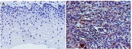
Fig. 1. Representative expression and distribution of CCN3 in normal cervical tissue and cervical cancer tissue (× 400). There was no or low expression of CCN3 in normal cervical tissue (A). In contrast, cervical cancer samples revealed intensive CCN3 staining, which was scored as positive where strong cytoplasmic staining was present (B)
Fig. 2. Western blot analysis of CCN3 in normal cer-vical tissue and cercer-vical cancer tissue. Western blot analysis was used to analyze CCN3 expression in human cervical cancer tissue and in corresponding normal tissue. The result showed that CCN3 level was significantly higher in cervical cancer tissue than in the corresponding normal tissue. gAPDH was the loading control. (C: normal cervical tissue, T: cervical cancer tissue)
Table 1. The correlation of CCN3 expression with clinicopathological parameters in 56 cervical cancer specimens
Parameters Total CCN3 P-value
Negative, n (%) Positive, n (%) Age (years)
< 40 23 12 (52.17) 11 (47.83) 0.472
≥ 40 33 14 (42.42) 19 (57.58)
grade
g1 19 11 (57.89) 8 (42.11) 0.441
g2 16 7 (43.75) 9 (56.25)
g3 21 8 (38.10) 13 (61.90)
Tumor size
< 2.5 cm 25 14 (56.00) 11 (44.00) 0.197
≥ 2.5 cm 31 12 (38.71) 19 (61.29)
FIgo disease stage
I–II 27 17 (62.96) 10 (37.04) 0.017*
III–IV 29 9 (31.03) 20 (68.97)
Lymph node
negative 30 19 (63.33) 11 (36.67) 0.006*
positive 26 7 (26.92) 19 (73.08)
Menses
Pre-menopause 32 15 (46.88) 17 (53.12) 0.938
Post-menopause 24 11 (45.83) 13 (54.17)
node metastasis were associated with CCN3 ex-pression (Table 1). Positive staining for CCN3 protein in patients < 40 and ≥ 40 years of age was 47.83% and 57.58%, respectively, which indicat-ed no significant difference between age groups in CCN3 positive staining with cervical cancer (P = 0.472). There were also no significant differ-ences in positive CCN3 protein staining in histo-logical g2 (56.25%) and g3 (61.90%) compared with g1 (42.11%) (P = 0.441). Meanwhile, CCN3 expression was not associated with tumor size (P = 0.197) or menses (P = 0.938). However, there was a higher percentage of positive CCN3 pro-tein staining in disease stages III and IV (68.97%) when compared with stages I and II (37.04%) (P = 0.017). There was also a significant differ-ence in the correlation of positive CCN3 pro-tein staining and lymph node invasion (negative lymph node invasion vs. positive lymph node in-vasion: 36.67% vs. 73.08%) (P = 0.006). These sults indicate that overexpression of CCN3 is re-lated to disease stage and lymph node invasion in cervical cancer.
CCN3 Protein Expression
Associated with Shorter Overall
Survival
The survival analysis of the 56 studied patients was performed using information available from the clinical follow-ups. At the end of follow-up, 26 patients were still alive; 27 had died; and 3 had
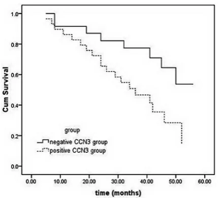
not reported for up, resulting in a follow-up rate of 94.64%. of the 53 evaluable patients, on-ly 8 of the 24 (33.33%) in the negative CCN3 stain-ing group had died of the disease, compared with 19 of the 29 (65.52%) in the positive CCN3 stain-ing group. CCN3 overexpression in patients with cervical cancer was related to reduced overall sur-vival time in a log-rank test (P = 0.021) (Fig. 3). These observations suggest that CCN3 overexpres-sion in cervical cancer patients is associated with reduced overall survival.
Discussion
Cervical cancer is the 3rd most common ma-lignancy in women and a major cause of morbid-ity and mortalmorbid-ity, particularly in developing coun-tries [9]. Even though radiotherapy, chemotherapy and surgery are used as standard treatment moda-lities for patients with cervical cancer, and can lead to consequent disease remission, the prognosis of patients remains unsatisfactory. Therefore, chara-cterizations of identifiable molecular markers should be of diagnostic, prognostic and therapeu-tic value in the management of cervical cancer.
CCN3 is a cysteine-rich protein that belongs to the CCN family of matricellular proteins [10, 11], which interact with the extracellular matrix and thereby regulate many cellular functions, includ-ing cell division, chemotaxis, apoptosis, adhesion, motility and ion transport [12–15]. In recent years, CCN3 has been shown to play an important role in tumorigenesis, including cancer cell proliferation, survival, adhesion and invasion [6, 7]. Perbal et al. found that elevated CCN3 expression in osteosar-coma significantly correlated with a worse prog-nosis, which suggests that assessment of CCN3 expression levels at the time of diagnosis may rep-resent a useful molecular tool for the early identifi-cation of patients with different prognoses [16]. It has also been demonstrated that CCN3 enhances the migration of chondrosarcoma cells [7]. Vallac-chi et al. [17] reported that cells with overexpres-sion of CCN3 by transfection showed increased adhesion to extracellular matrix proteins, where-as inhibition of CCN3 expression by siRNA de-creased adhesion to laminin and vitronectin; CCN3 overexpression was associated with mela-noma progression, including metastases and re-lapse by inducing increased expression of lam-inin and vitronectin integrin receptors α7β1 and αvβ5. Moreover, it has been reported that the most malignant prostate cancer cell line, PC-3, has the highest CCN3 expression [2, 18] and that CCN3 was overexpressed in human prostatic adenocarci-noma samples [18], suggesting that the expression
Fig. 3. overall survival curves for 53 patients with cer-vical cancer and different expression profiles of CCN3
of CCN3 is positively correlated with tumorigen-esis in prostate cancer. Recent data suggests that CCN3 impairs osteoblast and stimulates osteoclast differentiation to favor breast cancer metastasis to bone [19].
Although many studies have demonstrated the involvement of CCN3 expression in tumor biolo-gy, little is known about the expression of CCN3 in human cervical cancer. In this study, the au-thors initially aimed at analyzing the expression of CCN3 and its clinical significance in cervical can-cer, using qRT-PCR, immunohistochemistry and Western blotting analysis to describe the expres-sion and immunolocalization of CCN3 in cervi-cal carcinoma. It was found that CCN3 mRNA was overexpressed in cervical cancer tissue when compared with corresponding normal tissue. Fur-thermore, it was observed that CCN3 protein over-expression occurred in cervical cancer, whereas adjacent normal tissue showed little or no CCN3 expression. These results were also confirmed by Western blotting analysis. The study suggests that increased CCN3 levels may be closely associated with the pathogenesis of cervical cancer.
In order to investigate the clinical significance of CCN3 overexpression in cervical cancer, the authors evaluated the correlation between CCN3 and clini-copathological parameters, including prognosis. In all 56 patients, there were no significant differenc-es in positive staining for CCN3 protein in patients of different ages (P = 0.472) and with different his-tological grades (P = 0.441). Meanwhile, the expres-sion of CCN3 was not associated with tumor size
(P = 0.197) or with menses (P = 0.938). However, there was a higher percentage of positive CCN3 pro-tein staining in disease stages III–IV (68.97%) when compared with stages I–II (37.04%) (P = 0.017). There was also a significant difference in the corre-lation of positive CCN3 protein staining with lymph node invasion (negative lymph node invasion vs. positive lymph node invasion: 36.67% vs. 73.08%) (P = 0.006). These results indicate that overexpres-sion of CCN3 is related to the disease stage and to lymph node invasion in cervical cancer.
Using the Kaplan-Meier analysis, a compari-son of the survival curves of low vs. high expressers of CCN3 revealed a highly significant difference in human cervical cancer tissue (P = 0.021), which suggests that overexpression of CCN3 in cervical cancer patients is associated with a poorer prog-nosis and reduced overall survival (Fig. 3). These findings highlight CCN3 overexpression as a cer-vical cancer-specific event and suggest that CCN3 might be a reliable indicator of prognosis in cervi-cal cancer patients and represents a promising new target for treating cervical cancer.
This study indicates that CCN3 expression is increased in cervical cancer and is positively cor-related with a poor prognosis. The results demon-strate the importance of CCN3 in cervical carci-nogenesis, which suggests that CCN3 may play an important role as a biomarker for prognosis and as a therapeutic target in cervical cancer. However, further studies are necessary to elucidate the mo-lecular mechanisms of CCN3 in the pathogenesis of cervical cancer.
Acknowledgments. This work was supported by the National Natural Science Foundation of China (81202043, 81000258 and 81001444) and by a grant from the Revitalize and Defend the Key Talent’s Subsidy Project in Science and Education of Department of Public Health in Jiangsu Province (RC2011033).
References
[1] Forouzanfar MH, Foreman KJ, Delossantos AM, Lozano R, Lopez AD, Murray CJ, Naghavi M: Breast and cervical cancer in 187 countries between 1980 and 2010: a systematic analysis. Lancet 2011, 378, 1461–1484.
[2] Chen PC, Lin TH, Cheng HC, Tang CH: CCN3 increases cell motility and ICAM-1 expression in prostate cancer cells. Carcinogenesis 2012, 33, 937–945.
[3] Planque N, Perbal B: A structural approach to the role of CCN (CYR61/CTgF/NoV) proteins in tumourigenesis. Cancer Cell Int 2003, 3, 15.
[4] Zhang Y, Wang C: Nephroblastoma overexpressed (NoV/CCN3) gene: a paired-domain-specific PAX3-FKHR transcription target that promotes survival and motility in alveolar rhabdomyosarcoma cells. oncogene 2011, 30, 3549–3562.
[5] Huang CY, Lee CY, Chen MY, Tsai HC, Hsu HC, Tang CH: Nephroblastoma overexpressed gene (NoV) enhanc-es cell motility and CoX-2 upregulation of human osteosarcoma involvenhanc-es αvβ5 integrin, ILK and AP-1-dependent pathways. Biochem Pharmacol 2011, 81, 577–585.
[6] Benini S, Perbal B, Zambelli D, Colombo MP, Manara MC, Serra M, Parenza M, Martinez V, Picci P, Scotlandi K: In Ewing’s sarcoma CCN3(NoV) inhibits proliferation while promoting migration and invasion of the same cell type. oncogene 2005, 24, 4349–4361.
[7] Tzeng HE, Chen JC, Tsai CH, Kuo CC, Hsu HC, Hwang WL, Fong YC, Tang CH: CCN3 increases cell motility and MMP-13 expression in human chondrosarcoma through integrin-dependent pathway. J Cell Physiol 2011, 226, 3181–3189.
[9] Imesch P, Fink D: Cervical cancer. Ther Umsch 2011, 68, 545–552.
[10] Holbourn KP, Acharva KR, Perbal B: The CCN family of proteins: structure-function relationships. Trends Biochem Sci 2008, 33, 461–473.
[11] Perbal B: NoV (nephroblastoma overexpressed) and the CCN family of genes: structural and functional issues. Mol Pathol 2001, 54, 57–79.
[12] Perbal B: CCN proteins: multifunctional signalling regulators. Lancet 2004, 363, 62–64.
[13] Lin CG, Leu SJ, Chen N, Tebeau CM, Lin SX, Yeung CY, Lau LF: CCN3 (NoV) is a novel angiogenic regulator of the CCN protein family. J Biol Chem 2003, 278, 24200–24208.
[14] Lin CG, Chen CC, Leu SJ, Grzeszkiewicz TM, Lau LF: Integrin-dependent functions of the angiogenic inducer NoV (CCN3): implication in wound healing. J Biol Chem 2005, 280, 8229–8237.
[15] Chen CC, Lau LF: Functions and mechanisms of action of CCN matricellular proteins. Int J Biochem Cell Biol 2009, 41, 771–783.
[16] Perbal B, Zuntini M, Zambelli D, Lopez-Guerrero JA, Llombart-Bosch A, Scotlandi K, Picci P: Prognostic value of CCN3 in osteosarcoma. Clin Cancer Res 2008, 14, 701–709.
[17] Vallacchi V, Daniotti M, Ratti F, Di Stasi D, Deho P, De Filippo A, Tragni G, Balsari A, Carbone A, Rivoltini L, Parmiani G, Lazar N, Perbal B, Rodolfo M: CCN3/nephroblastoma overexpressed matricellular protein regulates integrin expression, adhesion, and dissemination in melanoma. Cancer Res 2008, 68, 715–723.
[18] Maillard M, Cadot B, Ball RY, Sethia K, Edwards DR, Perbal B, Tatoud R: Differential expression of the ccn3 (nov) protooncogene in human prostate cell lines and tissues. Mol Pathol2001, 54, 275–280.
[19] Ouellet V, Tiedemann K, Mourskaia A, Fong JE, Tran-Thanh D, Amir E, Clemons M, Perbal B, Komarova SV, Siegel PM: CCN3 impairs osteoblast and stimulates osteoclast differentiation to favor breast cancer metastasis to bone. Am J Pathol 2011, 178, 2377–2388.
Address for correspondence:
Chun Zhao
Nanjing Maternity and Child Health Hospital Affiliated to Nanjing Medical University Nanjing 210004
China
E-mail: [email protected] Conflict of interest: None declared Received: 26.02.2013