DOI: 10.7324/JAPS.2018.8904 ISSN 2231-3354
© 2018 Tharini Palani et al. This is an open access article distributed under the terms of the Creative Commons Attribution License -NonCommercial- ShareAlikeUnported License (http://creativecommons.org/licenses/by-nc-sa/3.0/).
*Corresponding Author
Dr. Rajeswary Hari, Department of Biotechnology, Dr. M.G.R. Educational & Research Institute, Maduravoyal, Chennai, India. E-mail: rajihar @ gmail.com
In vitro
and
In silico
Antigout arthritic activities of Ethanolic and
Aqueous stem extracts of
Cissus quadrangularis
– A TLR2 and TLR4
Receptor approach
Tharini Palani, Krishnan Shobha, Thirunavukkarasu P, Rajeswary Hari*
Department of Biotechnology Dr. M.G.R. Educational and Research Institute Deemed to be University, Chennai-95. Tamil Nadu, India.
ARTICLE INFO ABSTRACT
Article history: Received on: 05/07/2018 Accepted on: 19/08/2018 Available online: 30/09/2018
The in vitro anti-gouty arthritic activity of ethanolic and aqueous extracts of Cissus quadrangularis stem was evaluated in terms of inhibition of xanthine oxidase, proteinase enzyme, protein denaturation and membrane stabilization. The Monosodium Urate (MSU) up-regulated expression patterns of toll-like receptors namely TLR-2 and TLR-4 were analyzed. The molecular docking was performed to select the antagonistic phyto ligand for receptors since their activation leads to the destructive immunological reactions occurring in gouty arthritis. The in vitro anti-gout arthritic activities were estimated using standard protocols. The MSU crystal-induced toll-like receptors gene expression was analyzed. Molecular docking and LIGPLOT analysis were performed with the GCMS-derived phytoconstituents in the ethanolic extract of Cissus quadrangularis with the receptors to find the interactions. Colchicine was used as a positive drug in the study. Among the two extracts, ethanolic extract revealed better in-vitro anti-arthritic activity in the present investigation. Out of the GCMS derived 13 phytocomponents, based on docking score three main components namely Pentadecanoic acid, 14-methyl,-methyl ester, 10-Octadecenoic acid,methyl ester, 4-one,2-(3,4-dihydroxyphenyl)-2,3-dihydro-3,5,7-trihydroxy showed better antagonistic action in the LIGPLOT analysis. It may be concluded these three ligands may further develop as a potential anti-gouty arthritic drug.
Key words:
Anti gout, Monosodium urate, Toll-like receptor, Xanthine oxidase.
INTRODUCTION
Gout is a metabolic disorder with a worldwide distribution that is associated with an excess of circulating levels of uric acid (hyperuricemia). This results in the formation and deposition of monosodium urate crystals (MSU) in the joints causing inflammatory arthritis and severe pain. Hyperuricemia can be caused by impaired renal excretion or overproduction of uric acid due to overconsumption of purine-rich foods. Xanthine oxidase enzyme is responsible for the hyperuricemia and goutby catalyzing the oxidation of xanthine and hypoxanthine into uric acid (Unno et al., 2004). The risk for supersaturation occurs and the urate crystallizes when the plasma concentration of monosodium urate (MSU) exceeds its solubility (around 7 mg/dl) concentration due to impaired glomerular filtration. Recognition of the naked
MSU crystal by Toll-like receptor TLR2 and TLR4, which are normally involved in triggering the innate host defense responses to infectious pathogens, was recently discovered to be a primary trigger of the inflammatory and degenerative tissue reactions associated with gouty arthritis (Liu-Bryan et al., 2005; Liu-Bryan
et al., 2005). Nonsteroidal anti-inflammatory drugs (NSAIDs), as
well as systemic and intra-articular corticosteroids, are used to treat acute gout (Getting et al., 2002). In patients who cannot tolerate NSAIDs or systemic corticosteroids, oral colchicine is generally the next choice for primary therapy. But all these treatments have their own side effects such as gastrointestinal and liver toxicity.
leucorrhoea and syphilis (Yoganarsimhan, 2006). Siddha traditional medicine believes the use of this plant to heal broken bones, as an analgesic and a tonic (Mishra et al., 2010). The pathophysiology of the gouty arthritis is characterized by multiple steps, where a potential drug should not only act to reduce the production of uric acid but should also reduce the immune response by inhibiting the activation of the Toll-like receptor and its downstream processes leading to the degenerative reactions involved in gouty arthritis. With the above scenario, the present investigation evaluated the and anti-gout activity of Cissus quadrangularis stem extracts through in vitro and insilico analysis.
MATERIALS AND METHODS
Chemicals
The chemicals used in the present study such as Ethanol, Bovine Serum Albumin, Acetyl Salicylic Acid were obtained from SD Fine Chemicals Ltd. Xanthine oxidase from bovine milk was purchased from Sigma (×4500). Trypsin was purchased from SISCO Research Laboratories Pvt Ltd, Mumbai. The other chemicals used were of analytical grade.
Collection of plant material
The stem of Cissus quadrangularis was purchased from koyambdu market, Chennai, India and were authenticated by Dr.Sankaranarayanan, Deputy Director, captain Srinivasamorthy Siddha medical institute Chennai, India. The voucher specimen is also available in herbarium file of the same Centre.
Preparation of plant extract
The stem of Cissus quadrangularis (1 kg) was dried and ground into a coarse powder. The powder was extracted with 90% (v/v) ethanol at room temperature by cold maceration process
for three days. The resultant extract was filtered and the excess
solvent was removed by vacuum evaporator. The yield of the sample was calculated. Similarly, aqueous extract was prepared and the removal of water was carried out by freeze-drying and the yield of the sample is calculated. The prepared extracts were stored in the refrigerator until investigation.
In vitro anti-arthritic activity of gouty arthritis
The anti-arthritic activity of gouty arthritis was analyzed using standard established protocols. The in vitro Xanthine oxidase inhibitory activity, protein denaturation inhibition assay, membrane stabilization study, and Proteinase enzyme inhibition study were studied to assess the anti-arthritic activity of the plant extracts EECQ and AECQ. The activity of xanthine oxidase was calculated using spectrophotometer method by Owens and Johns 1999. The protein denaturation inhibition process was evaluated using Mizushima and Kobayashi, 1968 method. The membrane stabilization of RBC was carried out according to the studies by Sakat et al. (2010). The proteinase enzyme inhibitory studies were performed by the method of Oyedapo et al. (1995).
Determination of totalphenolic content
The total phenols present in the EECQ and AECQ extracts were calculated according to the methods of Slinkard and Singleton 1977. A quantity of 100 mg of the sample in 0.5 ml of
water is mixed with Folin–Ciocalteu’s reagent (diluted 1:10v/v) and 2 ml of Na2CO3 (7.5%, v/v) solution. The whole set up was incubated for 90 min at 30°C, and the resultant color was measured at 765 nm with results expressed as gallic acid equivalents. Determination of total flavanoid content
The total flavonoid content present in the extracts were calculated by the method of Zhishen et al. (1999). 100 mg of test drugs were dissolved in 2 ml of distilled water and 0.15 ml of sodium nitrite solution. 0.15 ml of aluminium chloride solution was added after 6 minutes and incubated for 6 minutes. To the mixture 2 ml of 4%, NaOH was added followed by the addition of water to bring the volume to 5 ml and incubated for 15 min, color absorbance was determined at 510 nm and results were expressed as catechin equivalents.
GC-MS analysis
For GC-MS analysis, the samples were injected into an HP-5 column (30 m × 0.25 mm i.d with 0.25 μm film thickness), Agilent technologies 6890 N JEOL GC Mate II GC-MS model. The following chromatographic conditions were used: Helium as a carrier gas, the flow rate of 1 mL/min; and the injector was operated at 200°C and column oven temperature was programmed as 50-250°C at a rate of 10°C/min injection mode. The Following MS conditions were used: ionization voltage of 70 eV; ion source temperature of 250°C; the interface temperature of 250°C; mass range of 50-600 mass units.
Monosodium urate crystals synthesis
MSU crystals were prepared according to Seegmiller et al., 1962.24 g of sodium hydroxide was solubilized in 200 ml of pyrogen-free double distilled water. 1 g of uric acid was added into the above solution and the pH was adjusted to 7.2 using 1N HCl. This solution was heated to 120°C for 6-hrs with gentle stirring. After incubation, the solution was left to cool at room temperature and kept at 4°C for overnight. The sedimented MSU crystals were collected by filtration and washed with absolute ethanol. Washed MSU crystals were dried under vacuum and sterilized by autoclaving at 121°C for 20 min. The sterilized crystals were prepared as 20 mg/ml stock in PBS and used for the assay. Cell treatment and quantitative Real-Time Polymerase Chain Reaction (RT-PCR) analysis of TLR4 and TLR2 expression
RNA was reverse transcribed by using the higher capacity cDNA reverse transcription kit (Applied Biosystems, CA, USA) and the mRNA expression was amplified by Quantitect SYBR1 PCR kit (QIAGEN, Valencia, CA, USA). Gene-specific primers for TLR4 and TLR2 expression were designed by referring to the NCBI/ primer-BLAST tool software which is depicted in the Table 1. The RT-PCR data was collected and analyzed on MxPro Software from Agilent Technologies. The fold change in gene expression levels of target genes was calculated with normalization to β-actin values using the 2-ΔΔCt comparative cycle threshold method. Molecular docking analysis
In silico molecular docking studies were conducted with the help of patch dock to identify the best ligand for TLR2 and TLR4 proteins from the Cissus quandrangularis phyto compounds. The three-dimensional structures of toll-like receptor 4 (TLR-4) (PDB ID: 5NAO)and toll-like receptor 2 (TLR-2) (PDB ID:2Z80) proteins were downloaded from protein data bank (http://www.rcsb. org/pdb/). The two-dimensional structures for GC-MS identified 13 phyto constituents from the Cissus quandrangularis were retrieved from PUB chemsite, and converted into three-dimensional structures using corina 3D converter. Using the patch dock server docking score regarding the ligand and protein interaction was identified. Using ligand showing appropriate docking score was selected and their further interactions with the proteins in terms of amino acid residues with the ligand and the active site were visualized and further confirmed using LIGPLOT. The docked results were saved as “pdb” file and binding affinity and molecular interaction between standard, test compounds and the receptor protein were visualized using PyMol Molecular Graphic System (Ver. 1.0) and Discovery Studio (Ver. 3.1) software, respectively The standard drug colchicine is used as a reference drug.
Statistical analysis
The values reported are Mean ± SE. The statistical analysis was carried out using analysis of variance (ANOVA)
followed by Dunnet’s ‘t’ test. The p values < 0.05 were considered significant.
RESULTS
Xanthine oxidase inhibitory assay
The Xanthine oxidase inhibitory activity of EECQ and AECQ were depicted in Table 2. Between these two extracts the ethanolic extract of Cissus quadrangularis shows higher the maximum inhibitory activity to xanthine oxidase starting from 50 µg concentration when compared to aqueous extract of Cissus quadrangularis. In the present study, the percentage of inhibition was almost similar to allopurinol the positive drug used.
Inhibition of protein denaturation
The potential of inhibition of protein denaturation was studied (Table 3) and better inhibition was seen for EECQ than AECQ and at the concentration to 500 µg and 1000 µg of EECQ maximum inhibition potential was noted which was higher than the inhibition percentage of Acetylsalicylic acid the positive control used.
Membrane stabilization study
The heat and hypotonic saline-induced damage to RBC membrane was stabilized by the plant extracts (Table 4) and were comparable with the positive drug Acetylsalicylic acid. The EECQ extract protected the RBC membrane than the AECQ.
Proteinase inhibition study
The ability of the EECQ and AECQ to inhibit proteinase enzyme was shown in the Table 5. Though both the extracts showed proteinase inhibitory activity, the inhibition was found to higher for EECQ than AECQ extracts. In the present investigation the, 200 µg to 1000 µg of EECQ showed higher inhibitory activity than the Acetylsalicylic acid which was the positive drug used in this study.
Table 1: Details of Primers used for TLR-2, 4 and β-actin.
Gene Primer Primer sequence Size of the amplicon (BP)
TLR-2 (NCBI Ref sequence NM_001318787.1) Forward GGCCAGCAAATTACCTGTGT 298 Reverse TTCTCCACCCAGTAGGCATC
TLR-4 (NCBI Ref sequence NM_138557.2) Forward TGAGCAGTCGTGCTGGTATC 167
Reverse CAGGGCTTTTCTGAGTCGT
β-actin (NCBI Ref sequence NM_001101) Forward CATCGAGCACGGCATCGTCA 211 Reverse TAGCACAGCCTGGATAGCAAC
Table 2: Effects of EECQ and AECQ on Inhibition of Xanthine Oxidase.
Conc of Extract (in µg) % Inhibition of EECQ % Inhibition of AECQ % Inhibition of Allopurinol
50 49.8 ± 1.41 a** 34.26 ± 0.9 b** 70.3 ± 0.7
100 56.8 ± 1.4 a** 48.9 ± 1.36 b** 72.0 ± 1.2
200 70.3 ± 1.05 a** 56.2 ± 0.75 b** 75.2 ± 0.76
500 83.5 ± 2.6 a ns 65.5 ± 1.45 b** 81.2 ± 1.15
1000 91.53 ± 1.35 a** 66.9 ± 1.2 b** 83.76 ± 1.49
Values are expressed in mean ± SD (n = 3), statistical significant test for comparison was done by ANOVA followed by Dunnet’s ‘t’ test. Comparison between a:
Table 3: Effects of EECQ and AECQ on Inhibition of Protein Denaturation.
Conc of Extract (in µg) % Inhibition of EECQ % Inhibition of AECQ % Inhibition of Acetyl Salicylic Acid
50 49.8 ± 1.41 a** 34.26 ± 0.9 b** 70.3 ± 0.7
100 56.8 ± 1.4 a** 48.9 ± 1.36 b** 72.0 ± 1.2
200 70.3 ± 1.05 a** 56.2 ± 0.75 b** 75.2 ± 0.76
500 83.5 ± 2.6 a ns 65.5 ± 1.45 b** 81.2 ± 1.15
1000 91.53 ± 1.35 a** 66.9 ± 1.2 b** 83.76 ± 1.49
Values are expressed in mean ± SD (n = 3), statistical significant test for comparison was done by ANOVA followed by Dunnet’s ‘t’ test. Comparison between a:
Acetylsalicylic acid vs. EECQ b: Acetylsalicylic acid vs. AECQ *p < 0.05, **p < 0.1 and ns: Non-Significant.
Table 4: Effects of EECQ and AECQ on membrane Stabilization.
Conc of Extract (in µg) % stabilization of EECQ % stabilization of AECQ % stabilization of Acetyl Salicylic Acid
50 50.2 ± 0.95 a** 36.1 ± 1 .06 b** 69.9 ± 1.3
100 61.9±1.58 a** 51.2 ± 1.10 b** 74.2 ± 0.95
200 71.5 ± 1.25 a** 58.6 ± 1.41 b** 80.23 ±1.0
500 78.33 ± 0.92 a** 63.9 ± 1.45 b** 82.8 ± 1.22
1000 82.6 ± 1.10 a ns 67.8 ± 2.4 b** 83.8 ± 1.5
Values are expressed in mean ± SD (n = 3), statistical significant test for comparison was done by ANOVA followed by Dunnet’s ‘t’ test. Comparison between a: Acetylsalicylic acid vs. EECQ b: Acetylsalicylic acid vs. AECQ *p < 0.05, **p < 0.1 and ns: Non-Significant.
Table 5: Effect of EECQ and AECQ on Proteinase Inhibition.
Conc. of Extract (in µg) % Inhibition of EECQ % Inhibition of AECQ % Inhibition of Acetyl salicylic acid
50 13.49 ± 0.70 a** 10.57 ± 0.84 b** 23.27 ± 0.67
100 26.56 ± 0.60 aa ns 20.44 ± 0.75 b** 27.40 ± 1.29
200 40.20 ± 1.19 a* 35.72 ± 0.79 b ns 37.56 ± 0.86
500 80.34 ± 0.72 a ns 72.52 ± 0.75 b** 79.56 ± 0.86
1000 92.69 ± 0.74 a** 86.31 ± 0.78 b** 89.28 ± 0.68
Values are expressed in mean ± SD (n = 3), statistical significant test for comparison was done by ANOVA followed by Dunnet’s ‘t’ test. Comparison between a:
Acetylsalicylic acid vs. EECQ b: Acetylsalicylic acid vs. AECQ *p < 0.05, **p < 0.1 and ns: Non-Significant.
GCMS analysis
In the present investigation, the ethanolic extract of
Cissus quadrangularis revealed better anti arthritic activity
than its aqueous extract. So the EECQ was subjected to GC-MS analysis which revealed the presence of 13 chemical components (Figure 1).
MSU effect on TLR-2 and TLR-4 gene expression
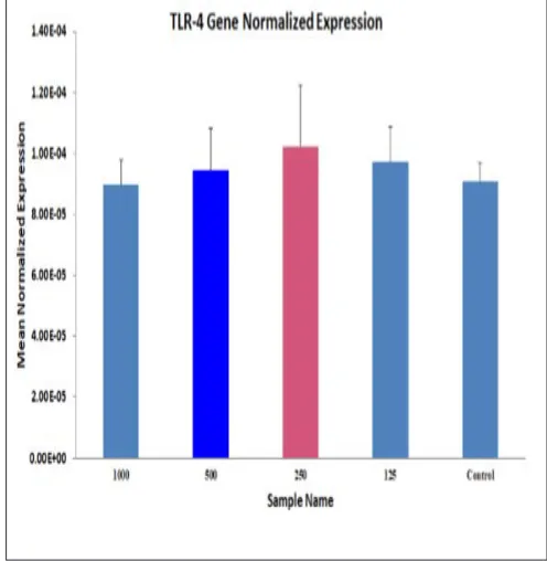
The effect of the MSU induced inflammatory response which is mediated by TLR-2 and TLR-4 receptors was studied using human peripheral blood mononuclear cells treated with monosodium urate crystals. The level of gene expression of these receptors with varying concentration of MSU crystals was studied using the RT-PCR method and depicted in Figure 2 and 3. In our present investigation, the expression pattern of TLR-2 was concentration-dependent that is at a lower concentration of MSU crystals such as 125 μg the gene expression was minimum and then gradually increases with increase in concentration and peaks at 1000 μg. Whereas TLR-4 gene expression was independent on the concentration of MSU and at lower concentration (125 μg)
itself it showed maximum gene expression. Molecular docking analysis
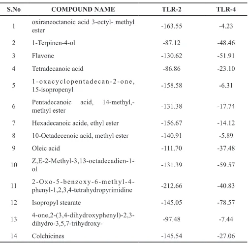
Table 6. Docking score values of compounds with TLR-2 and TLR-4.
S.No COMPOUND NAME TLR-2 TLR-4
1 oxiraneoctanoic acid 3-octyl- methyl ester -163.55 -4.23
2 1-Terpinen-4-ol -87.12 -48.46
3 Flavone -130.62 -51.91
4 Tetradecanoic acid -86.86 -23.10
5 1-oxacyclopentadecan-2-one, 15-isopropenyl -158.58 -6.31
6 Pentadecanoic acid, 14-methyl,-methyl ester -131.38 -17.74
7 Hexadecanoic acide, ethyl ester -156.67 -14.12 8 10-Octadecenoic acid, methyl ester -140.91 -5.89
9 Oleic acid -111.70 -37.48
10 Z,E-2-Methyl-3,13-octadecadien-1-ol -131.39 -59.57
11 2-Oxo-5-benzoxy-6-methyl-4-phenyl-1,2,3,4-tetrahydropyrimidine -212.66 -40.83
12 Isopropyl stearate -145.05 -78.57
13 4-one,2-(3,4-dihydroxyphenyl)-2,3-dihydro-3,5,7-trihydroxy- -97.48 -7.44
14 Colchicines -145.54 -27.06
DISCUSSION
In gout the uric acid in the form of monosodium urate crystals get deposited in the joints, synovial fluid and other tissues and causes an inflammatory and painful condition called as gouty arthritis (Albrecht et al., 2014). The inflammatory response in gout is characterized by multiple steps and complex interactions between various cell types which ultimately leads to the ulceration of the joint cartilage, marginal osteophytosis, and chronic inflammation of synovial membrane (Dalbeth and Haskard, 2005; Carado et al., 2006). The goals of gouty arthritis treatment are several fold. Initially, the drug should inhibit the overexpressed xanthine oxidase enzyme responsible for the production of uric acid. In the present investigation, the ethanolic extract of Cissus quadrangularis inhibited the xanthine
oxidase enzyme considerably. The presence of high content of phytosterol, calcium, ascorbic acid β-sitosterol, δ-amyrin, carotene and δ-amyronehave the ability to modify the metabolic and physiological effects as reported by Metha et al. (2001); Shirwaikar et al. (2003). These may be responsible for the xanthine oxidase inhibitory activity observed in the present study.
The binding of the MSU ligand as an agonist to TLR2 and TLR4 receptors in the neutrophils is the initial trigger to the immune response and inflammatory stimuli which orchestrate the multiple signaling pathways such as activating the NLRP3 inflammasome, resulting in processing and secretion of IL-1β, increased NF-kB activity, triggering the cytokines, such as interleukin-1 (IL-1), IL-6, IL-8,and tumor necrosis factor, matrix metalloproteinase (MMPs), and certain other mediators leading to the pathogenesis of gouty arthritis. (Haskard and Landis, 2002; Landis and Haskard, 2001). Similar to other forms of arthritis, there also the three basic interrelated processes like inflammation, synovial proliferation and joint tissue destruction that occurs in gouty arthritis. So the in vitro anti-arthritic activity in terms of inhibition of protein denaturation, membrane stabilization, and proteinase inhibition was studied for our plant extracts. The EECQ exhibited better anti gouty arthritic activity for membrane stabilization, inhibition of protease enzyme and protein denaturation. It is a well-known fact that the lysosomal membrane is similar to the RBC membrane and any constituent which protects the RBC membrane has the ability to protect the lysosomal membrane. This, in turn, inhibits the release of protein-degrading enzymes from the neutrophils at the inflammatory site and thereby tissue inflammation and damage (Chou CT, 1997). All types of inflammatory processes usually trigger directly or indirectly the different types of immunological mediators which subsequently induce the proteases and destruction of cells and tissues. Phytochemicals which specifically inhibit these inflammatory mediators have the protective effect as an anti-gout agent. According to Panthonget al. (2007), the analgesic and anti-inflammatory activity of the plant extract is due to the presence of flavonoids especially luteolin and by β-sitosterol. Delapureta et al. (2005), have stated that the influx of neutrophils, as well as reduction of enzymes like MPO in the inflamed tissue, can be reduced by the β-sitosterol present in the
Cissus quadrangularis extract.
Table 7: Interaction with the TLR-2 and TLR-4 proteins with phytochemicals of EECQ.
S. No Compound
TLR-2 amino acid binding site TLR-4 amino acid binding site
H bonding sites Hydrophobic contact sites H bonding sites Hydrophobic contact sites
1 acid, 14-methyl,-Pentadecanoic methyl ester
1 87 1 63
Arg155(A) Gln152(A), Leu176(A), Glu177(A), Glu178(A), Glu180(A). Arg289(B) Lys230(B), Arg234(B), Val259(B), Arg264(B), Ala271(B), Val316(B), Ser317(B)
2 acid, methyl ester 10-Octadecenoic
1 91
_
47
Arg155(A) Gln152(A), Thr174(A),Glu178(A), Glu180(A) Arg234(B), Arg264(B), Arg289(B), Ala291(B), Tyr296(B), Ser317(B), Asn339(B), Lys341(B), Lys362(B)
3
4-one,2-(3,4-
dihydroxyphenyl)-
2,3-ihydro-3,5,7-trihydroxy- 3 75 _ 62
Leu154(A), Arg155(A), Glu178(A).
Val156(A), Asp160(A),
Glu180(A), Ile181(A). Arg264(B), Leu293(B), Asp294(B), Tyr296(B), Thr319(B), Asn339(B), Lys341(B), Lys362(B).
4
Colchicines 2 102 3 95
Glu283(A),
Asp286(A). Phe256(A), Arg257(A), Asn258(A), Ser265(A), Asp285(A)
Asp294(B), Tyr296(B),
Asn339(B). Tyr292(B), Tyr295(B), Ser317(B).
Fig. 2: TLR-2 gene expression chart.
In the present study, we could able to conclude that there is an association between the MSU crystals and overexpression TLR 2 and TLR4 genes. The expression pattern was high for theTLR4 gene than the TLR 2 gene. So we also made an attempt to examine whether the phytochemicals present in the Cissus quadrangularis
stem extract could act as an antagonist to these transmembrane protein receptors (TLR 2 and TLR4) and thereby inhibit the down streaming inflammatory processes. Based on the GC-MS analysis of the ethanolic extract of the Cissus quadrangularis stem 14
compounds were subjected to patch dock method of molecular docking analysis. Three compounds namely Pentadecanoic acid,14-methyl,-methyl ester, 10-Octadecenoic acid,methyl ester, 4-one,2-(3,4-dihydroxyphenyl)-2,3-dihydro-3,5,7-trihydroxy showed better activation energy and docking score. These three compounds were further subjected for the ligand-protein interactions for the possible antagonist action for the TLR 2 and TLR4 trans-membrane receptor. This blocking of the TLR 2 and TLR4 transmembrane receptor could hinder the further immunological reactions and protect the joints from destruction and pain.
(A) (B)
(C) (D)
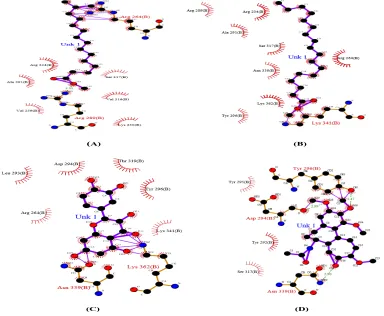
Fig. 4: Amino acid Interaction of TLR-2 protein with specific ligands. A) Pentadecanoic acid,14-methyl,-methyl ester. (B) 10-Octadecenoic acid, methyl ester. (C)
4-one,2-(3,4-dihydroxyphenyl)-2,3-dihydro-3,5,7-trihydroxy. (D) colchicines.
(A) (B)
(C) (D)
Fig. 5: Amino acid Interaction of TLR-4 protein with specific ligands. (A) Pentadecanoic acid,14-methyl,-methyl ester (B) 10-Octadecenoic acid, methyl ester. (C)
CONCLUSION
This knowledge will provide a new insight for the discovery of a single ligand which will be acting as an inhibitor for this TLR2 and TLR4 trans-membrane receptors thereby terminating the diverse immunochemical reactions leading to the pathogenesis of gouty arthritis. The single ligand may be used as a successful drug for the management of gouty arthritis which can replace the different types of drug which are at present in use for the treatment of gouty arthritis.
REFERENCES
Albrecht E, Waldenberger M, Krumsiek J, Evans AM, Jeratsch U, Breier M, et al. Metabolite profiling reveals new insights into the regulation of serum urate in humans. Metabolomics, 2014; 10:141–151.
Chou CT. The anti–inflammatory effect of Tripterygium wilfordii Hook F on adjuvant induced paw edema in rats and inflammatory mediators
release. Phytother Res, 1997; 11(2):152–54.
Corrado A, Onofrio FD, Santoro N, Melillo N, Cantatore FP. Pathogenesis, clinical findings and management of acute and chronic gout. Minerva Med, 2006; 97:495-509.
Dalbeth N, Haskard DO. Mechanism of inflammation in gout. Rheumatology, 2005; 44:1090-1096.
Delapureta R, Martinez DE, Ruiz GV. Effect of minor components of virgin olive oil on topical anti-inflammatory assays. Zetschrift Fur Naturforschong, 2000; 55:814-19.
Getting SJ, Christian HC, Flower RJ, Perretti M. Activation of melanocortin type 3 receptor as a molecular mechanism for adrenocorticotropic hormone efficacy in gouty arthritis. Arthritis Rheum, 2002; 46:2765-2775.
Haskard DO, Landis RC. Interactions between leukocytes and endothelial cells in gout: lessons from a self-limiting inflammatory response. Arthritis Res., 2002; 3:91.
Landis RC, Haskard DO. Pathogenesis of crystal-induced inflammation.Curr. Rheumatol. Rep, 2001; 3:36.
Liu-Bryan R, Pritzker K, Firestein GS, Terkeltaub R. TLR2 signaling in chondrocytes drives calcium pyrophosphate dehydrate and monosodium urate crystal-induced nitric oxide generation. J Immunol, 2005; 174:5016-5023.
Liu-Bryan R, Scott P, Sydlaske A, Rose DM, Terkeltaub R. Innate immunity conferred by toll-like receptors 2 and 4 and myeloid differentiation factor 88 expression is pivotal to monosodium urate monohydrate crystal-induced inflammation. Arthritis Rheum, 2005; 52:2936-2946.
Mehta M, Kaur N, Bhutani KK. Determination of marker constituents from Cissus quadrangularisLinn. and their quantitation by HPTLC and HPLC. Phytochem Anal, 2001; 12:91-5.
Mishra G, Srivastava S, Nagori BP. “Pharmacological and therapeutic activity of Cissus quadrangularis: An overview” International Journal of Pharm. Tech. Research, 2010; 2(2):1298-1310.
Mizushima Y, Kobayashi M. “Interaction of anti-inflammatory drugs with serum proteins especially with some biologically active proteins” J Pharm Pharmacol, 1968; 20:169-73.
Owen P, Johns T. Xanthine oxidase inhibitory activity of north eastern North American plant remedies used for gout, J Ethnopharmacol, 1999; 64:149.
Oyedapo, Famurewa AJ. “Anti-protease and membrane stabilizing activities of extracts of Fagra zanthoxiloides, Olax subscorpioides and Tetrapleura tetraptera”. Int J. pharmacogn, 1995; 33:65-9.
Panthong A, Wanicha S, Kanjanapothi D, Tawat T, Vichai R. Analgesic, anti-inflammatory and venotonic effects of Cissus quadrangularis Linn. Journal of Ethnophramacology, 2007; 110:264-70.
Sakat S, Juvekar AR, Gambhire MN. Inv itro antioxidant and anti-inflammatory activity of methanol extract of Oxalis corniculata Linn. Inter. J. Pharma Parmacol Sci, 2010; 2(1):146-55.
Seegmiller JE, Howel RR, Malaw ISTASE. The inflammatory reaction to urate. JAMA, 1962; 180:469-75.
Shirwaikar A, Khan S, Malini S. Antiosteoporotic effect of ethanol extract of Cissus quadrangularis Linn. on ovariectomized rat. J Ethnopharmacol, 2003; 89:245-50.
Slinkard J, Singleton VL. Total phenol analysis: automation and comparison with manual methods. Am J Enol Vitic, 1977; 28:49.
Unno T, Sugimoto A, Kakuda T. Xanthine oxidase inhibitors from the leaves of Lagerstroemia speciosa (L.) Pers. J. Ethnopharmacol, 2004; 93:391-395.
Yoganarsimhan SN. Medicinal plants of India. Cyber Media Bangalore 2, 2000; 2:136–137.
Zhishen J, Mengcheng T, Jianming W. The determination of flavonoid contents in mulberry and their scavenging effects on superoxide radicals. Food Chem, 1999; 64:555.
How to cite this article: