ISSN 0015–5659 www.fm.viamedica.pl
Address for correspondence: Dr. S.M. Zaki, Department of Anatomy and Embryology, Faculty of Medicine, Cairo University, Cairo, Egypt, e-mail: [email protected]
Stress-induced changes in the aged-rat adrenal
cortex. Histological and histomorphometric study
S.M. Zaki
1, 2, F.A. Abdelgawad
1, E.A.A. El-Shaarawy
1, R.A.K. Radwan
1, B.E. Aboul-hoda
11Department of Anatomy and Embryology, Faculty of Medicine, Cairo University, Cairo, Egypt 2Fakeeh College for Medical Sciences, Jeddah, Saudi Arabia
Received: 29 December 2017; Accepted: 1 March 2018]
Background: Stress exposure exerts direct effects on the morphology and func-tionality of the adrenal cortex. In addition, ageing effects growth, differentiation, apoptosis and cellularity of the cortex. The missing data is the combined effect of stress and ageing on the adrenal cortex. The aim of this study was to demonstrate the structural changes in the adrenal cortex following the exposure to stress in the adult and aged albino rats.
Materials and methods: Forty rats were divided into groups I and II (adult and senile). Each group was further subdivided into subgroups a and b (control and stressed). Light and electron microscopic studies were done. Area per cent of
collagen fibres (Masson’s trichrome-stained sections), number of proliferating
cells (optical density immunoreactivity in the Ki67 stained sections) and thickness of the three adrenal zones were also measured.
Results: Lamellar separation of the capsule with subcapsular spindle cell hyper-plasia and areas of ghost cells were observed in zona glomerulosa (ZG) and zona fasciculata (ZF) in group I-b. Separation and indentation of the capsule with its lamellar separation were observed in group II-a with the existence of multiple scattered degenerative foci in ZF and zona reticularis (ZR). Similar and aggressive was the architectural pattern of ZF in group II-b with the presence of areas of homogenous degeneration. The nuclei of ZG had marginated chromatin in group I-b and were pyknotic with deformed irregular outlines in group II-b. Multiple lysosomes and vacuolar degeneration mitochondria were also seen in group I-b. The nuclei of ZF were irregular with condensed marginated heterochromatin in group I-b, irregular with scattered chromatin in group II-a and indented with areas of chromatin destruction in group II-b. Mitochondria with disrupted cristae and cristolysis were also detected in group I-b. Numerous lipofuscin granules and dilated smooth endoplasmic reticulum were revealed in group II-b. The mean
collagen fibre area per cent and the mean number of the proliferating cells in group II-b were significantly higher by 39% and 23%. The thickness of ZG decre
-ased significantly by 20%in group I-b. Contrary, the thickness of both ZF and ZR
increased significantly by 10% in group I-b.
Conclusions: Histological alterations occurred in the adrenal cortex in response to stress, especially when coupled with the advance of age. This was accompanied by
increase in the area per cent of collagen fibres and increase in the mean number of
IntroductIon
Stress is a state of disturbed homeostasis that allows the living organisms to adapt to environmental pressure. Stress responses are mainly mediated by the activation of the hypothalamic-pituitary-adrenal axis [22]. The adrenal gland is the primary peripher-al endocrine gland in the stress response [16]. It is composed of two functionally discrete organs, the cortex which synthesizes steroid hormones that me-diate body homeostasis and chronic stress responses and the medulla which produces catecholamines that facilitate acute stress [27]. Stress exposure can exert direct effects on the morphology as well as the functionality of the adrenal gland [1].
Ageing of the human adrenal cortex presents several problems such as changes in growth, differ-entiation; apoptosis and cellular senescence affect the properties of this tissue over the lifespan [13]. The different layers of the adrenal cortex undergo changes with ageing, including a reduction in the thickness of the zona reticularis (ZR) and a relative increase of the thickness of the outer cortical zones. With the advance of age, the cortex begins to show microscopic nodular changes [3]. Spindle or fusi-form cell proliferation beneath the adrenal capsule is a common finding in mice with the advance of age [9].
The missing data is the combined effect of stress and ageing on the adrenal cortex. So, the aim of the present work was to demonstrate the histological, morphometric as well as the immunohistochemical changes occurring in the adrenal cortex following exposure to stress in the adult and aged albino rats.
MaterIalS and MetHodS
Animals
Forty Wister male albino rats were divided into two groups. Each group was further subdivided into two subgroups. The rats were sacrificed after 14 days. The study was approved by the Ethics Committee, Faculty of Medicine, Cairo University (431/2017). All animal procedures followed the ethical guidelines of
experimental animals. We followed the guidelines of the ethical standards of the National Institutes of Health guide for the care and use of Laboratory Animals (NIH Publications No. 8023, revised 1978).
Experimental design
Experimental design is shown in Table 1. Rats in subgroup a were housed separately in plastic re-strainers (6 cm wide × 12 cm long) for 2 h daily for 14 days [24].
Light microscopic examination
The specimens were obtained from the adrenal gland of each rat of all groups. They were kept in 10% formol saline, processed for paraffin sections of 5–7 µm thickness and mounted on glass slides for histo-pathological examination. The sections were stained with haematoxylin and eosin (H&E) stain to study the histological changes and stained with Masson’s trichrome stain to demonstrate the collagen fibres.
Immunohistochemical staining
The Ki67 antigen (a cell division marker) was used to detect cell proliferation in the adrenal cortex. Im-munohistochemistry for Ki67 antigen detects cells that are in late G1, S, G2, and M but not G0 phases of the cell cycle [5].
The technique was done according to the man-ufacturer’s instructions. The glands were mounted on polysine slides. Endogenous peroxidase activity was blocked by immersing sections in 3% hydrogen peroxide in methanol for 30 min in the dark at room temperature. The sections were then rehydrated and washed in phosphate buffered saline (PBS; 2–5 min). To unmask antigenic sites, sections were microwaved at full power for 4–5 min in 0.01 M sodium citrate buffer, pH 6.0, and then cooled and washed in PBS (5 min). The sections were treated with blocking serum (20% normal rabbit serum in 1× Tris-buff-ered saline [TBS], pH 7.6) for 30 min. Then, the sec-tions were treated with the primary anti Ki67 anti-Table 1. Experimental design
Group Rat’s age Number of rats
Group I (adult group) Subgroup a (control group) Reproductive senescence. Age of 10th month on the basis of our
animal house colony 10
Subgroup b (stressed group) 10
Group II (senile group) Subgroup a (control group) Post-senescence. Age of 21th month on the basis of our animal
house colony 10
body (Novocastra Lab Ltd.; NCL-Ki67-MM1) for 2 h. Biotinylated secondary antibody (rabbit anti-mouse IgG biotin, Dako E0354) was applied for 30 min. Then, the sections were treated with avidin-biotin-horserad-ish peroxidase solution (AB Complex/HRP kit; Dako K0355) for 30 min at room temperature. Finally, the sections were incubated for 3 min with 3,3’-diam-inobenzidine solution (DAB isopac, Sigma D9015), comprising 25 μL DAB (2 mg/mL) + 5.9 mL 0.2 MTBS + 1 μL 30% H2O2. After lightly counterstaining with haematoxylin (Vector, H3401), slides were rinsed, dehydrated through graded ethanol, cleared in xylene and mounted under coverslips with DPX mounting medium. The cells were considered positive for the Ki67 antigen when the nuclei were stained with a brownish colour [6].
Electron microscopic examination
Specimens from the adrenal cortex were fixed in 4% glutaraldehyde then washed in phosphate buffer and post-fixed in 1% osmium tetraoxide. Fixation was followed by dehydration and embedding in epoxy resins. Semithin sections (1 µm) were stained with toluidine blue. Ultrathin sections (50–60 nm) were stained with uranyl acetate and lead citrate. These sections were examined and photographed using a Joel, 100 CX II transmission electron mi-croscope.
Histomorphometric studies
Quantitative data were obtained using “Leica Qwin 500 C” image analyser computer system Ltd. (Cambridge, England). The following parameters were measured in different fields with fixed field area 4 × 104 µm2: area per cent of collagen fibres
(Masson’s trichrome-stained sections), the number of proliferating cells (optical density immunoreactivity in the Ki67 stained sections) and the thickness of the three adrenal zones.
Statistical analysis
Statistical analysis was performed using statis-tical package for the social sciences (SPSS) version 21.0 (IBM Corporation, Somers, NY, USA) statisti-cal software. The data were expressed as means ± standard deviation (SD). Statistical evaluation was done using one-way analysis of variance (ANOVA) followed by Bonferroni pairwise comparisons. Sig-nificance was considered when p-value was ≤ 0.05. The percentage of increase or decrease (difference)
of all study parameters were calculated per the fol-lowing formula:
Percentage of difference =
Mean difference value between two groups
× 100 Value of the compared group
reSultS
Histological section observations
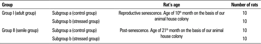
Zona glomerulosa (ZG) cells were arranged in clusters, zona fasciculata (ZF) cells were arranged in straight columns and ZR cells were arranged as anastomosing cords in group I-a (Fig. 1a). Lamellar separations of the capsule with areas of ghost cells were observed in ZG and ZF in group I-b (Fig. 1b, c). Separation and indentation of the capsule with its lamellar separation were observed in group II-a. In addition, the arrangement of ZG cells was disturbed with the existence of multiple scattered degenerative foci in ZF and ZR (Fig. 1d–f). Capsular separation with subcapsular spindle cell hyperplasia and disruption of the regular architectural pattern of ZF were seen in group II-b with the presence of areas of homogenous degeneration (Fig. 1g, h).
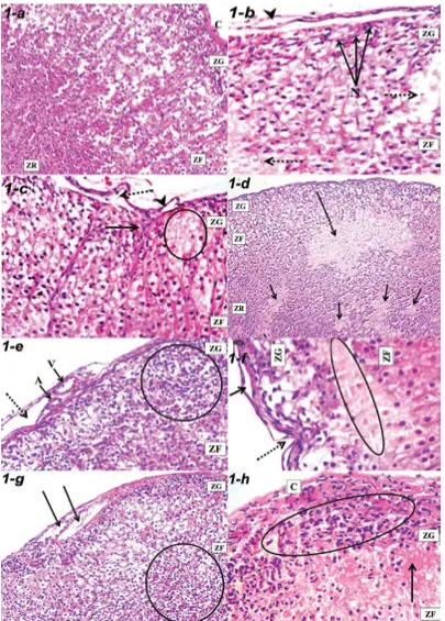
Spindle cell hyperplasia and vacuolated cell-are-as were detected in ZR in the group I-b (Fig. 2a, b). Scattered brown granules, wide sinusoids, and areas of degeneration in ZR were observed in group II-a (Fig. 2c, d). The disturbed arrangement of the ZR cells with engorged central adrenal veins and congested sinusoids were seen in group II-b (Fig. 2e, f).
Semithin sections findings
Thickening with lamellar separation of the capsule with irregular shape and arrangement of its fibro-blasts were observed in group II-b. ZG cells exhibited lipid droplet-loaded cytoplasm in the groups I-a, I-b, II-a. Their nuclei exhibited different shapes and sizes in group I-b, darkly stained in group II-a, pyknotic in group II-b (Fig. 3). The nuclei of ZF were rounded ve-sicular in groups I-a, II-a, while they were irregular in shape and darkly hyperchromatic in group II-b (Fig. 4).
Masson’s trichrome-stained sections and immunohistochemical study
Figure 2. Sections of the zona reticularis (ZR) and adjoining medulla (M) (H&E); a. Spindle cell hyperplasia (arrows) in a rat group I-b (×400).
b. An area of vacuolated cells (arrows) in a rat group I-b (×400). c. Scattered brown granules (arrows) and wide sinusoids (arrowheads) in a rat group II-a (×400). d. An area of degeneration (arrows) in a rat group II-a (×150). e. A disturbed arrangement (circle) of the ZR cells and an engorged central adrenal vein (arrow) in a rat group II-b (×150). f. A congested sinusoids (arrows) in a rat group II-b (× 400).
matin in group I-b and were pyknotic with deformed irregular outlines and disrupted nuclear envelops in group II-b. Multiple lysosomes and vacuolar degenera-tion mitochondria were also seen in group I-b (Fig. 8). The nuclei of ZF were regular and rounded in group I-a, irregular with condensed marginated het-erochromatin in group I-b, irregular with scattered chromatin in group II-a and indented with areas of chromatin destruction in group II-b. Mitochondria with disrupted cristae and discontinuity of their en-velopes and mitochondria with cristolysis and loss was thickened with lamellar separation and thickened
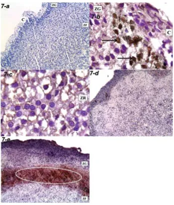
disrupted collagen bundles in group II-b. Dilated con-gested blood capillaries with extravasation of blood and exudation were also seen in group II-b (Figs. 5, 6). The immunoreaction of Ki67 was nuclear. It was absent in groups I-a, II-a, while it was positive in groups I-b, II-b (Fig. 7).
Ultrastructural remarks
chro-Figure 3. Semithin sections of the zona glomeru-losa (ZG) and zona fasciculata (ZF) (Toluidine blue ×1000); a. Capsular connective tissue fibres (dotted arrows), fibroblasts (white arrows) and
ZG cells (thick black arrows) with the cytoplasm loaded with lipid droplets (arrowheads) in a rat group I-a. Note a blood capillary (thin black arrow). b. Large swollen disrupted arrangement
fibroblasts (arrowheads). The nuclei of the ZG
cells exhibited different shapes and sizes (arrows), while the cytoplasm was loaded with lipid droplets (dotted arrows) in a rat group I-b.
c. A distended capillary (arrow) with folded en-dothelium (dotted arrows) in a rat group II-a.
d. Capsular separation (long arrow) and a mark-edly dilated blood vessel (short arrow). The ZG shows abundant lipid droplets (circles) and dark-ly stained nuclei (arrowheads) in a rat group II-a.
e. A lamellar separation of the capsule (arrows) and ZG with pyknotic nuclei (dotted arrow) in a rat group II-b. f. A marked thickening of the cap-sule with irregular shape and arrangement of its
fibroblasts (arrows) and collagen fibres (dotted
arrow) in a rat group II-b.
Figure 5. Collagen fibres in zona glomerulosa (ZG) and zona fasciculata (ZF) (Masson’s trichrome); a. A regular capsule (C), trabeculae of
col-lagen fibres (dotted arrow) and parallel arranged colcol-lagen fibres (arrows) perpendicular to the surface in a rat group I-a. Note blood sinusoids
(arrowheads) (×200). b. A thickened fibrous septum (arrow) and a disrupted area in the capsule (dotted arrow) in a rat group I-b (×200). c. Lamellar separation (dotted arrows) of the capsule (C) and irregularly dispersed collagen fibres (arrows) in a rat group II-a (×400). d. Accessory adrenocortical tissue (arrow) in a rat group II-a (×100). e. Thickened-capsule (C) with lamellar separation of its collagen
bundles (dotted arrows), thickened collagen bundles (white arrowheads), irregular collagen fibres (black arrowheads) and areas of exudate
(arrows) in a rat group II-b (×400). f. A thickened fibrous septum (arrows) and a thickened partially separated capsule (dotted arrow) in a rat
group II-b (×200).
of their envelopes were also detected in group I-b (Fig. 9).
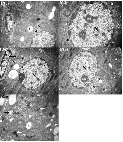
The absence of nuclear affection was perceived in the ZR cells of the different groups. Lipofuscin granules and dilated smooth endoplasmic reticulum were also observed in group II-b (Fig. 10).
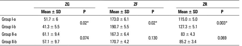
Morphometric measurements (Tables 2–4)
The mean collagen fibre area per cent of group II-b was 39%, significantly higher than that of group II-a. Using the optical density of the Ki67, the mean number of group II-b proliferating cells was signifi-cantly higher by 23% than that of group I-b.
The thickness of ZG decreased significantly by 20% in group I-b as compared to that of group I-a.
Contrary, the thickness of both ZF and ZR increased significantly by 10% in group I-b as compared to their respective of group I-a.
dIScuSSIon
Stress is a state of endangered homeostasis. The effect of stress on the adrenal cortex was evaluated as the adrenal gland is the chief stress responsive organ involved in the overall homeostasis. Various types of experimental stressors have been styled such as restraint, cold/heat, water immersion, water deprivation, noise, crowding and electric shock [10]. Among all these methods, restraint is the method that attains the least pain, suffering or distress to the animal.
Figure 8. Electron photomicrograph of the zona glomerulosa; a. A nucleus (N) containing euchromatin (Eu), clumps of peripheral heterochro-matin (Ht), prominent nucleolus (nu) and multiple elongated mitochondria (M) with tubulolamellar cristae in a rat group I-a (TEM ×20000).
b. A nucleus (N) with clogged marginated deeply stained chromatin distorting the nuclear contour. The cytoplasm contains multiple lyso-somes (arrows), mitochondria (M) with tubulolamellar cristae and mitochondria with vacuolar degeneration (thick arrow) in a rat group I-b (TEM ×20,000). c. A nucleus (N) with distinct nucleolus (nu), heterochromatin (Ht) and undulation of its nuclear envelope (black arrow) in a rat group II-a. Note lipid droplets (L) (TEM ×10,000). d. A pyknotic nucleus (N) with a deformed irregular outline (dotted arrow), dense clumps of clogged chromatin associated with disrupted nuclear envelope (thick arrow) in a rat group II-b. Note mitochondria (M),
desmo-somes (white arrows) and bundles of collagen fibres (black arrow) (TEM ×15,000).
The present study was confined to male rats since the adrenal gland of female rats undergo several chang-es such as the appearance and regrchang-ession of the X-zone [9] and the response to stress is a sex-specific due to a gender difference in the hormonal receptors [2].
Areas of subcapsular spindle cell hyperplasia were detected in the adult and senile stressed groups (groups I-b, II-b). These cellular clusters were locat-ed initially in the ZG and then extend centripetally between the underlying ZF cell cords. Several expla-nations had been suggested: a mast cells role [15], sex hormones association [20] or misspecifications of subcapsular mesenchymal stem cells that do not differentiate to enter the steroidogenic lineage [28]. Ghost cell areas, ghost nuclei, multiple pleomor-phic and pyknotic nuclei existed in ZG of the adult stressed group. The nuclei of ZG cells are extremely sensitive to stress [16]. Areas of vacuolated cells at the junctional zone between ZR and medulla existed
in this group. Stress induces pronounced changes at the interphase between ZR and medulla where con-nective tissue cords loosened, cell count decreased and cell-to-cell spaces extended [18]. These vacu-olated areas might denote an early stage of cystic degeneration [21].
A significant decrease in the thickness of the cor-tical zones was observed in the adult stressed group (group I-b) which may be related to shifting in hor-monal stress responsiveness [7].
Increase thickness, indentation with lamellar separation were constant findings of the capsule of the senile control group (group II-a). In addition, replacement of the capsular cellular elements with collagenous tissue was also detected; such replace-ment was suggested as a mechanism responsible for the age-dependent capsular changes [14].
imperfect repair of damage with the advance of age combined with compensatory overgrowth can lead to more and more abnormal tissue architecture [12]. In addition, increased vacuolation of the ZG cells reflect-ed a decrease in the secretory activity of ZG cells [25]. The three cortical zones normally have some degree of vacuolation; these vacuoles contain neutral lipid and cholesterol. The increased cortical vacuolation can be due to age-dependent drop in cholesterol biosynthesis or metabolism leading to accumulation of cholesterol and steroids [25].
Areas of degeneration with a cytoplasmic heter-ogeneity of the ZF cells was seen in group II-a which reflects different cell sensitivity to destructive factors and an increase in the subpopulation of newly formed adrenocortical cells [17]. Another notable finding in this group was the predominance of lipofuscin-like inclusions seen in the cytoplasm of the ZR cells. Lipo-fuscin is a biomarker of ageing and its progressive
accumulation is one of the most prominent changes with ageing [14]. Deposition of lipofuscin pigment is most probably due to decreased metabolic activities in the cells [14]. It accumulates gradually in lysosomes of post-mitotic tissue and it is not removed by deg-radation or exocytosis [4].
Accessory adrenocortical tissue was also encoun-tered in group II-a. This tissue is composed of normal cortical tissue, in which ZG and ZF are notable while the medullary tissue is not normally associated [11]. The accessory cortical tissues undertake the same ageing changes as the main body of the adrenal [11].
Figure 10. Electron photomicrograph of the zona reticularis; a. A euchromatic nucleus (N) with a nuclear envelope (arrowhead), a profile of smooth endoplasmic reticulum (SER) (S), lipid droplets (L) and a spindle-shaped fibroblast (arrow) in a rat group I-a (TEM ×10,000). b. A rounded euchromatic nucleus (N) possessing double nucleoli (nu) and mitochondria (M) with numerous tubular cristae in a rat group I-b (TEM ×15,000). c. a euchromatic nucleus (N), SER (S), multiple mitochondria (M) and few lipid droplets (L) in a rat group II-a. Note an elongated endothelial nucleus (black arrow) (TEM ×10,000). d. A euchromatic nucleus (N) with a distinct nucleolus (nu), mitochondria (M), dilated SER (S), lysosomes (white arrows), and Golgi body (black arrow) in a rat group II-b (TEM ×10,000). e. Few lipid droplets (L), numerous lipofuscin granules (white arrows), dilated SER (black arrow) and free ribosomes (arrowheads) in a rat group II-b (TEM ×20,000).
Table 2. Area per cent of collagen fibres
Groups Mean ± SD P
Group I-a 31.6 ± 4.7
0.09
Group I-b 38.7 ± 0.9
Group II-a 41.4 ± 3.7
0.02*
Group II-b 57.8 ± 2.2
*Significant (p ≤ 0.05); SD — standard deviation
Table 3. Optical density of Ki67 immunoreactivity in the stressed groups
Groups Mean ± SD P
Group I-b 67.46 ± 5.17
0.02*
Group II-b 83.05 ± 1.63
engorgement of the central adrenal veins. The mor-phological changes under stress conditions adrenals developed against a background of haemodynamic changes that involved all layers of the adrenal cortex where the sinusoids and capillaries were dilated and plethoric [18].
Immunoreactivity to Ki67 was detected only in the stressed groups (groups I-b, II-b) as the cellular turnover in the adrenal cortex occurs only to ac-commodate the physiological needs or in response to experimental manipulations [23]. The pattern of distribution of immunoreactivity varied; in the adult stressed group, the reactivity was located in the subcapsular region, while in the senile stressed group it was confined to the junctional zone be-tween ZG and ZF. The stem/progenitor cells exist in the adrenal capsule, subjacent cortex, and jux-tamedullary region. These cells proliferate, differen-tiate, and repopulate in the cortex centripetally or centrifugally [23].
The ultrastructural remodelling of the adrenals of the stressed groups was mainly manifested as chang-es in the constituents involved in steroid hormone biosynthesis, namely, the lipid droplets, mitochon-dria, and smooth endoplasmic reticulum. The lipid droplets were seen packed together. These packed lipid droplets represent packets of steroid hormones awaiting release by exocytosis [8]. Other changes correlate with adrenal stimulation was the increasing number and size of mitochondria with more infolding of their inner membranes and their transformation to a more lamellar membrane pattern.
Another ultrastructural feature encountered in the senile stressed group was the degenerative changes of mitochondrial in the form of cristolysis, disrup-tion of mitochondrial envelopes with the presence of intra-mitochondrial electron-dense inclusions [17]. The number of secondary lysosomes increased in this group which indicated autophagocytosis in the adrenocortical cells [19].
The senile stressed rats (group II-b) showed evi-dent nuclear changes. The ZG cells exhibited nuclei with pyknosis, highly deformed irregular outlines and dense clumps of clogged chromatin. The ZF cells also showed highly indented bilobed nuclei with the undulation of the nuclear envelopes, elec-tron-lucent spots and focal areas of chromatin de-struction. These features of nuclear degeneration could reflect the superimposed effect of stress when coupled with ageing. The senile rats which were already exposed to the deleterious effect of ageing, showed marked deterioration upon stress exposure. Age prompted such effects through the affection of the hypothalamic-adrenocorticotropic-adrenal axis which may be confounded by multiple factors, including stress [26].
concluSIonS
In conclusion, histological alterations occurred in the adrenal cortex in response to stress, especially when coupled with the advance of age. This was accompanied by proliferative changes.
referenceS
1. Bali A, Jaggi AS. Preclinical experimental stress studies: protocols, assessment and comparison. Eur J Pharmacol.
2015; 746: 282–292.
2. Balog M, Miljanović M, Blažetić S, et al. Sex-specific chronic stress response at the level of adrenal gland modified
sexual hormone and leptin receptors. Croat Med J. 2015;
56(2): 104–113.
3. Becker KL. Principles and practice of endocrinology and metabolism. 3rd edn. Philadelphia: Lippincott Williams & Wilkins. 2001.
4. Brunk UT, Terman A. Lipofuscin-mechanisms of age-related
accumulation and influence on cell function. Free Radic
Biol Med. 2002; 33(5): 611–619.
5. Chang SP, Mullins JJ, Morley SD, et al. Transition from organogenesis to stem cell maintenance in the mouse
adrenal cortex. Organogenesis. 2011; 7(4): 267–280.
6. da Silva Bi, Lopes-Costa PV, dos Santos AR, et al. Evaluation of Ki-67 antigen expression in the zona reticularis of the adrenal cortex of female rats in persistent estrus. Hum
Reprod. 2009; 24(3): 705–709.
Table 4. Thickness of the three adrenal zones
ZG ZF ZR
Mean ± SD P Mean ± SD P Mean ± SD P
Group I-a 51.7 ± 6
0.02* 173.0 ± 6.1 0.02* 115.0 ± 5.0 0.003*
Group I-b 41.3 ± 5.5 190.7 ± 5.5 127.3 ± 5.1
Group II-a 61.1 ± 9.4
0.074 167.3 ± 6.4 0.130 83 ± 4.3 0.069
Group II-b 57.1 ± 9.7 170.7 ± 4.2 85.2 ± 3.4
7. Foilb AR, Lui P, Romeo RD. The transformation of hormonal stress responses throughout puberty and adolescence. J Endocrinol. 2011; 210(3): 391–398.
8. Gannouni N, Mhamdi A, El May M, et al. Morphological changes of adrenal gland and heart tissue after varying duration of noise exposure in adult rat. Noise Health.
2014; 16(73): 416–421.
9. Greaves P. Histopathology of preclinical toxicity studies: interpretation and relevance in drug safety evaluation.
4th edn. Amsterdam: Elsevier AP. 2012.
10. Hayashi T, Ikematsu K, Abe Y, et al. Temporal changes of the adrenal endocrine system in a restraint stressed mouse and possibility of postmortem indicators of prolonged
psy-chological stress. Leg Med (Tokyo). 2014; 16(4): 193–196.
11. Hedrich HJ, Bullock GR. The laboratory mouse. Amsterdam;
Oxford: Elsevier Academic Press. 2004.
12. Hornsby PJ. Aging of the human adrenal cortex. Sci Aging
Knowledge Environ. 2004; 2004(35): re6, doi: 10.1126/ sageke.2004.35.re6, indexed in Pubmed: 15342924. 13. Hornsby PJ. Adrenarche: a cell biological perspective.
J Endocrinol. 2012; 214(2): 113–119, doi:
10.1530/JOE-12-0022, indexed in Pubmed: 22573830.
14. Janjua MZ, Khan MY. Age related changes in the rat
adre-nal cortex. J Pak Med Assoc. 1992; 42(4): 89–94.
15. Kim JS, Kubota H, Nam SY, et al. Expression of cytokines and proteases in mast cells in the lesion of subcapsular cell hyperplasia in mouse adrenal glands. Toxicol Pathol. 2000; 28(2): 297–303.
16. Koko V, Djordjeviae J, Cvijiae G, et al. Effect of acute heat stress on rat adrenal glands: a morphological and
stere-ological study. J Exp Biol. 2004; 207(Pt 24): 4225–4230.
17. Koldysheva EV, Lushnikova EL. Ultrastructural reorganiza-tion of rat adrenal cortex after whole body hyperthermia.
Bull Exp Biol Med. 2008; 145(5): 650–655.
18. Koldysheva EV, Lushnikova EL, Nepomnyashchikh LM. Mor-phogenesis of adaptation and compensatory reactions in
mouse adrenals during restitution after thermal exposure.
Bull Exp Biol Med. 2005; 140(4): 464–467.
19. Koldysheva EV, Lushnikova E, Proskuriakova IS. [Ultrastruc-tural reorganization of the adrenal cortex during hypoxia and its correction by nerobolil in rats]. Biull Eksp Biol Med. 1999; 127(5): 588–592.
20. Krachulec J, Vetter M, Schrade A, et al. GATA4 is a crit -ical regulator of gonadectomy-induced adrenocort-ical tumorigenesis in mice. Endocrinology. 2012; 153(6): 2599–2611.
21. Laast VA, Larsen T, Allison N, et al. Distinguishing cystic degeneration from other aging lesions in the adrenal cortex
of Sprague-Dawley rats. Toxicol Pathol. 2014; 42(5): 823–9.
22. McEwen BS. Physiology and neurobiology of stress and adaptation: central role of the brain. Physiol Rev. 2007;
87(3): 873–904, doi:10.1152/physrev.00041.2006,
in-dexed in Pubmed: 17615391.
23. Pihlajoki M, Dörner J, Cochran RS, et al. Adrenocortical zonation, renewal, and remodeling. Front Endocrinol (Lausanne). 2015; 6: 27, doi:10.3389/fendo.2015.00027,
indexed in Pubmed: 25798129.
24. Pérez M, Terreros G, Dagnino-Subiabre A. Long-term ω-3 fatty acid supplementation induces anti-stress effects and improves learning in rats. Behav Brain Funct. 2013; 9: 25. 25. Sahota PS. Toxicologic pathology: nonclinical safety
as-sessment. Boca Raton CRC Press. 2013.
26. Veldhuis JD, Sharma A, Roelfsema F. Age-dependent and gender-dependent regulation of hypothalamic-adreno-corticotropic-adrenal axis. Endocrinol Metab Clin North
Am. 2013; 42(2): 201–225.
27. Wass JA, Stewart PM. xford textbook of endocrinology and diabetes. 2nd edn. Oxford: Oxford University Press. 2011. 28. Yates R, Katugampola H, Cavlan D, et al. Adrenocortical de-velopment, maintenance, and disease. Curr Top Dev Biol. 2013; 106: 239–312, doi:10.1016/B978-0-12-416021-7.