PRIMARY RESEARCH
Antitumor activity of arsenite
in combination with tetrandrine against human
breast cancer cell line MDA-MB-231 in vitro
and in vivo
Bo Yuan
1,2*†, Mingjiang Yao
1,3†, Xiao Wang
1†, Ai Sato
2, Ayane Okazaki
1, Hana Komuro
1, Hideki Hayashi
1,
Hiroo Toyoda
2, Xiaohua Pei
4, Xiaomei Hu
3, Toshihiko Hirano
5and Norio Takagi
1Abstract
Background: Triple-negative breast cancer (TNBC) is one of the most difficult subtypes of breast cancer to treat
due to its aggressive, metastatic behavior, and a lack of a targeted therapy. Trivalent arsenic derivatives (arsenite, AsIII)
with remarkable clinical efficacy in acute promyelocytic leukemia has been demonstrated to exhibit inhibitory effect against breast cancer cells. To provide novel insight into the development of new therapeutic strategies, antitumor activity of AsIII and tetrandrine (Tetra), a Chinese plant-derived alkaloid, against the TNBC cell line MDA-MB-231 in vitro
and in vivo was investigated.
Methods: Cytotoxicity was evaluated using cell viability, lactate dehydrogenase leakage and cell cycle assay.
Altera-tions of genes related to cell proliferation and death were analyzed using western blotting. In vivo antitumor activity of AsIII alone or in combination with Tetra was studied using MDA-MB-231 xenografts in nude mice.
Results: Synergistic cytotoxic effects of two drugs were observed in the cells. In vivo study also showed that
co-administration of AsIII and Tetra significantly reduced tumor volume and weight, directly supporting its in vitro
antitu-mor activity. No deaths and reduction of body-weight were observed after a long-term co-administration, indicating its good tolerability. S-phase arrest associated with the upregulation of FOXO3a, p27 along with decreased Cyclin D1 expression was observed in the cells treated with the combined regimen. A substantial upregulated p21 expression and downregulated phospho-FOXO3a and Cyclin D1 expression was observed in the tumor tissues of mice co-admin-istered with AsIII and Tetra. Autophagy induction was observed in the combination treatment in vitro and in vivo. The
addition of wortmannin, a potent autophagy inhibitor, significantly rescued MDA-MB-231 cells from their cytotoxicity of AsIII and Tetra.
Conclusions: S-phase arrest, autophagic and necrotic cell death contribute to the cytocidal effects of the combined
regimen of AsIII and Tetra. Considering our previous study showing synergistic cytotoxic effects of the combined
regimen in estrogen receptor-positive breast cancer cell line MCF-7, these results suggest that development of the combination regimen of AsIII plus Tetra may offer many benefits to patients with different types of breast cancer.
Keywords: Arsenite, Tetrandrine, MDA-MB-231 cells, Cell death, Combination therapy
© The Author(s) 2018. This article is distributed under the terms of the Creative Commons Attribution 4.0 International License (http://creat iveco mmons .org/licen ses/by/4.0/), which permits unrestricted use, distribution, and reproduction in any medium, provided you give appropriate credit to the original author(s) and the source, provide a link to the Creative Commons license, and indicate if changes were made. The Creative Commons Public Domain Dedication waiver (http://creat iveco mmons .org/ publi cdoma in/zero/1.0/) applies to the data made available in this article, unless otherwise stated.
Open Access
*Correspondence: yuanbo@toyaku.ac.jp
†Bo Yuan, Mingjiang Yao and Xiao Wang contributed equally to this work 1 Department of Applied Biochemistry, School of Pharmacy, Tokyo University of Pharmacy & Life Sciences, 1432-1 Horinouchi, Hachioji, Tokyo 192-0392, Japan
Background
Breast cancer is the most common cancer among women worldwide and persists as one of the leading causes of cancer-related deaths in women. Three distinct bio-markers including the estrogen receptor (ER), progester-one receptor (PR), and human epidermal growth factor receptor-2 (HER2) are used to determine the appropriate breast cancer therapy [1]. Tamoxifen (Nolvadex®) and
trastuzumab (Herceptin®) have already been
success-fully applied to patients diagnosed with ER-positive and HER2-positive breast cancer, respectively [2]. Among dif-ferent types of breast cancer, triple negative breast can-cer (TNBC; ER, PR, and HER2-negative breast cancan-cer), accounting for approximately 10–20% of breast cancer cases, is one of the most difficult subtypes of breast can-cer to treat due to its aggressive, metastatic behavior, and a lack of a targeted therapy [1]. Therefore, novel thera-peutic strategies are urgently needed as most patients with TNBC relapse with distant metastases, and hormo-nal therapies and HER2-targeted agents are ineffective in this group of tumors.
Administration of trivalent arsenic derivatives (arsen-ite, AsIII) such as arsenic trioxide (As
2O3) has
demon-strated a remarkable efficacy in the treatment of relapsed and refractory acute promyelocytic leukemia (APL) patients. Several research groups including us have con-ducted detailed pharmacokinetic studies of AsIII in APL
to optimize its treatment [3–6]. We have also investi-gated the effects of AsIII using a unique in vitro system
comprising primary cultured chorion and amnion cells prepared from human fetal membranes, and demon-strated that aquaporin 9 and multidrug resistance-asso-ciated protein 2 are functionally involved in controlling arsenic accumulation in these normal cells, which then contribute to differential sensitivity to AsIII
cytotoxic-ity between these cells [7]. These findings may provide a new insight into clinical applications of As2O3, and
bet-ter therapeutic protocols [8]. At the same time, the suc-cessful clinical efficacy of AsIII in the treatment of APL
patients has encouraged further studies on its potential treatment applications for other malignancies, including solid tumors [8, 9]. In fact, AsIII has been demonstrated
to exhibit inhibitory effects against breast cancer cells [10, 11], raising the possibility of repositioning arsenic compounds to treat patients with breast cancer.
Recently, we demonstrated a clear cytotoxic effect of AsIII against ER-positive human breast cancer cell line
MCF-7, and further clarified that tetrandrine (Tetra), a bis-benzylisoquinoline alkaloid isolated from the root of
Stephania tetrandra S. Moore, significantly enhanced the cytotoxicity of AsIII in a synergistic manner [12]. QT
pro-longation is known as a major complication in AsIII
ther-apy [8], closely related to the intracellular [Ca2+] overload
induced by AsIII [13], Tetra, on the other hand, has been
demonstrated to serve as a calcium channel antagonist significantly decreasing intracellular [Ca2+] within ven-tricular cells [14]. Therefore, we suggested that the com-bination regimen of AsIII and Tetra may be expected not
only to achieve improved efficacy of AsIII in the treatment
with ER-positive breast cancer, but also overcome its adverse cardiac effects secondary to Tetra functioning as calcium channel blocker. However, the antitumor activ-ity of AsIII in combination with Tetra against TNBC cell
line MDA-MB-231 in vitro and in vivo has not yet been investigated.
Cell cycle arrest as well as autophagic cell death has been considered as the major underlying mechanisms of action of most anticancer drugs [11, 15–19]. The cell cycle is known to be precisely regulated by a number of vital molecules known as cyclin-dependent kinases (CDKs) and CDK inhibitors such as p21 Waf1/Cip1 (p21) and p27 Kip1 (p27) [11, 20, 21]. Forkhead box transcrip-tion factor (FOXO3a), which is considered to be involved in the development of breast cancer and may also serve as its prognostic marker [22], has been linked to the regu-lation of genes involving multiple cellular processes such as cell cycle, invasion, and cell death [21–24]. FOXO3a is also known to be targeted for degradation by phospho-rylation [25, 26]. Phosphorylation of FOXO3a will results in its nuclear export and thereby consequent degrada-tion, and consequently interfered with its function as tumor suppressor [25, 26]. Upregulation of p21 and p27 associated with the increased FOXO3a expression has been demonstrated to be responsible for G0/G1 cell cycle
arrest of MCF-7 [12], while their alterations has also been implicated in S-phase arrest in various types of cancer cells including another TNBC cell line Hs578T [27–30]. These differential cell cycle responses may be attributed to different cell types and/or genetic and phenotypic diversity of cancer cells. However, whether and how these molecules contribute to the potential cytotoxic effects induced by the combination of AsIII and Tetra against
MDA-MB-231 in vitro and in vivo remain to be seen. In this study, antitumor activity of AsIII in
combina-tion with Tetra against the TNBC cell line MDA-MB-231 in vitro and in vivo was investigated by focusing on cell cycle arrest and autophagic cell death. Key regulatory molecules associated with the cell cycle and death were investigated to further elucidate cytotoxic mechanisms.
Materials and methods Materials
Sodium arsenite (NaAsO2, AsIII) and tetrandrine (Tetra)
serum (FBS) was purchased from Nichirei Biosciences (Tokyo, Japan). Dulbecco’s modified Eagle’s medium (DMEM), phenazine methosulfate (PMS) and dime-thyl sulfoxide (DMSO) were obtained from Wako Pure Chemical Industries (Osaka, Japan). Wortmannin, a potent autophagy inhibitor, propidium iodide (PI), ribo-nuclease A (RNaseA) and 2,3-bis(2-methoxy-4-nitro-5-sulfophenyl)-5-[(phenylamino)carbonyl]-2H-tetrazolium hydroxide (XTT) were purchased from Sigma-Aldrich (St. Louis, MO, USA). Tetrandrine hydrochloride injec-tion and arsenious acid and sodium chloride injecinjec-tion were obtained from Jiangxi Yintao Pharmaceutical Co., Ltd. (Jiangxi, China) and Heilongjiang Harbin Pharma-ceutical Co., Ltd. (Heilongjiang, China), respectively for the in vivo study. Can Get Signal® Immunoreaction Enhancer Solution were purchased from Toyobo Co., Ltd. (Osaka, Japan).
Cell culture and treatment
MDA-MB-231 cells were obtained from the American Type Culture Collection (ATCC, Manassas, VA, USA). Cells were cultured in DMEM medium supplemented with 10% heat-inactivated FBS and 100 U/ml of penicillin and 100 µg/ml of streptomycin in a humidified 5% CO2
atmosphere at 37 °C. The cells were seeded in 96-well plates (Iwaki, Tokyo, Japan) at a density of 1 × 10⁴ cells/
well in 0.1 ml medium and cultivated for 24 h. Cultures in sixplicate were treated with various concentrations of AsIII and Tetra, alone or in combination.
Cell viability assay
The cytotoxicity of AsIII and Tetra, alone or in
combi-nation, to MDA-MB-231 cells was measured by XTT dye-reduction assay according to the method previously described with slight modifications [7, 12]. Briefly, after treatment with AsIII and Tetra, alone or in
combina-tion, for 48 h, XTT and PMS were added into each well at final concentrations of 0.2 mg/ml and 1 mM, respec-tively. After incubation at 37 °C for 4 h, the plates were mixed and the absorbance at 450 nm was measured with a microplate reader (EMax Plus®, Molecular Devices, CA, USA). The relative cell viability was expressed as the ratio of the absorbance of each treatment group against those of the corresponding untreated control group. Data are shown as mean ± standard deviation (SD) from more than three independent experiments. The IC50 values of
AsIII and Tetra were calculated using GraphPad Prism®
6 software. In order to evaluate whether the two drugs generated synergistic, antagonistic, or additive effects, a combination index (CI) was determined as reported previously, using the computer software ComboSyn (Combosyn Inc. NJ, USA) for drug combinations and for general dose–effect analysis, which was developed by
Chou [31, 32]. The effect of the combination treatment was defined as a synergistic effect if CI < 1, an additive effect if CI = 1 or an antagonistic effect if CI > 1 [12, 15].
Antitumor activity of AsIII alone or in combination
with Tetra in MDA‑MB‑231 mouse xenografts
In vivo antitumor activity of AsIII alone or in combination
with Tetra was studied using human breast cancer nude mouse xenograft model. 5-week-old female immunodefi-cient BALB/c nude mice were obtained from Japan SLC, Inc. (Shizuoka, Japan) and housed at 23 ± 1 °C in a room with a constant humidity of 55 ± 5% and a regular 12-h light/12-h dark cycle for several days. MDA-MB-231 cells [1 × 10⁷, suspended in 0.1 ml phosphate-buffered saline
(PBS)] were then injected subcutaneously into the right flank of each mouse. Tumors (visualized as a small nod-ule with approximate size of 20–30 mm3 at the sites of
injection) appeared approximately 5–7 days later after injection, and the mice were randomly divided into four groups (n = 5) according to body weight and tumor size using SPSS 21.0 software, and given the following treat-ments: vehicle-control (treated with PBS); AsIII alone
(2 mg/kg/day); Tetra alone (20 mg/kg/day); AsIII (2 mg/
kg/day) + Tetra (20 mg/kg/day). The mice were admin-istered i.p. as described above once a day for 10 weeks, respectively. The tumor size was measured every day in two perpendicular dimensions with vernier cali-pers, and the tumor volume (TV) (mm3) was calculated
by the formula reported previously [33]: TV = length (mm) × width2 (mm2) × 0.5. The body weights were
also measured every day and were used as an indicator of systemic toxicity of the treatment. Throughout the experiment, the mice had free access to food and water according to the National Institute of Health Guide for the Care and Use of Laboratory Animals and the Guid-ance for Experimental Animal Care issued by the Prime Minister’s Office of Japan. At the end of the treatment, all animals were sacrificed, and the tumors were removed, photographed and weighed. Tumors were fixed in 4% paraformaldehyde (PFA) in 0.1 M phosphate buffer or frozen in liquid nitrogen for further analysis. The study was approved by the Committee of Animal Care and Welfare of Tokyo University of Pharmacy and Life Sciences.
Cell cycle analysis
After treatment with the indicated concentrations of AsIII
in 70% (v/v) cold ethanol and kept at − 20 °C for at least 4 h. Cell pellets were then washed twice with PBS after centrifugation and incubated with 0.25% Triton-X 100 for 5 min on ice. After centrifugation and washing with PBS, cells were resuspended in 500 µl of PI/RNase A/PBS (5 µg/ml of PI and 0.1% RNase A in PBS) and incubated for 30 min in the dark at room temperature. A total of 10,000 events were acquired and Diva software and Mod-Fit LT™ Ver.3.0 (Verity Software House, ME, USA) were used to calculate the number of cells at each G0/G1 and S
phase fraction.
Lactate dehydrogenase (LDH) assay
Since the combined treatment of the relatively low con-centration of 10 µM AsIII and 4 µg/ml Tetra achieved
appropriate level of the cytocidal effect in MDA-MB-231 cells, the above-mentioned concentrations were used to conduct LDH assay. After treatment with 10 µM AsIII
and 4 µg/ml Tetra, alone or in combination, for 48 h, LDH leakage from cells was measured using the LDH-Cytotoxic Test Wako kit (Wako Pure Chemical Indus-tries, Osaka, Japan) according to the method previously described with slight modifications [7, 12]. Briefly, cul-ture supernatants were collected by centrifugation at 2500 rpm for 5 min at 4 °C. Non-treated cells were lysed in culture medium containing 0.2% Tween 20, and mixed aggressively using a vortex mixer, followed by the cen-trifugation at 12,000×g for 10 min and the cell lysate was
used as the positive control. Culture medium served as the negative control. Culture supernatants were collected then diluted 16-fold with PBS and 50 μl of the diluted solution was transferred into wells of a 96-well plate. LDH activities were determined by adding 50 μl of ‘sub-strate solution’ from the kit, followed by incubation at room temperature for 30 min. The reaction was stopped by the addition of 100 μl of ‘stopping solution’ and the absorbance at 560 nm was measured with a microplate reader (Safire, Tecan, Switzerland). Cell damage was cal-culated as a percentage of LDH leakage from damaged cells using the following formula:
where Sup, NC, P and NCT refer to the absorption of the culture supernatant, negative control, positive con-trol and culture medium containing 0.2% Tween 20, respectively. In order to evaluate the correlation between necrosis and autophagy induction, cells were treated with 0.25 μM wortmannin for 30 min prior to the treatment with 10 μM As and 4 μg/ml Tetra, alone or in combina-tion, in the presence or absence of 0.25 μM wortmannin for an additional 48 h, followed by LDH leakage assay as described above.
LDH leakage(%)=(Sup−NC)/(P−NCT)×100
Western blot analysis
For preparation protein samples, cell pellets (approxi-mately 1–2 × 10⁶ cells per 110 μl Laemmli buffer) and
tumor tissues (at a ratio of approximately 1 g of tis-sue per 10 ml Laemmli buffer) obtained from MDA-MB-231 mouse xenografts were suspended in Laemmli buffer containing 100 mM DTT, 2 μg/ml leupeptin, 2 μg/ml aprotinin, 1 μg/ml pepstatin, 1 mM PMSF. The suspensions of cells and tumor tissues were sonicated using a sonicator (Qsonica, LLC, CT, USA) with 10 short burst of 2 s followed by intervals of 2 s for cool-ing. The suspensions were kept at all times in an ice bath. Sonicated cells and tumor tissues were heated in 95 °C for 5 min, and then centrifuged at 13,000g for 15 min at 4 °C. Protein concentrations of the superna-tant were determined according to Bradford’s method using the protein assay dye reagent (Bio-Rad, CA, USA) according to the manufacturer’s instructions, and using BSA as the standard. Western blot analysis was carried out according to the methods previously described [35]. Briefly, after separation of proteins on a sodium dode-cyl sulfate (SDS) polyacrylamide gel electrophoresis, followed by transferring to a polyvinylidene difluoride (PVDF) membrane (Millipore Corp, MA, USA), protein bands were detected using the following primary anti-bodies and dilution ratios: mouse anti-human β-actin (1:5000 dilution; cat. no. A-5441; Sigma-Aldrich, MO, USA); rabbit anti-human phospho-FOXO3a (Ser253) (1:1000 dilution; cat. no. 9466) and FOXO3a (1:1000 dilution; cat. no. 2497), rabbit anti-human p27 (1:1000 dilution; cat. no. 2552), mouse anti-human p21 (1:1000 dilution; cat. no. 2946), rabbit anti-human Cyclin D1 (1:1000 dilution; cat. no. 2978), rabbit anti-human phospho-AMPKα1 (Ser485) (1:1000 dilution; cat. no. 2537) and AMPKα (1:1000 dilution; cat. no. 2532), rab-bit anti-human phospho-mTOR (Ser2448) (1:1000 dilu-tion; cat. no. 5536) and mTOR (1:1000 diludilu-tion; cat. no. 2983), rabbit anti-human Beclin-1 (1:1000 dilution; cat. no. 3495), rabbit anti-human LC3 (1:1000 dilution; cat. no. 12741) (Cell Signaling Technology, MA, USA). Blot-ted protein bands were detecBlot-ted with respective horse-radish peroxidase-conjugated secondary antibody and an enhanced chemiluminescence (ECL) Western blot analysis system (Amersham Pharmacia Biotech, Buck-inghamshire, UK).
Statistical analysis
Results
In vitro and in vivo growth inhibition of human breast cancer cell line MDA‑MB‑231 by AsIII and Tetra, alone
or in combination
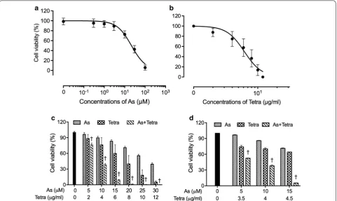
A significant decrease in cell viability was observed in a dose-dependent manner in MDA-MB-231 cells after treatment with various concentrations of AsIII or Tetra
alone for 48 h (Fig. 1a, b), and the IC50 values were
19.2 ± 2.6 µM and 6.2 ± 0.2 µg/ml for AsIII and Tetra
treatment, respectively. Next, two-drug combination in constant ratio were designed according to the median-effect method of Chou [31, 32] to evaluate if the two drugs generated synergistic, antagonistic, or additive cytotoxic effects against the cells. As shown in Fig. 1c, the combined treatment was significantly more cytotoxic than either drug alone (p < 0.05). The values of combina-tion index (CI) were < 1, indicating the two drugs worked in a synergistic manner (Table 1). Based on the above-described IC50 values of each drug, relatively low
dif-ferent concentrations of each drug (5, 10 and 15 µM for AsIII, and 3.5, 4, and 4.5 µg/ml for Tetra) were designed
to generate appropriate efficacy of drug combination in order to clarify the mechanisms underlying the cyto-cidal effect of the combination of AsIII and Tetra in detail.
Again, synergistic cytotoxic effects of the two drugs were confirmed (Fig. 1d and Table 2).
The antitumor activity of AsIII alone or in
combina-tion with Tetra was further evaluated in vivo with MDA-MB-231 human breast cancer xenograft model. As shown
Fig. 1 Cytotoxic effect of AsIII and Tetra, alone or in combination, against human breast cancer cell line MDA-MB-231. Cell viability was determined
by XTT assay after the treatment with various concentrations of AsIII alone (0.3, 1, 3, 10, 30 and 100 µM) (a), Tetra alone (2, 4, 6, 8, 10 and 12 µg/ml)
(b), their combination in constant ratio (5 µM AsIII+ 2 µg/ml Tetra, 10 µM AsIII+ 4 µg/ml Tetra, 15 µM AsIII+ 6 µg/ml Tetra, 20 µM AsIII+ 8 µg/ml Tetra,
25 µM AsIII+ 10 µg/ml Tetra, and 30 µM AsIII+ 12 µg/ml Tetra) (c), or combination treatment with relatively low concentrations (5 µM AsIII+ 3.5 µg/
ml Tetra, 10 µM AsIII+ 4 µg/ml Tetra, 15 µM AsIII+ 4.5 µg/ml Tetra) (d) for 48 h. Relative cell viability was calculated as the ratio of the absorbance
at 450 nm of each treatment group against those of the corresponding untreated control group. Data are shown as the means and SD from more than three independent experiments. †p < 0.05 vs. each alone. As, AsIII; Tetra, tetrandrine; As + Tetra, AsIII+ tetrandrine
Table 1 CI values of AsIII at concentrations in combination
with Tetra in MDA-MB-231 cells
As (μM) Tetra (μg/ml) Fa CI value
5 2 0.2352 0.89233
10 4 0.6149 0.87988
15 6 0.9077 0.60981
20 8 0.9878 0.33548
25 10 0.9947 0.29544
in Fig. 2a, a reduction in tumor volume was observed in the mice treated by a single administration of AsIII or
Tetra alone, and further strengthened by their combina-tion. Compared to vehicle-control group, tumor volume was reduced by 35.8, 37.2, and 51.6% for AsIII (2 mg/kg/
day) alone, Tetra (20 mg/kg/day) alone, and their com-bination, respectively, at the end of experiments (week 10 point). Consistently, treatment with AsIII and Tetra,
alone or in combination, also led to reduction of tumor
weight. Tumor weights were 1.47 ± 0.76, 1.07 ± 0.56, 0.77 ± 0.29 and 0.69 ± 0.29 g in the vehicle-control group, AsIII alone, Tetra alone and co-administration groups at
week 10 after implantation, respectively (Fig. 2c, d). Of note, no alteration in the body weight was observed in mice between vehicle-control group and treatment group regardless of the long-term administration of either drug alone or their combination (Fig. 2b), indicating the com-bined treatment was well tolerated by all mice.
Effects of AsIII and Tetra, alone or in combination,
on the cell cycle profiling and the expression level of cell cycle related‑proteins in MDA‑MB‑231 cells
To explore whether cell cycle arrest is involved in the cytotoxic effect of AsIII and Tetra, cell cycle analysis was
performed using flow cytometry. As shown in Fig. 3a, c, after treatment with various concentrations of AsIII and
Tetra, alone or in combination, for 48 h, a significant Table 2 CI values of AsIII at concentrations in combination
with Tetra in MDA-MB-231 cells
As (μM) Tetra (μg/ml) Fa CI value
5 3.5 0.47567 0.83571
10 4 0.62086 0.87539
15 4.5 0.94673 0.36602
Fig. 2 Antitumor activity of AsIII alone and in combination with Tetra in MDA-MB-231 mouse xenografts. The experiment was carried out using
human breast cancer nude mice implanted subcutaneously with 1 × 10⁷ MDA-MB-231 cells. The administration of AsIII and Tetra, alone or in
Fig. 3 Effects of AsIII and Tetra, alone or in combination, on the cell cycle profiling and the expression level of cell cycle related-proteins in
MDA-MB-231 cells. a–d After treatment with various concentrations of AsIII (5, 10 and 15 µM), and Tetra (3.5, 4 and 4.5 µg/ml), alone or in
combination (5 µM AsIII+ 3.5 µg/ml Tetra, 10 µM AsIII+ 4 µg/ml Tetra), for 48 h, cell cycle profiling was performed by FACSCanto flow cytometer
as described under “Materials and methods”. Analyzed data and profiles for each G0/G1 and G2/M phase using Diva software and ModFit LT™ ver.
3.0. are shown in the gray area. Cells at S phase are shown as shaded area. A representative FACS histogram from three separate experiments is shown. Significant difference between control and treatment with AsIII and Tetra, alone or in combination, are shown (*p < 0.05, ‡p < 0.01, §p < 0.001, †p < 0.0001 vs. control, #p < 0.05, $p < 0.01 vs. AsIII alone; &p < 0.05, ¶p < 0.01 vs. Tetra alone). e Representative image of the expression profile of each
protein is shown from three independent experiments. The densitometry of protein bands was analyzed using a program, NIH ImageJ 1.52a. The values under each image represent the ratios between each key molecule and β-actin protein expression levels, which were further compared with those of control group (untreated cells). As, AsIII; Tetra, tetrandrine. Since enough cells cannot be collected in the group treated with 15 µM AsIII in
increase in the number of cells in S phase was induced by AsIII alone and Tetra alone. Furthermore, a significant
increase in the number of cells in S phase was induced by the combined treatment in comparison to that induced by either drug alone. Concomitantly, a significant decrease in the number of cells in G0/G1 phase was also
observed (Fig. 3a, b). Almost no alteration was observed in the number of cells in G2/M phase was observed
(Fig. 3a, d). As shown in Fig. 3e, in comparison to control group, the expression of FOXO3a was clearly upregulated by the combined treatment of 10 μM AsIII and 4 μg/ml
Tetra, although almost no alteration was observed in the cells treated with AsIII and Tetra, each alone. The
expres-sion level of p27 was increased by the highest concen-trations of AsIII (15 μM AsIII) and Tetra alone compared
to the control, and a modest but clear enhancement in its expression was observed in the combined treatment group, especially in the combined regimen of 10 μM AsIII
and 4 μg/ml Tetra. Although only a modest decrease in the expression level of Cyclin D1 was observed when treated with AsIII and Tetra, each alone, a substantial
decrease in its expression was confirmed in the com-bined treatment group.
Enhanced LDH release in MDA‑MB‑231 cells treated with AsIII combined with Tetra
The release of LDH provides an accurate measure of the cell membrane integrity and cell viability [7, 12]. After treatment with 10 μM AsIII and 4 μg/ml Tetra, alone or
in combination, for 48 h, LDH leakage analysis was thus performed to examine whether the treatments affected cell membrane integrity. As shown in Fig. 4, a non-signif-icant increase in the LDH leakage was observed in MDA-MB-231 cells treated with either AsIII or Tetra alone
compared to the control. A significant enhancement in the LDH leakage was further observed in the combined treatment group, indicating the involvement of necrosis in the cytotoxic effect of AsIII and Tetra.
Effect of wortmannin on the cytotoxicity of the combined treatment of AsIII and Tetra in MDA‑MB‑231 cells
Since induction of autophagy by various anticancer drugs has been suggested to be a potential therapeutic strategy for cancer including breast cancer [17, 36, 37], a potent autophagy inhibitor, wortmannin was used to investigate whether the induction of autophagy contrib-uted to the combined treatment-induced cytotoxicity. As shown in Fig. 5, a significant decrease in cell viabil-ity was observed in MDA-MB-231 cells after treatment with 10 μM AsIII combined with 4 μg/ml Tetra for 24 h
(Fig. 5a) and 48 h (Fig. 5b), respectively. The addition of either 0.25 or 1.0 μM wortmannin, however, significantly rescued cell viability from 39.5 ± 5.4 to 71.0 ± 8.3% and
65.0 ± 13.8%, respectively, for 24 h, and from 15.5 ± 10.0 to 44.0 ± 18.1% and 32.0 ± 13.1%, respectively, for 48 h. It was also confirmed that cell viability was almost not altered by wortmannin alone in the cells. These results indicated the involvement of autophagic cell death in the cytotoxicity of the combination of AsIII and Tetra. In
order to explore a correlation of autophagy with necrosis, the influence of wortmannin on the alterations of LDH leakage in the cells treated with a combination of AsIII
and Tetra was evaluated. No any influence of wortman-nin on LDH leakage (Fig. 4) was observed, indicating that autophagy and necrosis independently contribute to the cytotoxicity of the combination of AsIII and Tetra.
Activation of autophagic pathway in MDA‑MB‑231 cells treated with AsIII and Tetra, alone or in combination
As shown in Fig. 6, the expression level of LC3, an autophagic marker, was dramatically upregulated by Tetra. Although a relatively low concentration of AsIII
(5 and 10 μM) did not affect the expression level of LC3, a clear increase in its expression level was observed when treated with the highest concentration of 15 μM AsIII. Consistent with the reduction of cell viability
induced by 10 μM AsIII in combination with 4 μg/ml
Tetra, and its restoration by the addition of wortman-nin (Fig. 6), an obvious upregulation of LC3 expression was observed in the combined treatment in compari-son to the treatment with either drug alone. In order to evaluate the mechanisms responsible for the signaling pathway activating autophagy, the expression of a num-ber of autophagy-related proteins was evaluated. Simi-lar to the alterations of the expression levels of LC3, the expression levels of phosphorylated of AMP-activated
Fig. 4 Enhanced LDH release in MDA-MB-231 cells treated with AsIII combined with Tetra. After treatment with 10 μM AsIII and 4 μg/
ml Tetra, alone or in combination, in the presence or absence of 0.25 μM wortmannin, for 48 h, LDH leakage was measured using the LDH-Cytotoxic test kit as described under “Materials and methods”. Significant difference between control and treatment with AsIII and
protein kinase (AMPK) (phospho-AMPK), a key energy sensor and an upstream promoter of autophagy induc-tion [23], was modestly upregulated by Tetra and the
highest concentrations of 15 μM AsIII. Again, compared
to the treatment with either 10 μM AsIII or 4 μg/ml Tetra
alone, a large enhancement in the expression of phospho-AMPK was observed in their combination treatment. Although treatment with either AsIII or Tetra alone,
except for 15 μM AsIII, almost did not affect the
expres-sion levels of total-AMPK, its enhanced expresexpres-sion level was further confirmed in the cells treated with the com-bination of 10 μM AsIII and 4 μg/ml Tetra. The alteration
of the expression levels of phosphorylated mammalian target of rapamycin (phospho-mTOR) and total-mTOR demonstrated an almost opposite behavior, showing a downregulation of their expression in the treated cells as compared to controls, especially in the combined treat-ment of 10 μM AsIII and 4 μg/ml Tetra. These results
indicating that AMPK-mediated mTOR deactivation is involved during the autophagy induction. The expres-sion levels of Beclin-1, an autophagic mediator which is deleted in 50% of breast tumors [19], were modestly but clearly upregulated in the cells treated with Tetra alone, but not by AsIII alone. Notably, the increase in the
expres-sion of Beclin-1 by 4 μg/ml Tetra was slightly strength-ened by the addition of 10 μM AsIII.
Expression profile of autophagy and cell cycle
arrest‑related proteins in MDA‑MB‑231 mouse xenografts treated with AsIII and Tetra, alone or in combination
As shown in Fig. 7, the expression level of LC3 was modestly and clearly upregulated by either AsIII or Tetra
alone. Consistent with the in vitro study, the upregula-tion was further strengthened by their co-adminis-tration. The expression levels of phospho-AMPK and total-AMPK were slightly increased by either AsIII Fig. 5 Effect of wortmannin on the cytotoxicity of the combined treatment of AsIII and Tetra. After the treatment with 10 μM AsIII combined with
4 μg/ml Tetra in the presence or absence of 0.25 μM and 1.0 μM wortmannin, respectively, for 24 h (a) and 48 h (b), cell viability was determined by XTT assay as described under “Materials and methods”. Relative cell viability was calculated as the ratio of the absorbance at 450 nm of each treatment group against those of the corresponding untreated control group. †p < 0.0001 vs. control; ∂p < 0.05, £p < 0.0001 vs. AsIII+ Tetra. As, AsIII;
Tetra, tetrandrine
Fig. 6 Expression profile of autophagy-related proteins in MDA-MB-231 cells treated with AsIII and Tetra, alone or in
combination. After treatment with various concentrations of AsIII
and Tetra, alone or in combination, for 48 h, the expression profile of autophagy-related proteins was analyzed using western blot as described in “Materials and methods”. Representative image of the expression profile of each protein is shown from three independent experiments. The densitometry of protein bands was analyzed using a program, NIH ImageJ 1.52a. The values under each image represent the ratios between each key molecule and β-actin protein expression levels, which were further compared with those of control group (untreated cells). As, AsIII; Tetra, tetrandrine. Since enough cells cannot
be collected in the group treated with 15 µM AsIII in combination
or Tetra alone, and a further increase in its expres-sion level was observed in the co-administered group. A slight increase in the expression levels of FOXO3a was observed in the both single-drug treatment and co-administration group. At the same time, the expres-sion of phospho-FOXO3a (p-FOXO3a) was promi-nently downregulated by Tetra alone, although only a modest increase in its expression level was observed in AsIII-treatment group. Intriguingly, the modest
increase in the expression of p-FOXO3a induced by AsIII was successfully corrected by the addition of
Tetra. Of note, in line with the in vitro study showing that S-phase arrest was induced by AsIII or Tetra alone,
and further strengthened by their combination in MDA-MB-231 cells (Fig. 3), the expression levels of p21 were prominently induced by the co-administration of AsIII and Tetra. Concomitantly, a substantial decrease
in the expression levels of Cyclin D1 was confirmed in the co-administered group, similar to the phenomena
observed in MDA-MB-231 cells treated with AsIII
com-bined with Tetra (Fig. 3e).
Discussion
Results from this study clearly demonstrated that Tetra significantly enhanced the cytotoxicity of AsIII against
TNBC cell line MDA-MB-231 in a synergistic manner as evidenced by the XTT assay (Fig. 1 and Tables 1, 2). In agreement with our observations, Tetra has been shown to augment the cytocidal effects of chemotherapeutic agents, including AsIII, in different types of solid tumor
cells [15, 38]. In addition, our in vivo study demonstrated that compared to vehicle-control group, tumor growth delay was observed after a single administration of AsIII
or Tetra (Fig. 2). Co-administration of AsIII and Tetra
further significantly reduced tumor volume and tumor weight by more than 50% (Fig. 2), directly supporting the in vitro antitumor activity of AsIII in combination
with Tetra. The discrepancy in the antitumor activity
Fig. 7 Expression profile of autophagy and cell cycle arrest-related proteins in MDA-MB-231 mouse xenografts treated with AsIII and Tetra, alone or
in combination. The experiment was carried out using human breast cancer nude mice implanted subcutaneously with 1 × 10⁷ MDA-MB-231 cells.
The administration of AsIII and Tetra, alone or in combination, and the preparation of tumor tissues and their protein samples were carried out as
of AsIII and Tetra between in vitro and in vivo might be
attributed to the distribution of the two drugs in differ-ent organs including tumor tissue of the mice bearing MDA-MB-231 cells xenografts, since the metabolism and distribution of the two drugs in vivo are considered to be more complex than in vitro. Further investigation into a correlation between the distribution of the two drugs and their anti-tumor activity in the MDA-MB-231 breast cancer xenograft model is ongoing in our laboratory.
More importantly, remarkable tolerance to the long-term co-administration of AsIII with Tetra was confirmed
in all mice as evidenced by the fact that the treatment did not cause death among the mice and decrease their body weight. In line with our findings, Tetra has been dem-onstrated to potentiate the antitumor activity of doxo-rubicin without a significant increase in toxicity in mice bearing the multidrug resistance (MDR) MCF-7/adr cell xenografts [39]. Tetra combined with daunorubicin, etoposide and cytarabine has also been used to treat the patients with refractory and relapsed acute myeloid leu-kemia in a multicenter clinical trial. Of 36 patients, 16 (44%) achieved complete remission, 9 (25%) achieved partial remission, and no increase of side effects was observed [40]. In conventional therapy for patients with breast cancer, estrogen receptor is the necessary mol-ecule requisite for the treatment with the antiestrogen tamoxifen [24]. Recently, we have demonstrated similar synergistic cytotoxic effects of AsIII and Tetra against
ER-positive human breast cancer cell line MCF-7 [12]. On the other hand, TNBC cell line MDA-MB-231 is an estrogen independent cell line that does not depend on estrogen for growth and survival [41]. Taking these previ-ous results and our observations into account, we suggest that Tetra can be a useful combination anticancer agent to enhance the therapeutic effect of AsIII for patients with
different types of breast cancers regardless of their estro-gen dependency.
We next demonstrated that a clear S-phase arrest along with a significant decrease in the number of cells in G0/G1 phase was observed simultaneously in
MDA-MB-231 cells treated with either drug alone (Fig. 3). A significant enhancement of S-phase arrest was further observed in the combined treatment, indicating that the cytotoxic effects of AsIII in combination with Tetra
appeared to be due to their ability to induce S-phase arrest. Our findings are also supported by previous studies showing that arsenic induced cell cycle arrest in the S phase in various types of cancer cell lines such as human malignant melanoma cell line and breast cancer cell line [42, 43]. FOXO3a has been implicated in cell cycle arrest leading to growth inhibition via upregula-tion of p21, p27 and downregulaupregula-tion of Cyclin D1 in vari-ous cancers [21–24]. Of note, upregulation of p21 and
p27, and downregulation of Cyclin D1 has been closely associated with S-phase arrest in various types of cancer cells including another human TNBC cell line Hs578T [27–30]. In agreement with these previous reports, a clear upregulation of the expression level of FOXO3a and p27 along with decreased Cyclin D1 expression was observed in MDA-MB-231 cells treated with AsIII
com-bined with Tetra (Fig. 3). We also demonstrated a slight increase in the expression levels of FOXO3a in both single-drug treated and co-administered mice (Fig. 7). More importantly, the modest increase in the expression of p-FOXO3a induced by AsIII was successfully corrected
by the addition of Tetra (Fig. 7), suggesting that the two drugs worked coordinately to downregulate phosphoryl-ated FOXO3a in tumor tissue, consequently maintained its function as tumor suppressor. Similar to the in vitro study, our in vivo study demonstrated that a substantial upregulated p21 expression and downregulated Cyclin D1 expression was also observed in the co-administered mice (Fig. 7). Collectively, our results suggest that antitu-mor activity of the combined treatment is partially attrib-uted to S-phase arrest associated with the upregulation of FOXO3a, p21, p27 along with the downregulation of p-FOXO3a and Cyclin D1 in MDA-MB-231 cells in vitro and in vivo.
We further demonstrated enhanced LDH release in MDA-MB-231 cells treated with AsIII in combination
with Tetra (Fig. 4), suggesting the involvement of necro-sis in the mechanisms of action for the combined treat-ment. Similarly, a previous report demonstrated that AsIII
induced necrosis through a regulated, Bcl-xL-sensitive mitochondrial pathway in an acute promyelocytic leuke-mia NB4 cell line [44]. Induction of autophagic cell death by various chemotherapeutic agents has also considered as a potential therapeutic strategy for cancer [17, 36, 37]. In the current study, the cytotoxicity of combining AsIII
with Tetra was partially but significantly abrogated by the addition of wortmannin, a potent autophagy inhibi-tor [45, 46] (Fig. 5). The activation of autophagy signal-ing pathway was further confirmed in both vitro and in vivo study, as evidenced by the striking increase in the expression levels of LC3, an autophagic marker [17, 36,
37], along with the activation of the autophagic pathway involving a number of important molecules including phospho-AMPK, phospho-mTOR and Beclin-1 (Figs. 6
and 7). In line with these current findings, Liu et al. [36] and Wang et al. [47] have demonstrated that Tetra func-tions as a potent agonist for cell autophagy in numerous cancer cells including breast cancer cells. In addition, autophagy has also been demonstrated to partially con-tribute AsIII-triggered cytocidal effects in human glioma
and necrosis induction, autophagic cell death also con-tributes to the antitumor activity of the combined regi-men of AsIII plus Tetra. Although correlation between
autophagy and necrosis has been suggested in various cancer cells [49], results from the current study suggest that the signaling pathway of autophagy and necrosis is independently activated to contribute to the cytotoxic-ity of the combined treatment based on the fact that the addition of wortmannin showed no influence on the LDH release induced by the combing treatment (Fig. 4).
Recently, an interesting phenomenon referred to as oncology drug repositioning, in which certain drugs con-ventionally used to treat non-malignant diseases exhibit anticancer effects, has been reported [50]. Study of drug repositioning further suggests that conventional drugs can have antitumor therapeutic effects by activating/ suppressing autophagy [50]. Tetra is extensively refer-enced in the Chinese Pharmacopoeia for its use in the Chinese medicinal system as an analgesic and diuretic agent and also in the treatment of hypertension [51]. Col-lectively, considering high cost associated with antican-cer drug development, AsIII as well as Tetra, which have
long been successfully used in clinic, can probably serve as promising candidates for the development of the novel therapeutic strategies of drug repositioning targeting autophagy to induce breast cancer cell death.
Conclusions
Our results suggest that Tetra can be a useful combina-tion anticancer agent to enhance therapeutic effect of AsIII, and that development of the combination regimen
of AsIII plus Tetra may offer many benefits to patients
with different types of breast cancers. Besides a con-tribution of S-phase arrest, autophagic cell death and necrosis seemed to independently contribute to the cyto-cidal effects of the combined regimen of AsIII and Tetra.
Recently, we demonstrated that enhanced intracellular arsenic accumulation (As[i]) along with synergistic cyto-toxicity was observed in MCF-7 cells treated with AsIII
combined with Tetra or Ko134, an inhibitor of breast cancer resistance protein (BCRP), suggesting that Tetra or the BCRP inhibitor probably intervened in the occur-rence of resistance to arsenic therapy by enhancing the As[i] via modulation of multidrug efflux transporters [12]. In addition, CYP3A4, a major P450 in humans, is involved in the metabolism of half of all currently used drugs including AsIII [52] and docetaxel, one of the most
commonly used chemotherapeutics for breast cancer [53]. Therefore, further studies about the effect of Tetra on AsIII pharmacokinetics need to be launched in order
to provide direct evidence for their clinical use.
Abbreviations
AsIII: trivalent arsenic (arsenite); Tetra: tetrandrine; p21: p21 Waf1/Cip1; p27: p27 Kip1; FOXO3a: forkhead box transcription factor 3a; phospho-FOXO3a: phos-phorylated FOXO3a; TNBC: triple negative breast cancer; ER: estrogen receptor; HER2: epidermal growth factor receptor-2; LDH: lactate dehydrogenase; As[i]: intracellular arsenic accumulation; BCRP: ABCG2/breast cancer resistance pro-tein; AMPK: phosphorylated AMP-activated protein kinase; phospho-mTOR: phosphorylated mammalian target of rapamycin.
Authors’ contributions
BY, MY and XW contributed equally to this study. BY conceived and designed the study, and drafted the manuscript. MY, XW and AS performed the XTT assay. XW and MY also performed cell cycle analysis, western blot analysis, LDH assay and in vivo study. AO and HK analyzed the partial western blot analysis. HH, HT, XP, XH, TH and NT assisted interpretation of the results with BY. All authors read and approved the final manuscript.
Author details
1 Department of Applied Biochemistry, School of Pharmacy, Tokyo University of Pharmacy & Life Sciences, 1432-1 Horinouchi, Hachioji, Tokyo 192-0392, Japan. 2 Department of Clinical Molecular Genetics, School of Pharmacy, Tokyo University of Pharmacy & Life Sciences, 1432-1 Horinouchi, Hachioji, Tokyo 192-0392, Japan. 3 XiYuan Hospital, China Academy of Chinese Medical Sciences, Beijing 100091, People’s Republic of China. 4 The Third Affiliated Hospital of Beijing University of Traditional Chinese Medicine, Beijing 100029, People’s Republic of China. 5 Department of Clinical Pharmacology, School of Pharmacy, Tokyo University of Pharmacy & Life Sciences, 1432-1 Horinouchi, Hachioji, Tokyo 192-0392, Japan.
Acknowledgements Not applicable.
Competing interests
The authors declare that they have no competing interests.
Availability of data and materials
All data generated or analyzed during this study are included in this published article.
Consent for publication Not applicable.
Ethics approval and consent to participate
The study was approved by the Committee of Animal Care and Welfare of Tokyo University of Pharmacy and Life Sciences (No. P16-80).
Funding
This work was supported by The Japan Society for the Promotion of Science (JSPS) KAKENHI Grant to Bo Yuan (Grant Numbers 26460233) (Grant Numbers 17K08465).
Publisher’s Note
Springer Nature remains neutral with regard to jurisdictional claims in pub-lished maps and institutional affiliations.
Received: 14 May 2018 Accepted: 9 August 2018
References
1. Carey L, Winer E, Viale G, Cameron D, Gianni L. Triple-negative breast cancer: disease entity or title of convenience? Nat Rev Clin Oncol. 2010;7(12):683–92.
2. Sutherland S, Miles D, Makris A. Use of maintenance endocrine therapy after chemotherapy in metastatic breast cancer. Eur J Cancer. 2016;69:216–22.
blood and bone marrow from an acute promyelocytic leukemia patient. J Hematol Oncol. 2012;5(1):1.
4. Kiguchi T, Yoshino Y, Yuan B, Yoshizawa S, Kitahara T, Akahane D, Gotoh M, Kaise T, Toyoda H, Ohyashiki K. Speciation of arsenic trioxide penetrates into cerebrospinal fluid in patients with acute promyelocytic leukemia. Leuk Res. 2010;34(3):403–5.
5. Shen ZX, Chen GQ, Ni JH, Li XS, Xiong SM, Qiu QY, Zhu J, Tang W, Sun GL, Yang KQ, Chen Y, Zhou L, Fang ZW, Wang YT, Ma J, Zhang P, Zhang TD, Chen SJ, Chen Z, Wang ZY. Use of arsenic trioxide (As2O3) in the treatment of acute promyelocytic leukemia (APL): II. Clinical efficacy and pharmacokinetics in relapsed patients. Blood. 1997;89(9):3354–60. 6. Yoshino Y, Yuan B, Miyashita SI, Iriyama N, Horikoshi A, Shikino O, Toyoda
H, Kaise T. Speciation of arsenic trioxide metabolites in blood cells and plasma of a patient with acute promyelocytic leukemia. Anal Bioanal Chem. 2009;393(2):689–97.
7. Yoshino Y, Yuan B, Kaise T, Takeichi M, Tanaka S, Hirano T, Kroetz DL, Toy-oda H. Contribution of aquaporin 9 and multidrug resistance-associated protein 2 to differential sensitivity to arsenite between primary cultured chorion and amnion cells prepared from human fetal membranes. Toxi-col Appl PharmaToxi-col. 2011;257(2):198–208.
8. Yuan B, Yoshino Y, Kaise T, Toyoda H. Application of arsenic trioxide therapy for patients with leukaemia. In: Sun H, editor. Biological chemistry of arsenic, antimony and bismuth. Chichester: John Wiley Sons, Ltd.; 2010. p. 263–92.
9. Dilda PJ, Hogg PJ. Arsenical-based cancer drugs. Cancer Treat Rev. 2007;33(6):542–64.
10. Liu W, Gong Y, Li H, Jiang G, Zhan S, Liu H, Wu Y. Arsenic trioxide-induced growth arrest of breast cancer MCF-7 cells involving FOXO3a and IkappaB kinase beta expression and localization. Cancer Biother Radiopharm. 2012;27(8):504–12.
11. Wang X, Gao P, Long M, Lin F, Wei JX, Ren JH, Yan L, He T, Han Y, Zhang HZ. Essential role of cell cycle regulatory genes p21 and p27 expres-sion in inhibition of breast cancer cells by arsenic trioxide. Med Oncol. 2011;28(4):1225–54.
12. Yao M, Yuan B, Wang X, Sato A, Sakuma K, Kaneko K, Komuro H, Okazaki A, Hayashi H, Toyoda H, Pei X, Hu X, Hirano T, Takagi N. Synergistic cytotoxic effects of arsenite and tetrandrine in human breast cancer cell line MCF-7. Int J Oncol. 2017;51(2):587–98.
13. Zhang Y, Dong Z, Jin L, Zhang K, Zhao X, Fu J, Gong Y, Sun M, Yang B, Li B. Arsenic trioxide-induced hERG K(+) channel deficiency can be rescued by matrine and oxymatrine through up-regulating transcription factor Sp1 expression. Biochem Pharmacol. 2013;85(1):59–68.
14. Liu QY, Karpinski E, Pang PK. Tetrandrine inhibits both T and L cal-cium channel currents in ventricular cells. J Cardiovasc Pharmacol. 1992;20(4):513–9.
15. Chen Y, Li P, Yang S, Tong N, Zhang J, Zhao X. Tetrandrine enhances the anticancer effects of arsenic trioxide in vitro. Int J Clin Pharmacol Ther. 2014;52(5):416–24.
16. Kon A, Yuan B, Hanazawa T, Kikuchi H, Sato M, Furutani R, Takagi N, Toyoda H. Contribution of membrane progesterone receptor alpha to the induc-tion of progesterone-mediated apoptosis associated with mitochondrial membrane disruption and caspase cascade activation in Jurkat cell lines. Oncol Rep. 2013;30(4):1965–70.
17. Wang X, Qi W, Li Y, Zhang N, Dong L, Sun M, Cun J, Zhang Y, Lv S, Yang Q. Huaier extract induces autophagic cell death by inhibiting the mTOR/S6K pathway in breast cancer cells. PLoS ONE. 2015;10(7):e0131771. 18. Yuan B, Okusumi S, Yoshino Y, Moriyama C, Tanaka S, Hirano T, Takagi N,
Toyoda H. Delphinidin induces cytotoxicity and potentiates cytocidal effect in combination with arsenite in an acute promyelocytic leukemia NB4 cell line. Oncol Rep. 2015;34(1):431–8.
19. Chen N, Karantza-Wadsworth V. Role and regulation of autophagy in cancer. Biochim Biophys Acta. 2009;1793(9):1516–23.
20. Mei L, Chen Y, Wang Z, Wang J, Wan J, Yu C, Liu X, Li W. Synergistic anti-tumour effects of tetrandrine and chloroquine combination therapy in human cancer: a potential antagonistic role for p21. Br J Pharmacol. 2015;172(9):2232–45.
21. Lin CH, Chang CY, Lee KR, Lin HJ, Chen TH, Wan L. Flavones inhibit breast cancer proliferation through the Akt/FOXO3a signaling pathway. BMC Cancer. 2015;15:958.
22. Jiang Y, Zou L, Lu WQ, Zhang Y, Shen AG. Foxo3a expression is a prognos-tic marker in breast cancer. PLoS ONE. 2013;8(8):e70746.
23. Chiacchiera F, Simone C. The AMPK-FoxO3A axis as a target for cancer treatment. Cell Cycle. 2010;9(6):1091–6.
24. Taylor S, Lam M, Pararasa C, Brown JE, Carmichael AR, Griffiths HR. Evaluat-ing the evidence for targetEvaluat-ing FOXO3a in breast cancer: a systematic review. Cancer Cell Int. 2015;15(1):1.
25. Fu Z, Tindall DJ. FOXOs, cancer and regulation of apoptosis. Oncogene. 2008;27(16):2312–9.
26. Wolfe AR, Debeb BG, Lacerda L, Larson R, Bambhroliya A, Huang X, Bertucci F, Finetti P, Birnbaum D, Van Laere S, Diagaradjan P, Ruffell B, Trenton NJ, Chu K, Hittelman W, Diehl M, Levental I, Ueno NT, Woodward WA. Simvastatin prevents triple-negative breast cancer metastasis in pre-clinical models through regulation of FOXO3a. Breast Cancer Res Treat. 2015;154(3):495–508.
27. Huang Z, Wang L, Chen L, Zhang Y, Shi P. Induction of cell cycle arrest via the p21, p27-cyclin E, A/Cdk2 pathway in SMMC-7721 hepatoma cells by clioquinol. Acta Pharm. 2015;65(4):463–71.
28. Li YG, Ji DF, Zhong S, Liu PG, Lv ZQ, Zhu JX, Chen JE, Chen HP. Polysac-charide from Phellinus linteus induces S-phase arrest in HepG2 cells by decreasing calreticulin expression and activating the P27kip1-cyclin A/ D1/E-CDK2 pathway. J Ethnopharmacol. 2013;150(1):187–95.
29. Zheng W, Han S, Jiang S, Pang L, Li X, Liu X, Cao M, Li P. Multiple effects of Xihuang pill aqueous extract on the Hs578T triple-negative breast cancer cell line. Biomed Rep. 2016;5(5):559–66.
30. Zhong S, Ji DF, Li YG, Lin TB, Lv ZQ, Chen HP. Activation of P27kip1-cyclin D1/E-CDK2 pathway by polysaccharide from Phellinus linteus leads to S-phase arrest in HT-29 cells. Chem Biol Interact. 2013;206(2):222–9. 31. Chou TC. Theoretical basis, experimental design, and computerized
simulation of synergism and antagonism in drug combination studies. Pharmacol Rev. 2006;58(3):621–81.
32. Chou TC. Drug combination studies and their synergy quantification using the Chou-Talalay method. Cancer Res. 2010;70(2):440–6. 33. Chiu HW, Lin JH, Chen YA, Ho SY, Wang YJ. Combination treatment with
arsenic trioxide and irradiation enhances cell-killing effects in human fibrosarcoma cells in vitro and in vivo through induction of both autophagy and apoptosis. Autophagy. 2010;6(3):353–65.
34. Kikuchi H, Yuan B, Yuhara E, Takagi N, Toyoda H. Involvement of histone H3 phosphorylation through p38 MAPK pathway activation in casticin-induced cytocidal effects against the human promyelocytic cell line HL-60. Int J Oncol. 2013;43(6):2046–56.
35. Yuan B, Ohyama K, Takeichi M, Toyoda H. Direct contribution of induc-ible nitric oxide synthase expression to apoptosis induction in primary smooth chorion trophoblast cells of human fetal membrane tissues. Int J Biochem Cell Biol. 2009;41(5):1062–9.
36. Liu T, Men Q, Wu G, Yu C, Huang Z, Liu X, Li W. Tetrandrine induces autophagy and differentiation by activating ROS and Notch1 signaling in leukemia cells. Oncotarget. 2015;6(10):7992–8006.
37. Zhang L, Shamaladevi N, Jayaprakasha GK, Patil BS, Lokeshwar BL. Polyphenol-rich extract of Pimenta dioica berries (Allspice) kills breast cancer cells by autophagy and delays growth of triple negative breast cancer in athymic mice. Oncotarget. 2015;6(18):16379–95.
38. Wei J, Liu B, Wang L, Qian X, Ding Y, Yu L. Synergistic interaction between tetrandrine and chemotherapeutic agents and influence of tetrandrine on chemotherapeutic agent-associated genes in human gastric cancer cell lines. Cancer Chemother Pharmacol. 2007;60(5):703–11.
39. Fu LW, Zhang YM, Liang YJ, Yang XP, Pan QC. The multidrug resistance of tumour cells was reversed by tetrandrine in vitro and in xenografts derived from human breast adenocarcinoma MCF-7/adr cells. Eur J Cancer. 2002;38(3):418–26.
40. Xu WL, Shen HL, Ao ZF, Chen BA, Xia W, Gao F, Zhang YN. Combination of tetrandrine as a potential-reversing agent with daunorubicin, etopo-side and cytarabine for the treatment of refractory and relapsed acute myelogenous leukemia. Leuk Res. 2006;30(4):407–13.
41. Meena R, Kumar S, Kumar R, Gaharwar US, Rajamani P. PLGA-CTAB cur-cumin nanoparticles: fabrication, characterization and molecular basis of anticancer activity in triple negative breast cancer cell lines (MDA-MB-231 cells). Biomed Pharmacother. 2017;94:944–54.
•fast, convenient online submission •
thorough peer review by experienced researchers in your field • rapid publication on acceptance
• support for research data, including large and complex data types •
gold Open Access which fosters wider collaboration and increased citations maximum visibility for your research: over 100M website views per year •
At BMC, research is always in progress.
Learn more biomedcentral.com/submissions
Ready to submit your research? Choose BMC and benefit from:
43. Pozo-Molina G, Ponciano-Gomez A, Rivera-Gonzalez GC, Hernandez-Zavala A, Garrido E. Arsenic-induced S phase cell cycle lengthening is associated with ROS generation, p53 signaling and CDC25A expression. Chem Biol Interact. 2015;238:170–9.
44. Scholz C, Wieder T, Starck L, Essmann F, Schulze-Osthoff K, Dorken B, Dan-iel PT. Arsenic trioxide triggers a regulated form of caspase-independent necrotic cell death via the mitochondrial death pathway. Oncogene. 2005;24(11):1904–13.
45. Francois A, Marchal S, Guillemin F, Bezdetnaya L. mTHPC-based photody-namic therapy induction of autophagy and apoptosis in cultured cells in relation to mitochondria and endoplasmic reticulum stress. Int J Oncol. 2011;39(6):1537–43.
46. Jia T, Zhang L, Duan Y, Zhang M, Wang G, Zhang J, Zhao Z. The differential susceptibilities of MCF-7 and MDA-MB-231 cells to the cytotoxic effects of curcumin are associated with the PI3K/Akt-SKP2-Cip/Kips pathway. Cancer Cell Int. 2014;14(1):126.
47. Wang H, Liu T, Li L, Wang Q, Yu C, Liu X, Li W. Tetrandrine is a potent cell autophagy agonist via activated intracellular reactive oxygen species. Cell Biosci. 2015;5:4.
48. Chiu HW, Ho SY, Guo HR, Wang YJ. Combination treatment with arsenic trioxide and irradiation enhances autophagic effects in U118-MG cells through increased mitotic arrest and regulation of PI3K/Akt and ERK1/2 signaling pathways. Autophagy. 2009;5(4):472–83.
49. Feng R, Wang SY, Shi YH, Fan J, Yin XM. Delphinidin induces necrosis in hepatocellular carcinoma cells in the presence of 3-methyladenine, an autophagy inhibitor. J Agric Food Chem. 2010;58(7):3957–64. 50. Yoshida GJ. Therapeutic strategies of drug repositioning targeting
autophagy to induce cancer cell death: from pathophysiology to treat-ment. J Hematol Oncol. 2017;10(1):67.
51. Bhagya N, Chandrashekar KR. Tetrandrine—a molecule of wide bioactiv-ity. Phytochemistry. 2016;125:5–13.
52. Noreault TL, Kostrubsky VE, Wood SG, Nichols RC, Strom SC, Trask HW, Wrighton SA, Evans RM, Jacobs JM, Sinclair PR, Sinclair JF. Arsenite decreases CYP3A4 and RXRalpha in primary human hepatocytes. Drug Metab Dispos. 2005;33(7):993–1003.