Open Access
Research
Influence of the atrio-ventricular delay optimization on the intra left
ventricular delay in cardiac resynchronization therapy
Christoph Melzer*
1, Fabian Knebel
1, Bruno Ismer
2, Hansjürgen Bondke
2,
Christoph A Nienaber
2, Gert Baumann
1and Adrian C Borges
1Address: 1Universitätsmedizin Berlin, Medical Clinic for Cardiology, Angiology, Pulmology, Charité Campus Mitte, Germany and 2University of
Rostock, Clinic for Internal Medicine, Rostock, Germany
Email: Christoph Melzer* - [email protected]; Fabian Knebel - [email protected]; Bruno Ismer - [email protected]; Hansjürgen Bondke - [email protected]; Christoph A Nienaber - [email protected]@med.uni-rostock.de; Gert Baumann - [email protected]; Adrian C Borges - [email protected]
* Corresponding author
Abstract
Background: Cardiac Resynchronization Therapy (CRT) leads to a reduction of left-ventricular dyssynchrony and an acute and sustained hemodynamic improvement in patients with chronic heart failure. Furthermore, an optimized AV-delay leads to an improved myocardial performance in pacemaker patients. The focus of this study is to investigate the acute effect of an optimized AV-delay on parameters of dyssynchrony in CRT patients.
Method: 11 chronic heart failure patients with CRT who were on stable medication were included in this study. The optimal AV-delay was defined according to the method of Ismer (mitral inflow and trans-oesophageal lead). Dyssynchrony was assessed echocardiographically at three different settings: AVDOPT; AVDOPT-50 ms and AVDOPT+50 ms. Echocardiographic assessment included 2D-and M-mode echo for the assessment of volumes 2D-and hemodynamic parameters (CI, SV) 2D-and LVEF and tissue Doppler echo (strain, strain rate, Tissue Synchronisation Imaging (TSI) and myocardial velocities in the basal segments)
Results: The AVDOPT in the VDD mode (atrially triggered) was 105.5 ± 38.1 ms and the AVDOPT in the DDD mode (atrially paced) was 186.9 ± 52.9 ms. Intra-individually, the highest LVEF was measured at AVDOPT. The LVEF at AVDOPT was significantly higher than in the AVDOPT-50setting (p
= 0.03). However, none of the parameters of dyssynchrony changed significantly in the three settings.
Conclusion: An optimized AV delay in CRT patients acutely leads to an improved systolic left ventricular ejection fraction without improving dyssynchrony.
Background
Asynchronous myocardial contraction in heart failure is associated with poor prognosis. Recent studies have shown an acute and sustained hemodynamic
improve-ment after biventricular pacing (BVP), reversal of LV-remodelling, an increased quality of life, a reduction of symptoms of heart failure, and an improvement of exer-cise tolerance [1-7].
Published: 26 January 2006
Cardiovascular Ultrasound 2006, 4:5 doi:10.1186/1476-7120-4-5
Received: 23 December 2005 Accepted: 26 January 2006
This article is available from: http://www.cardiovascularultrasound.com/content/4/1/5
© 2006 Melzer et al; licensee BioMed Central Ltd.
The optimization of the AV delay in DDD pacemaker patients is generally recommended and is performed in clinical practice. A variety of invasive and non-invasive methods were assessed in the past [8-15]. Recent studies have shown that also in CRT patients, invasively (dP/dt) [16-19] and non-invasively measured hemodynamic parameters (stroke volume) [20,21] are modified accord-ing to the programmed AV delay. A hemodynamically optimal AV delay can be defined.
Ismer's method of AV delay optimization [22] is validated for biventricular as well as right ventricular DDD pacing.
Tissue Doppler Imaging (TDI) is an evaluated tool in clin-ical practice to identify myocardial dyssynchrony. TDI (including strain and strain rate) imaging measures regional wall motion velocities and can accurately quan-tify regional left ventricular function [24].
Strain measures compression and distension of myocar-dial segments ("deformation imaging") and strain rate imaging expresses strain changes per time interval [25]. TSI (Tissue Synchronization Imaging) utilizes color-coded time-to-peak tissue Doppler velocities and visualizes seg-ments of dyssynchrony in real-time by superimposing these temporal motion data on 2D echo images. [26,27].
These new techniques could potentially improve patient selection and guidance of implantation and programming of the devices for BVP. There is a variety of methods to determine dyssynchrony as summarized elsewhere [28].
There are no published data on the correlation of param-eters of dyssynchrony and programming of the optimal AV interval. Aims of our study were therefore to investi-gate the influence of an optimized AV delay determined by the method of Ismer et al. [22] on dyssynchrony. Table 1: Patient characteristics
Age(mean ± SD) 63.2 ± 11.7
Gender (m/f) (n/%) 7(63.6)/4(36.4%)
Coronary artery disease (n/%) 4 (36.4%)
Dilated Cardiomyopathy (n/%) 7 (63.6%)
Left-ventricular ejection fraction (mean ± SD) 27.3% ± 11.9
Interval in months between stress testing and ICD implantation (months) (mean ± SD) 11.9 ± 12.9 location of the CS – electrode
lateral 6 (54%)
posterolateral 4 (36.4%)
anterolateral 1 (9%)
diabetes mellitus (n/%) 6 (54%)
medication
ACE inhibitors (n/%) 9 (82%)
ARB (n/%) 2 (18%)
Beta-blockers (n/%) 10 (91%)
Digitalis (n/%) 8 (73%)
Diuretics (n/%) 11 (100%)
spironolactone (n/%) 8 (73%)
ARB = Angiotensin-receptor blockers; CS = coronary sinus
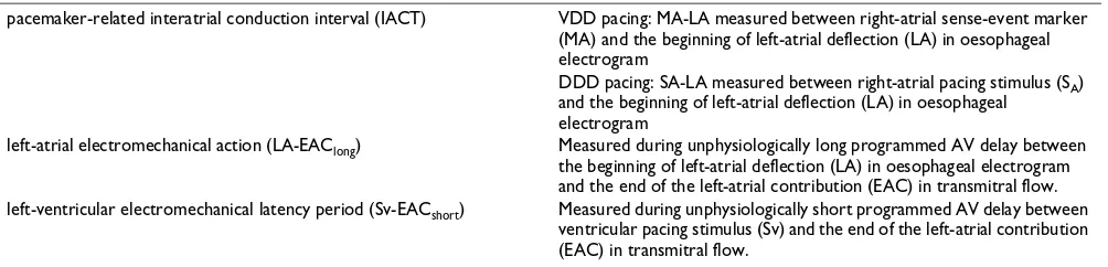
Table 2: Measurement of the components of the optimal AV delay according to Ismer et al. [22]
pacemaker-related interatrial conduction interval (IACT) VDD pacing: MA-LA measured between right-atrial sense-event marker (MA) and the beginning of left-atrial deflection (LA) in oesophageal electrogram
DDD pacing: SA-LA measured between right-atrial pacing stimulus (SA) and the beginning of left-atrial deflection (LA) in oesophageal
electrogram
left-atrial electromechanical action (LA-EAClong) Measured during unphysiologically long programmed AV delay between the beginning of left-atrial deflection (LA) in oesophageal electrogram and the end of the left-atrial contribution (EAC) in transmitral flow. left-ventricular electromechanical latency period (Sv-EACshort) Measured during unphysiologically short programmed AV delay between
Methods
Patients
11 chronic heart failure patients of our clinic were included in this study. All patients had a biventricular ICD (pre-implantation NYHA III-IV, EF < 35%, QRS width > 120 ms). Clinical characteristics are demonstrated in Table 1. Patient exclusion criteria were as follows: atrial fibrillation, pacemaker malfunction and oesophageal dis-eases, NYHA IV, prosthetic mitral valve replacement.
AV delay: components and optimization
For the AV delay optimization we used the method pro-posed by Ismer et al [22].
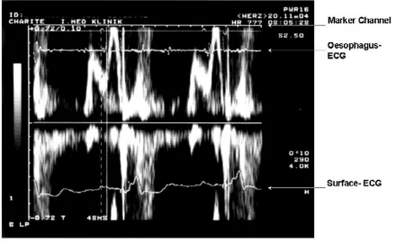
This approach needs the placement of a bi-polar oesopha-geal electrode to provide a filtered left-atrial electrogram (LAE). We applied a 5F oesophagus electrode (Osypka TO2/5F, order no. TA12991101, Rheinfelden, Germany). Filtered oesophageal electrogram and telemetric real-time pacemaker markers provided by the programmer's ana-logue output were superimposed on the display of trans-mitral flow velocity on the Doppler-echo system (Figure 1). The simultaneous recording of transmitral flow, the left atrial oesophageal electrogram and the real-time sense-event markers, allow determining the components of the optimal AV delay (Table 2, Figure 1 and 2).
Based on these measurements, optimal AV delays were calculated for VDD triggered) and DDD (atrial-paced) mode using the equations:
AVDOPT VDD = MA-LA + LA-EAClong - Sv-EACshort
and
AVDOPT DDD = SA-LA + LA-EAClong - Sv-EACshort
Echocardiograhy
Echocardiography to assess dyssynchrony was performed subsequently under three pacemaker settings: optimal AV delay (AVDOPT), optimal AV delay minus 50 ms (AVDOPT -50), optimal AV delay plus 50 ms (AVDOPT+50).
Echocardiography was performed on the Vivid 5 and Vivid 7 Dimension (GE Vingmed Ultrasound, Horton, Norway) machines. The TDI and strain analysis were per-formed in an off-line work station. The LVEF was assessed by area-length method in the apical four chamber view. The CI and the SV were calculated from the systolic veloc-ities measured by PW-Doppler in the aortic outflow tract. Strain rate, tissue Doppler velocities were measured in the basal segments of the apical four-, three- and two-cham-ber views.
Statistics
Values are expressed as mean ± standard deviation (SD). Groups were compared by parametric or non-parametric tests (t-tests and Wilcoxon-Mann-Whitney tests, respec-tively). Statistical significance was assumed at a value of P < 0.05. Statistical analysis was performed with the SPSS 12 software package (SPSS; Chicago, Ill, USA).
Results
Optimal AV delay
In all patients, we could define an optimal AV delay in the VDD and the DDD modes respectively. The AVDOPT in VDD mode was 105.5 ± 38.1 ms and the AVDOPT in the DDD pacing mode was 186.9 ± 52.9 ms. The results are summarized in Table 3. As expected, the mean optimal AV delay was lower in the VDD than in the DDD mode. Assessment of the left-atrial electromechanical action = LA-EAClong
Figure 2
Assessment of the left-atrial electromechanical action = LA-EAClong. LA = left atrial deflection (see oesophagus- ECG).
EAClong = the end of the A-wave in an unphysiologically long AV-intervall. In this particular patient the LA-EAClang is 160 ms
Measurement of the IACT in the VDD - Mode = MA-LA Figure 1
Echocardiography was performed subsequently under three pacemaker settings: AVDOPT, AVDOPT-50,
AVDOPT+50. All patients had continuous biventricular stimulation even under AVDOPT+50.
2D and TDI echocardiography
The LVEF with AVDOPT was 28% (± 12%), with an AVDOPT-50 20% (± 7%, p= 0.03 compared to AVDOPT), with an AVDOPT+50 23% (± 7%, p = 0.11 compared to AVDOPT). The heart rate did not change significantly in the
different settings (AVDOPT: 65,4/min, AVDOPT-50: 65,6/ min, AVDOPT+50: 65,8 ms). The hemodynamics (SVI, CI, LVEF) and the TDI derived data are listed in Table 4. There was no significant difference of the amount of segments with dyssynchrony in TSI in the three settings. The
maxi-mal delay in the basal segments in the apical two-, three-and four-chamber views measured by TSI three-and strain did not differ in the AVDOPT, AVDOPT+50 and AVDOPT-50 set-ting.
Discussion
Optimal AV delay
To date, Ismer's method for the optimal AV delay was applied to patients with DDD pacemakers and normal left ventricular function [22,23]. This is the first study to assess the optimal AV delay by Ismer's method in patients with reduced left ventricular function. In our CRT patients, an optimal AV delay according to Ismer's method could be defined. This is the only method that allows separate measurement of the three AV-delay com-ponents: i.e., the pacemaker-related interatrial conduction time, the atrial electromechanical action, and the left-ventricular latency period. The benefits of this method, however, are offset by the necessity for placement of an oesophageal electrode. This requirement explains why only a few medical centres have applied this method in clinical practice and in most cases for purposes of scien-tific investigation only.
Our results concerning the AVDOPT in the VDD mode (105.5 ± 38.1 ms) are in agreement with the results of other studies on AVDOPT in CRT patients: Butter [16] determined an AVDOPT of 100 ms in 30 patients, Auric-chio [17] an AVDOPT of 112 ± 33 ms in 41 patients and Kass [18] an AVDOPT of 125 ± 49 ms. A study that was recently published by Porciani [29] found an AVDOPT
dur-ing simultaneous biventricular pacdur-ing of 97 + 27 ms.
In the literature, there are no published data on AVDOPT in DDD mode. Therefore, our AVDOPT in DDD mode of 186.9 ± 52.9 ms cannot be compared to other studies.
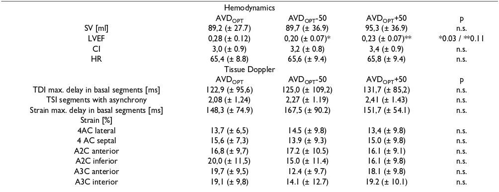
Table 4: Hemodynamic and Tissue Doppler Echocardiography parameters in the AVDOPT, AVDOPT-50 and AVDOPT+50 modes.
Hemodynamics
AVDOPT AVDOPT-50 AVDOPT+50 p
SV [ml] 89,2 (± 27.7) 89,7 (± 36.9) 95,3 (± 36.9) n.s.
LVEF 0,28 (± 0.12) 0,20 (± 0.07)* 0,23 (± 0.07)** *0.03 / **0.11
CI 3,0 (± 0.9) 3,2 (± 0.8) 3,4 (± 0.9) n.s.
HR 65,4 (± 8.8) 65,6 (± 9.4) 65,8 (± 9.4) n.s.
Tissue Doppler
AVDOPT AVDOPT-50 AVDOPT+50 p
TDI max. delay in basal segments [ms] 122,9 (± 95,6) 125,0 (± 109,2) 131,7 (± 85,2) n.s. TSI segments with asynchrony 2,08 (± 1,24) 2,27 (± 1.19) 2,41 (± 1.43) n.s. Strain max. delay in basal segments [ms] 148,3 (± 74.9) 167,5 (± 90.2) 151,7 (± 54.1) n.s.
Strain [%]
4AC lateral 13,7 (± 6,5) 14.5 (± 9.8) 13,4 (± 9.8) n.s.
4 AC septal 15,6 (± 7,3) 13.9 (± 9.3) 15.0 (± 9.8) n.s.
A2C anterior 16,8 (± 9,7) 17.2 (± 10.5) 16.1 (± 9.1) n.s.
A2C inferior 20,0 (± 11,5) 15.0 (± 11.4) 16.1 (± 9.8) n.s.
A3C anterior 19,7 (± 9,5) 12.4 (± 9.7) 18.1 (± 9.8) n.s.
A3C interior 19,1 (± 9,8) 14.1 (± 12.7) 19.2 (± 10.1) n.s.
Table 3: AVDOPT VDD = optimal AV delay for atrially triggered (VDD) and atrially paced (DDD) modes
patient AVDOPT VDD AVDOPT DDD
1 60 172
2 78 154
3 96 204
4 92 132
5 92 144
6 122 174
7 136 216
8 64 128
9 84 180
10 168 252
11 168 300
105,5 ± 38,1 ms 186,9 ± 52,9 ms
Hemodynamics
Intra-individually, the patients had the best LVEF under optimal AV-delay compared to the +50 and -50 ms set-tings. The LVEF is significantly higher in the AVDOPT set-ting than in the AVDOPT -50 setting. Obviously the formation of "cannon waves" seen with a shorter AV inter-val (AVDOPT -50) had a more negative hemodynamic
effect than the diastolic mitral regurgitation seen with longer AV delays (AVDOPT +50). The hemodynamically unfavourable effects of "cannon waves" are described since the beginning of pacemaker therapy and are also termed "pacemaker syndrome". It is generally accepted that an adequate pacemaker programming can avoid this [30]. Toda et al. [31] could show in his studies that the mean LVEF in AVDOPT is higher than in prolonged AV
delays. However, he found no significant difference.
Dyssynchrony
Changes of dyssynchrony can be seen immediately, as seen in studies that have examined on/off comparisons in CRT patients [32]. However, an optimized AV interval does not change the markers of dyssynchrony. The reason for the improved hemodynamic situation under AVDOPT
seems to be the better left ventricular filling and not the altered dyssynchrony.
Limitations
This study included only a small number of patients. There was no follow-up examination of the patients.
Conclusion
This study confirmed that an optimized AV delay improves the left ventricular ejection fraction. Acutely, the optimized AV delay does not influence left ventricular dyssynchrony. Whether a long-term AVDOPT leads to changes in left ventricular dyssynchrony via an improved LVEF and reverse remodelling can only be speculated. This has to be addressed in future studies with a long-term observation interval.
Abbreviations
AVDOPT optimal AV delay
AVDOPT-50 optimal AV delay -50 ms
AVDOPT+50 optimal AV delay + 50 ms
CRT Cardiac Resynchronization Therapy
DCM Dilated Cardiomyopathy
EMD Electromechanical Delay
IVMD Inter-ventricular mechanical delay
LBBB Left Bundle Branch Block
SRI strain rate imaging
TDI Tissue Doppler Imaging
TSI Tissue Synchronization Imaging
VDD atrially triggered mode
DDD atrailly paced mode
EAC the end of the A-wave
LVEF Left ventricular ejection fraction
Competing interests
The author(s) declare that they have no competing inter-ests.
Authors' contributions
CM and FK have equally contributed to this publication. CM, BI, FK and ACB have designed and performed the study and have written the manuscript. HJB, CAN and GB have participated in the study design and coordination and have helped to draft the manuscript. All authors read and approved the final manuscript.
References
1. Bristow MR, Saxon LA, Boehmer J, Krueger S, Kass DA, De Marco T, Carson P, DiCarlo L, DeMets D, White BG, DeVries DW, Feldman AM, Comparison of Medical Therapy Pacing and Defibrillation in Heart Failure (COMPANION) Investigators: Cardiac Resynchro-nization Therapy with or without an Implantable Defibrilla-tor in Advanced Chronic Heart Failure. N Eng J Med 2004, 350:2140-2150.
2. Linde C, Leclercq C, Rex S, Garrigue S, Lavergne T, Cazeau S, McK-enna W, Fitzgerald M, Deharo JC, Alonso C, Walker S, Braunschweig F, Bailleul C, Daubert JC: Long-term benefits of biventricular pacing in congestive heart failure: results from the MUltisite STimulation in cardiomyopathy (MUSTIC) study. J Am Coll Cardiol 2002, 40:111-118.
3. Abraham WT, Fisher WG, Smith AL, Delurgio DB, Leon AR, Loh E, Kocovic DZ, Packer M, Clavell AL, Hayes DL, Ellestad M, Trupp RJ, Underwood J, Pickering F, Truex C, cAtee P, Messenger J, MIRACLE Study Group: Multicenter InSync Randomized Clinical evalua-tion. Cardiac resynchronization in chronic heart failure. N Engl J Med 2002, 346:1845-1853.
4. Cazeau S, Leclercq C, Lavergne T, Walker S, Varma C, Linde C, Gar-rigue S, Kappenberger L, Haywood GA, Santini M, Bailleul C, Daubert JC, Multisite Stimulation in Cardiomyopathies (MUSTIC) Study Inves-tigators: Effects of multisite biventricualr pacing in patients with heart failure and intraventricular conduction delay. N Engl J Med 2001, 344:873-880.
5. Saxon LA, De Marco T, Schafer J, Chatterjee K, Kumar UN, Foster E: VIGOR Congestive Heart Failure Investigators. Effects of long-term biventricular stimulation for resynchronization on echocardiographic measures of remodeling. Circulatio
2002, 105:1304-1310.
Publish with BioMed Central and every scientist can read your work free of charge "BioMed Central will be the most significant development for disseminating the results of biomedical researc h in our lifetime."
Sir Paul Nurse, Cancer Research UK
Your research papers will be:
available free of charge to the entire biomedical community
peer reviewed and published immediately upon acceptance
cited in PubMed and archived on PubMed Central
yours — you keep the copyright
Submit your manuscript here:
http://www.biomedcentral.com/info/publishing_adv.asp
BioMedcentral 7. Cleland JG, Daubert JC, Erdmann E, Freemantle N, Gras D,
Kappen-berger L, Tavazzi L, Cardiac Resynchronization-Heart Failure (CARE-HF) Study Investigators: The effect of cardiac resynchronization on morbidity and mortality in heart failure. N Engl J Med 2005, 352(15):1539-1549.
8. Eugene M, Lascault G, Frank R, Fontaine G, Grosgogeat Y, Teillac A: Assessment of the optimal atrio-ventricular delay in DDD paced patients by impedance plethysmography. Eur Heart J
1989, 10:250-255.
9. Kindermann M, Fröhlig G, Doerr T, Schieffer H: Optimizing the AV delay in DDD pacemaker patients with high degree AV block: mitral valve doppler versus impedance cardiography.
PACE 1997, 20:2453-2462.
10. Ovsyshcher IE: Toward physiological pacing: optimization of cardiac hemodynamics by AV delay adjustment. PACE 1997, 20:861-865.
11. Ishikawa T, Sumita S, Kimura K, Kikuchi M, Kosuge M, Kuji N, Endo T, Sugano T, Sigemasa T, Kobayaski I, Tochikubo I, Usui T: Predic-tion of optimal atrioventricular delay in patients with implanted DDD pacemakers. PACE 1999, 22:1365-1371. 12. Von Knorre GH, Petzsch M, Ismer B: Approximation of optimal
atrioventricular delay in DDD pacemaker patients with atri-oventricular block by oesophageal electrocardiography (abstract). Eur Heart J 1996, 17(Supplement):487.
13. Ritter Ph, Dib JC, Lelievre T: Quick determination of the opti-mal AV delay at rest in patients paced in DDD mode for complete AV block. (abstract). Eur J CPE 1994, 4(2):A163. 14. Ritter P, Padeletti L, Gillio-Meina L, Gaggini G: Determination of
the optimal atrioventricular delay in DDD pacing. Compari-son between echo and peak endocardial acceleration meas-urements. Europace 1999, 1:126-30.
15. Occhetta E, Rognoni G, Perucca A, Aina F, Magnani A, Francalacci G, Rossi P: The functional and hemodynamic benefits of auto-matic atrioventricular interval delay in permanent atrial syn-chronized pacing. G Ital Cardiol 1993, 23:877-886.
16. Butter C, Auricchio A, Stellbrink C, Fleck E, Ding J, Yu Y, Huvelle E, Spinelli J, Pacing Therapy for Chronic Heart Failure II Study: Effect of resynchronization therapy stimulation site on the systolic function of heart failure patients. Circulation 2001, 104:3026-3029.
17. Auricchio A, Stellbrink C, Sack S, Block M, Vogt J, Bakker P, Huth C, Schondube F, Wolfhard U, Bocker D, Krahnefeld O, Kirkels H, Pacing Therapies in Congestive Heart Failure (PATH-CHF) Study Group: Long-term clinical effect of hemodynamically optimized car-diac resynchronization therapy in patients with heart failure and ventricular conduction delay. J Am Coll Cardiol 2002, 39:2026-2033.
18. Kass D, Chen CH, Curry C, Talbot M, Berger R, Fetics B, Nevo E: Improved left ventricular mechanics from acute VDD pacing in patients with dilated cardiomyopathy and ventricular con-duction delay. Circulation 1999, 99:1567-1573.
19. Auricchio A, Stellbrink C, Block M, Sacks S, Vogt J, Bakker P, Klein H, Kramer A, Ding J, Salo R, Tockmann B, Pochet T, Spinelli J: Effect of pacing chamber and atrioventricular delay on acute systolic function of paced patients with congestive heart failure. Cir-culation 1999, 99:2993-3001.
20. Stellbrink C, Breithardt OA, Diem B, Franke A, Pochet T, Salo R, Aur-icchio A: Acute effects of multisite pacing with different AV delays on diastolic and systolic function in congestive heart failure (abstract). PACE 1999, 22:829.
21. Meluzin J, Novak M, Mullerova J, Krejci J, Hude P, Eisenberger M, Dusek L, Dvorak I, Spinarova L: A fast and simple echocardio-graphic method of determination of the optimal atrioven-tricular delay in patients after bivenatrioven-tricular stimulation.
Pacing Clin Electrophysiol 2004, 27:8-64.
22. Ismer B, von Knorre GH, Voß W, Körber T: Definition of the opti-mal atrioventricular delay by simultaneous measurement of electrocardiographic and doppler-echocardiographic parameters. Prog Biomed Res 2003, 7:116-120.
23. Ismer B, von Knorre G, Voss W, Grille W, Klenke G, Kamesh Pulya, Koglek W, Suntinger A, Luessow H: Exercise induced sympa-thetic influences do not change interatrial conduction times in VDD and DDD pacing. PACE 1996, 19:1786-1790.
24. Borges AC, Kivelitz D, Walde T, Reibis RK, Grohmann A, Panda A, Wernecke KD, Rutsch W, Hamm B, Baumann G: Apical tissue tracking echocardiography for characterization of regional
left ventricular function: comparison with magnetic reso-nance imaging in patients after myocardial infarction. J Am Soc Echocardiogr 2003, 3:254-262.
25. Mele D, Pasanisi G, Heimdal A, Cittanti C, Guardigli G, Levine RA, Sutherland G, Ferrari R: Improved recognition of dysfunction-ing myocardial segments by longitudinal strain rate versus velocity in patients with myocardial infarction. J Am Soc Echocardiogr 2004, 4:313-321.
26. Gorcsan J, Kanzaki H, Bazaz R, Dohi K, Schwartzman D: Usefulness of Echocardiografic Tissue Synchronization Imaging to Pre-dict Acute Response to Cardiac Resynchronization Therapy.
Am J Cardiol 2004, 93:1178-1181.
27. Yu CM, Zhang Q, Fung JW, Chan HC, Chan YS, Yip GW, Kong SL, Lin H, Zhang Y, Sanderson JE: A novel tool to assess systolic asynchrony and identify responders of cardiac resynchroni-zation therapy by tissue synchroniresynchroni-zation imaging. J Am Coll Cardiol 2005, 45(5):677-684.
28. Knebel F, Reibis RK, Bondke HJ, Witte J, Walde T, Eddicks S, Bau-mann G, Borges AC: Tissue Doppler echocardiography and biv-entricular pacing in heart failure: patient selection, procedural guidance, follow-up, quantification of success.
Cardiovasc Ultrasound 2004, 2(1):17.
29. Porciani MC, Dondina C, Macioce R, Demarchi G, Pieragnoli P, Musilli N, Colella A, Ricciardi G, Michelucci A, Padeletti L: Echocardio-graphic examination of atrioventricular and interventricular delay optimization in cardiac resynchronization therapy. Am J Cardiol 2005, 95:1108-1110.
30. Schuller H, Brandt J: The pacemaker syndrome: old and new causes. Clin Cardiol 1991, 14:336-340.
31. Toda N, Ishikawa T, Nozawa N, Kobayashi I, Oghial H, Miyamoto K, Sumita S, Kimura K, Umemura S: Doppler index and plasma level of atrial natriuretic hormone are improved by optimizing atrioventricular delay in atrioventricular block patients with implanted DDD pacemakers. PACE 2001, 24:1660-1663. 32. Breithardt OA, Sinha AM, Schwammenthal E, Bidaoui N, Markus KU,