PRIMARY RESEARCH
Epithelial to mesenchymal transition
induces stem cell like phenotype in renal cell
carcinoma cells
Mamta Singla
1, Ajay Kumar
1, Amanjit Bal
2, Subhendu Sarkar
3and Shalmoli Bhattacharyya
1*Abstract
Background: Metastatic dissemination of solid tumors is often initiated by reactivation of an embryonic develop-ment program, epithelial-to-mesenchymal-transition (EMT). EMT has been associated with acquiring invasiveness and resistance to conventional therapies. However, the precise role of EMT during renal cell carcinoma is still debatable and is under investigation. In this context, our study is designed to evaluate the role of cyclosporine (CsA) and trans-forming growth factor-β (TGFβ) administration in inducing EMT-like state in renal carcinoma cells. We also studied the associated phenotypic changes which may lead to tumor metastasis.
Methods: The morphological changes in renal cell carcinoma cells (A498) treated with TGF-β/CsA were observed by microscopy. Atomic force microscope was used to evaluate the changes in elasticity of cells treated with TGF-β/CsA. The expression of mesenchymal and chemoresistance genes were checked by RT-PCR. Assays for migration, invasion, sphere formation ability and expression of cancer stem cell-like phenotypes were done to evaluate the metastatic potential of these cells. Lineage specific differentiations were also done to determine the acquisition of stem-cell like phenotype.
Results: Our results showed that treatment with TGF-β/CsA led to loss of epithelial characteristics and gain of mesenchymal phenotype in vitro. Changes in shape and elastic properties of the cancer cells favoured metastatic progression, increased tumorisphere formation and invasiveness post treatment. We also observed higher expression of chemoresistance and stemness markers in EMT-induced cells. These cells also differentiated to various lineages like osteoblasts, adipocytes, neural and hepatic cells when induced with the respective differentiation media.
Conclusion: We concluded that TGF-β/CsA treatment led to acquisition of EMT-like cancer stem cells phenotype that enhanced local invasion and dissemination of renal carcinoma cells. This subpopulation of cells with EMT-like phenotype a can provide a better perception of the metastatic process. This can provide an in vitro system for testing pharmaceuticals for modulating EMT as a viable strategy within the therapeutic armamentarium for RCC patients. The results of our findings also suggest that CsA directly induced EMT like changes in epithelial cell which may be respon-sible for the potential risk of malignancy in transplant patients.
Keywords: Renal cell carcinoma, Cyclosporine A, Transforming growth factor-beta, Epithelial to mesenchymal transition, Chemoresistant gene, Cancer stem cells
© The Author(s) 2018. This article is distributed under the terms of the Creative Commons Attribution 4.0 International License (http://creativecommons.org/licenses/by/4.0/), which permits unrestricted use, distribution, and reproduction in any medium, provided you give appropriate credit to the original author(s) and the source, provide a link to the Creative Commons license, and indicate if changes were made. The Creative Commons Public Domain Dedication waiver (http://creativecommons.org/ publicdomain/zero/1.0/) applies to the data made available in this article, unless otherwise stated.
Background
Primary neoplasia is treatable, but metastasis pose a major threat to cancer patients. It is very important to
understand the mechanism of metastases, especially to cure advanced cancers. Growing evidences suggest that the process that aids in the progression toward invasive and metastatic cancer is associated with the reactiva-tion of an embryonic development program, epithelial-to-mesenchymal-transition (EMT) [1]. It is a complex process that regulates changes in cell morphology and
Open Access
*Correspondence: shalmoli2007@yahoo.co.in
1 Department of Biophysics, Postgraduate Institute of Medical Education
& Research, Chandigarh, India
mechanical properties of the cells. Modulation of elas-tic properties of cells during transition allows the cells to detach from their neighbours and the underlying basement membrane to facilitate their easy migration and invasion during metastasis [2]. During the process of tumor progression, a small fraction of cancer cells acquires self-renewal potential like stem cells and are referred as cancer stem cell (CSC). A reciprocal relation-ship between CSC and EMT might have a role in tumor progression. EMT is critical for acquisition of invasive behaviour and CSC properties, therefore effective thera-pies can be developed against metastatic cells via inter-ference with the CSC/EMT differentiation program.
The association of carcinogenesis with organ transplan-tation is found to be complicated. CsA, an immunosup-pressive agent is used during the organ transplant has been linked for cancer progression by cell autonomous mechanism [3]. There is increasing evidence that immu-nosuppressive agents like CsA may have unique onco-genic profiles [4]. A recent case study has shown that cyclosporine treatment to a psoriasis patient, led to mul-tiple metastases [5]. CsA has also been shown to induce phenotypic changes in keratinocytes that can increase cell invasiveness and unregulated tumor growth [6]. CsA has also been associated with the promotion and stimula-tion of cellular proliferastimula-tion in normal human fibroblasts [7]. However, the precise mechanism underlying the tum-origenic effect of CsA remains largely obscure though CsA is known to increase the level of TGF-β which is strongly involved in the regulation of EMT in cancer [8]. Higher production of TGF-β is positively associated with tumor aggressiveness and poor prognosis [9, 10]. How-ever, there is no direct evidence that CsA may induce malignancy or cause increase in invasiveness or CSC-like phenotype. We designed our study on A-498 cells which is considered a “classical” clear cell renal carcinoma line with VHL mutation belonging to the NCI-60 panel. Clear cell carcinoma is the most prevalent subtype of RCC and is therefore widely used in cancer research. In this study, we observed that CsA could cause EMT in A498 cells leading to morphological change, increased invasive-ness and stem cell-like phenotype. Thus, it is important to consider the possibility of malignant transformation during treatment planning in patients. The results of our study indicate that EMT plays a role in metastatic pro-gression, drug resistance during RCC and targeting EMT may represent an effective strategy to combat the disease.
Materials and methods Cell culture
In-vitro studies were done with A498 cell line obtained from NCCS, Pune, India and grown in the DMEM-high glucose (Dulbecco’s modified eagle’s medium) containing
10% FBS (Fetal Bovine Serum) and antibiotics (pen-strep, 1U/ml) at 37 °C at 5% CO2 in humidified incubator.
Cyclosporine-A was obtained from Sigma Aldrich (USA).
In vitro induction of EMT
5 × 10⁴ cells were seeded per well of 12-well plates and
allowed to grow overnight. Next day, these cells were incubated with TGF-β (5 ng/ml) or CsA (10 μM) sepa-rately for another 48 h in the DMEM containing 10% FBS.
Light microscopy
The morphological changes due to EMT induction were confirmed by observing under bright field microscope (Nikon).
Atomic force microscopy
A multimode 8 atomic force microscopy (AFM) system (Bruker AXS) was used to image the control and treated cells and perform mechanical measurements on them. A large scanner having a maximum x–y scan range of 125 × 125 µm and a z-limit of 5 µm was employed to scan the surface. Measurements were done in tapping mode using a cantilever tip (SCANASYST-AIR) having a force constant of 0.4 N/m, prior to measurements, the canti-lever was calibrated using the thermal vibration method. DMT modulus was calculated by using the load force and adhesion data. The reduced Young’s modulus (E) in this model was obtained by fitting the retract curve accord-ing to the equation Ftip= 43E
√
Rd3
+Fadh. Here, Ftip is
the force on the tip, Fadh is the adhesive force, R is the tip
radius and d are the tip-sample separation.
Staining with acridine orange (AO)
A498 cells (4 × 10³/well) were treated with CsA for 48 h.
After treatment, live cells were washed with PBS and stained with AO (1 µg/ml) for 15 min. Cells were again washed in PBS and visualized using Fluorescent micro-scope (Olympus 1X51) under excitation and emission wavelengths of 488/532 nm.
Immunocytochemistry
For immunofluorescence study, the cells were incu-bated with the human EMT 3-color immunocytochem-istry kit (SC026) as per manufacturer’s instructions and observed under confocal microscope (Olympus). Images were acquired using FV1000 software at-10× and 20× magnification.
Real time polymerase chain reaction (PCR)
Total RNA was isolated from the cells using Trizol rea-gent (Invitrogen) and reverse-transcribed using cDNA synthesis kit (Bio-Rad) per manufacturer’s protocol. Real-time PCR analysis was performed on a Roche real-Real-time PCR system using the Power SYBR Green PCR Master Mix (Roche, Foster City, CA). The expression value of each gene was normalized against the amount of β-actin and calculated by the ΔΔCt method. Details of the primer
sequence are given in Table 1.
Migration and transwell invasion assay
The cells were cultured up to 80–90% confluency and scratched with a pipette tip to create a uniform wound. Migratory ability of the cells was evaluated from the time taken in filling the open space created by scratch under inverted microscope. The wound area closure was calcu-lated using the T-Scratch software developed by the Kou-moutsakos group, Zurich [11].
For invasion assay, Matrigel™ (BD Biosciences, 1 mg/ ml) was added to 24 well cell culture inserts (8 µm). Cells were seeded on the upper chamber of the inserts in serum free media. Media containing 10% FBS was added in the lower chamber and incubated for 48 h at 37 °C in a humidified 5% CO2 incubator. The migratory cells
pre-sent on the lower surface were stained with 0.1% crystal violet and photographed.
Colony formation assay
Single cell suspensions (1000 cells/well) were seeded on 6 well tissue culture plate in DMEM containing 10% FBS without any additional coating [12]. After 14 days of cul-ture, the colonies were washed with phosphate buffer saline (PBS), fixed in methanol, and stained with 0.1% crystal violet. The stained colonies were observed under the microscope and photographed.
Tumorisphere assay
Cells treated with and without cyclosporine (CsA) were cultured using DMEM-Ham’s F12 (1:1; Sigma) contain-ing bFGF (basic fibroblast growth factor; 10 ng/ml), B27 (0.5%), EGF and 0.4% bovine serum albumin (BSA). These cells could grow for 7 days in low adherence plates (six well, Corning). Photography was done after 7 days to observe the tumorisphere formation ability of cells in presence or absence of CsA.
Differentiation assays
The control and CsA treated cells were induced for dif-ferentiation to osteogenic, adipogenic and neural lineage using suitable differentiation media.
For osteogenic differentiation, cells were incubated in alpha-minimal essential medium (α-MEM) complete medium (CM), supplemented with 0.01 mM dexameth-asone disodium phosphate, 1.8 mM monopotassium phosphate (KH2PO4) and 5 mM β-glycerophosphate.
Media was changed every 3rd day. At the end of 21st day, the cells were stained with 1% Alizarin Red S (AR-S) for osteogenic differentiation.
For adipogenic differentiation, cells were exposed to adipogenic media made of αMEM containing isobu-tyl-methylxanthine (IBMX, 0.5 mM), dexamethasone (1 μM), insulin (10 μM), and indomethacin (200 μM). At the end of 21st day, the cells were subjected to oil red O staining for the presence of intracellular lipid droplets.
For neural differentiation, cells were incubated in neu-robasal medium (Life technologies) pen/strep (1%), sup-plemented with B27, 20 ng/ml bFGF and EGF, N2 and G5 supplement. After 21st day, cells were fixed and incu-bated with anti-human neurofilament antibody (NFM, 1:50) (Sigma Aldrich), followed by FITC labelled second-ary antibody (Sigma) and observed under fluorescence microscope (Nikon).
For hepatic differentiation, cells were incubated in αMEM containing ITS + (Invitrogen, 50 mg/ml) pre-mix, EGF (epidermal growth factor; Invitrogen, 2 ng/ ml), dexamethasone (0.5 μM) and HGF (hepatocyte growth factor, Sigma, 20 ng/ml) during induction phase (14 days). The maturation medium contained supple-ments like Oncostatin M (Sigma, 20 ng/ml), ITS and dexamethasone. Analysis of hepatocytes was done by low
Table 1 The list of primers used for Real time PCR studies
S. no Gene name Sequence
1 β-Actin F 5′CAAGAGATGGCCACGGCTGCT3′
R 5′TCCTTCTGCATCCTGTCGGCA3′
2 Vimentin F 5′TCTACGAGGAGGAGATGCGG3′
R 5′GGTCAAGACGTGCCAGAGAC3′
3 Snail F 5′GAAAGGCCTTCAACTGCAAA3′
R 5′TGACATCTGAGTGGGTCTGG3′
4 Slug F5′ATTCGGACCCACACATTACCTTG3′
R5′TGGAGAAGGTTTTGGAGCAGTTT3′
5 Twist F5′TGAGCAAGATTCAGACCCTCA3′
R 5′ATCCTCCAGACCGAGAAGG3′
6 ABCG2 F5′-CTGAGATCCTGAGCCTTTGG-3′
R5′-TGCCCATCACAACATCATCT-3′
7 MDR2 F5′-GCCTGGCAGCTGGAAGACAAATAC-3′
density lipoprotein assay kit (LDL assay kit-Abcam) as per instructions provided with kit. Fluorescent markers used in the kit include DyLight™ 550 and LDL receptor antibody-for LDL uptake and receptor distribution in the cells respectively.
Statistical analysis
All experiments were performed at least in triplicates and were repeated three times. Data was reported as mean ± SD. All statistical analyses were performed using one-way ANOVA and paired t test.
Results
Induction of EMT in vitro
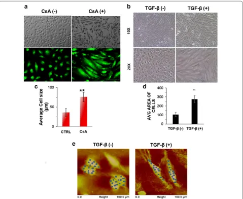
A498 cells were treated with CsA (10 μM)/TGF-β (5 ng/ ml) in separate set of experiments and morphological changes were observed at 48 h. Light microscopy images showed disruption of cell to cell junctions and distinct change to spindle shaped morphology in the cells post CsA/TGF-β treatment (Fig. 1a, b). The Additional file 1: Fig. S1 show that cells progressively acquire the spindle shaped morphology on CsA treatment. Acridine orange staining post 48 h of CsA treatment in A498 cells showed fibroblast like cell shape when observed under fluores-cence microscope (Fig. 1a). A significant increase in the average cell size was observed after exposing the cells to CsA as calculated by NIS elements software of the Nikon microscope (Fig. 1c). A significant increase in the aver-age cell size was again observed after exposing the cells to TGF-β as calculated by ImageJ software. (Figure 1d).
To further confirm the biophysical changes in cellular characteristics after EMT induction, we employed atomic force microscopy (AFM) in both control and treated cells. The topography and modulus images obtained are shown in Fig. 1e for the control as well as the treated samples. Topography images clearly indicate that the treated cells become elongated as compared to the control ones. The increase in size of the treated cells in the longitudinal direction was about four times as compared to the con-trol cells. DMT modulus values were extracted from the shaded regions in the modulus images. Corresponding regions are shown as shaded regions in the topography images. Average moduli of the regions were calculated for both the samples. It was found that the modulus of treated cells is less than 25% of that of the control cells.
Increase in expression of mesenchymal genes in EMT induced cells
CsA treated A498 cells showed significantly higher expression of EMT regulators like snail, slug, twist as well as vimentin (2.5- to fourfold) (Fig. 2a). We observed that the ABC transporter, ABCG2 and multidrug resist-ance gene, MDR1 were also upregulated to a significant
level (Fig. 2a). Immunocytochemistry results showed that expression of epithelial markers like E-cadherin and pan-cytokeratin decreased in cells treated with CsA with con-comitant rise in the expression of vimentin and α-SMA (Fig. 2b). The EMT signature was also confirmed through immunofluorescence analysis where a higher expression of vimentin and snail were observed in both TGF-β and CsA treated cells (Fig. 3a, b).
Metastatic potential of EMT induced cell A498 cells
Metastasis is characterized by increased invasiveness and migratory ability, so we further analysed whether the CsA and TGF-β treated cells acquired invasive behaviour associated with metastatic cells. Maximum cell migra-tion was observed after 24 h treatment at 10 µM con-centration of CsA. Quantification of the open wound area showed that cells treated with CsA migrated faster from both edges of the scratch and filled the gap area in lesser time compared to the control cells (Fig. 4a, b). The Matrigel invasion assay showed that CsA and TGF-β treatment significantly increased the ability of cells to degrade the Matrigel and invade to the opposite side. Crystal violet staining on the lower surface of the poly-ethylene terephthalate (PET) membrane showed higher number of invaded cells following CsA and TGF-β treat-ment (Fig. 5a). Both CsA and TGF-β treated cells showed higher proliferative capacity as confirmed by the colony formation assay (Fig. 5b).
Stem cell like properties in EMT induced cells
oil red o stain and no significant change in deposition of oil droplets was observed between EMT induced cells and control cells (Fig. 7a). We also observed an increase in density and average size of neurospheres on day 7 in the plate containing EMT induced cells (Fig. 7b, c).
Discussion
Epithelial–mesenchymal transition (EMT) is an impor-tant process during initiation and progression of the metastatic cascade. EMT pathway can have therapeutic
implications in cancer treatment. The tendency to metastasize and resistance to chemotherapy is the main cause of low survival in most of the malignancies includ-ing RCC. It was recently hypothesized that drug resist-ance, disease progression, and recurrence are mediated by stem cell-like cancer cells also referred to as cancer stem cells/tumor-initiating cells (CSCs/TICs) [13, 14]. The phenotypic plasticity displayed by CSCs may arise in the process of and/or undergo EMT. Isolating the CSC population in each tumor type is a major challenge due
Fig. 1 Morphological change in A498 cells. a Light microscopy images of A498 cells treated with CsA (10 µM) for 48 h. Magnification ×10. Scale bar—20 µm. Upper panel—bright field images of A498 cell morphology with or without CsA treatment. Lower panel—CsA treated A498 cells, stained with acridine orange and visualized under fluorescent microscope in a blue filter. Magnification—×10, ×20. b Change in cell shape with TGF-β treatment: A498 cells were treated with or without TGF-β (5 ng/ml) for 48 h. Bright field images showing change in morphology after treat-ment and cells acquire spindle shaped morphology. Magnification: ×10 and ×20. c Graphical representation of average cell size in control and CsA treated A498 cells. Statistical comparison represents average cell size in minimum 10 fields. Data is represented as mean ± SD. P value *< 0.05, **< 0.
to its small subpopulation. CD 105 was identified as a biomarker for renal CSCs [15] and another study sug-gested that it can also serve as a functional target for therapeutic intervention [16]. Subsequent gene profil-ing and pathway analysis of CD105 high cells showed high activation of TGF-beta in these cells [17]. TGF beta is an inducer of EMT and promotes tumorigenesis and metastasis by EMT [18–20]. Previously, it was demon-strated that exposure to a clinically relevant dose of the immunosuppressant agent CsA, led to an EMT in human proximal tubular cells through TGF-β [21]. In this con-text, we pharmacologically induced EMT in RCC cell line A498 with CsA and TGF-β. We observed that the bio-logical behaviour of TGF-β and CsA treated cells were consistent with that of mesenchymal cells, including a change in morphological features, lower adhesion and higher in vitro migratory capacity. The treatment caused higher expression of EMT regulators like snail, slug, twist as well as vimentin along with deceased expression of E cadherin. These modulations in cellular phenotype cause increased motility, invasiveness and resistance to apopto-sis of cancer cells [22]. There is loss of E cadherin pro-tein during EMT to occur and promote metastasis [23]. The loss of E-cadherin is an integral step during the EMT process and a key feature of metastatic cells. Without the tight adherent junctions which bind the tissues together, individual cells are free to migrate, a crucial requirement for cancer metastasis. The shift from cytokeratin inter-mediate filaments to vimentin and expression of other mesenchymal markers like snail, slug and twist suggested the occurrence of an EMT-like event in cells treated with TGF-β/CsA. The morphology of the cancer progenitor
cells changes during the process of EMT. Rearrangement of the cytoskeleton is an essential feature during EMT. Expression of vimentin, which is an important constitu-ent of the cytoskeleton, goes up in many types of cancer during EMT and in the stromal cells of several cancers [24].
The adaptation for invasiveness and metastasis in can-cer cells involve mechanical softening and modification in interactions with ECM. These modifications enable the cancer cells to migrate from their primary site to invade another secondary site. Young’s modulus is an excellent representative of cell’s elasticity/stiffness and evalua-tion of this modulus by atomic force microscopy offers an excellent approach to correlate the malignancy and deformity of cancer cells [25, 26]. A decreased Young’s modulus (< 25%) in case of EMT induced cells as com-pared to control cells clearly reflects a softening in EMT induced cells, an adaptation for invasion and metastasis.
It has been known that there is an intricate relationship between cell shape and function. So, a change in the cell after induction of EMT can be extrapolated to changes in cellular physiology due to interplay of genes and pro-teins. Cells that are characterized as invasive and meta-static are typically able to invade matrigel matrix which mimics the cell basement membrane [27]. The increased migration and stronger ability to invade through matrigel further indicated that these cells of epithelial origin have acquired metastatic phenotype. The cells treated with TGF-β and CsA also showed higher colony formation signifying an increased proliferative potential compared to bulk A498 cells. We have identified several changes in both protein and mRNA expression indicative of EMT in
TGF-β and CsA treated cells. The tumorispheres forma-tion by A498 cells post CsA treatment, further confirmed that these cells have attained an EMT-like phenotype. The formation of tumorispheres in non-adherent cul-ture is a useful functional approach to enrich the poten-tial subpopulation of CSC. The sphere forming cells are reported to show higher proliferation and self-renewal ability [28]. Self-renewal of undifferentiated stem cells is mainly regulated by octamer binding transcription fac-tor 4 (Oct-4) protein [29]. Thus, our observation of an increase in the oct4 expression in CsA treated cell further highlights the fact that these cells acquire a stem cell-like phenotype. A study by Takahashi and Yamanaka reported
that kruppel-like factor 4 (KLF4) is indispensable for maintenance of stem cells [30]. Fang et al. also elucidated that consistent overexpression of KLF4 led to increase in population of the cancer stem cells [31]. mRNA expres-sion of KLF4 was found to be upregulated in TGF-β/CsA treated cells. Sphere formation ability and increase in the expression of Oct-4 in CsA treated cells may account for an increase in stemness and differentiation potential of these cells.
The ability for osteogenic, hepatogenic and neural dif-ferentiation depicts the multilineage potential of EMT cells. There was significant rise in expression of each of osteogenic, hepatogenic and neural phenotype in CsA
Fig. 4 EMT induced cells are more migratory. a The migration ability of CsA treated A498 cells and control untreated cells were measured by wound healing assay after 6 and 24 h of wound induction in a 12 well plate. Photos were taken at 0, 6 and 24 h. Magnification—×4. b The healing rate was quantified by measurement of the gap size with the T-scratch assay software (open software at http://www.cse-lab.ethz.ch/)
Fig. 5 EMT induced cells are more invasive and have high colony forming ability. a Transwell invasion assay. 1 × 10⁵ cells were seeded on Matrigel
treated cells compared to control during lineage specific induction for differentiation. The ability for multilineage differentiation by EMT induced cells implies the acqui-sition of stem cell-like characteristics by these cells. The expression of oil red O in the control cells may be due to the fact that the clear cell RCC is reported to contain lipid droplets and histologically resemble adipocytes [32].
EMT phenomenon is reported to be associated with drug resistance in many tumors. In our study, we observed higher expression of ATP-binding cassette sub-family G member 2 (ABCG2) and multidrug resist-ance protein 1 (MDR1) genes in CsA treated cells. In accordance to our study, Saxena et al. demonstrated that EMT transcription factors cause multidrug resistance by upregulation of ABC transporters. Furthermore, these authors reported that ABC transporters harbour many binding sites for EMT inducing transcription factors, like Snail, and FOXC2, Twist etc. which ultimately induce EMT by enhancing the activity at promoter site of ABC transporters [33].
The machinery of EMT seems to be connected directly to formation and maintenance of CSCs leading to tumor metastases, drug resistance and recurrence [34]. Our results suggest that a mesenchymal-like phenotype
following CsA/TGF-β treatment is due to EMT. Walsh et al. have demonstrated that administration of CsA to animal model of cutaneous squamous cell carcinoma alters the phenotype to an invasive and aggressive tumor-type by enhancing EMT through the TGFβ1 signaling pathway [35]. These cells behave as de-differentiated stem cells isolated from normal or neoplastic cells. Previ-ously, Mani et al. induced EMT in human, non-tumori-genic mammary epithelial cells using TWIST or SNAIL transcription factors. They observed that EMT in breast cancer cells generated a subpopulation of cells with prop-erties of stem cells which may be responsible for drug resistance and recurrence [36]. This small subpopulation of cells within the tumor bulk was categorised as the can-cer stem cells. These may serve as an in vitro platform for improving the present clinical therapy of cancer since cells become invasive and resistant to apoptosis after acquiring mesenchymal phenotype. We show that the pharmacologically induced EMT cells of A498 behave as cancer stem-like cells (Fig. 8). In some cases, CSCs have been identified as independent predictor of progression-free survival and overall survival [37, 38]. These cells may therefore serve as an in vitro platform for improving the present clinical therapy of cancer since cells become
Fig. 6 EMT induced cells acquire stem cell-like phenotype: a immunofluorescence show expression of the marker for pluripotency i.e. Oct-4 in CsA (10 µM) treated A498 cells. Magnification ×10. Scale bar—20 µm. b Agarose gel electrophoresis (2%) showing expression of the stemness gene KLF4 in A498 cells with or without the treatment of CsA (8, 10 μM) and β-actin was used as housekeeping gene. c Tumorisphere formation assay: Bright field image showing tumorisphere formation by CsA treated cells grown on non-adherent surface in serum free media. Magnification ×10. d
invasive and resistant to apoptosis after acquiring mes-enchymal phenotype. As observed in our study, many clinical studies also detected the presence of EMT molec-ular markers in the CSCs. Breast cancer patients derived CSCs showed decreased expression of epithelial markers and increased mesenchymal markers [39–42]. In another study, Twist1 expression in breast cancer patients corre-lated with early distant relapse [43]. CSCs isolated from hepatocellular carcinoma (HCC) patients with metastasis showed higher expression of Snail1 transcripts compared to patients with no metastasis [44]. One caveat of the studies involving CSCs is that, the CSCs constitute a very minor fraction of the total cancer cells and hence may be missed out during isolation in vivo [45–47]. Thus, there is a need to explore the various methods to generate a CSC-like population in vitro and study the potential util-ity of CSCs in targeting the metastatic spread and cancer recurrence.
Our results suggest that CsA and TGF-β induced EMT like changes may help to study the mechanisms regulat-ing drug resistance and invasiveness. Data on patients under CsA treatment have suggested that long-term CsA may lead to an increased susceptibility to certain tumours and tumor metastasis [48]. Present study endorses the view of risk of malignancy in transplant patients and pro-vides evidence that cyclosporine can enhance cancer in renal transplant patients and can make it more aggres-sive. Rate of complete response in patients with meta-static RCC treated with targeted agents is very low and, better understanding of the pathogenic mechanisms of RCC brings hope for new strategies. Thus, targeting the pathways that promote EMT may lead to novel therapeu-tic strategies for the prevention and treatment of cancer, especially following organ transplantation.
Conclusion
Our study delineates the precise role of EMT in RCC carcinogenesis and metastasis. The expression profile and other biological properties suggest that EMT can be pharmacologically induced in vitro by CsA or TGF-beta and these EMT-induced cells acquire CSC-like pheno-type. The results of our study support the rationale for future EMT-directed therapeutic approaches for RCC patients.
Abbreviations
EMT: epithelial to mesenchymal transition; CsA: cyclosporine A; TGF-β: trans-forming growth factor beta; CSC: cancer stem cell; hEGF: human epidermal growth factor; bFGF: basic fibroblast growth factor; NFM: neurofilament; Oct-4: Octamer binding transcription factor-4; ABCG2: ATP binding cassette sub-family G member 2; MDR1: multidrug resistance protein 1.
Authors’ contributions
MS and AK performed the experimental study and data collection. MS, AB and SB analysed and interpreted the data. MS and SS performed and interpreted the AFM studies. MS and SB wrote and reviewed the manuscript. AK, AB and SS revised the manuscript and gave the material support. SB conceived and supervised the whole project. All authors read and approved the final manuscript.
Additional file
Additional file 1: Fig. S1. Time dependent change in cell morphology with CsA: the A498 cells were treated with CsA for 6, 12 and 24 h and the morphological change was compared with corresponding untreated con-trol to assess EMT. The result was confirmed by the expression of various epithelial and mesenchymal markers at the selected time and dose.
Author details
1 Department of Biophysics, Postgraduate Institute of Medical Education &
Research, Chandigarh, India. 2 Department of Histopathology, Postgraduate
Institute of Medical Education & Research, Chandigarh, India. 3 Department
of Physics, Indian Institute of Technology, Roopnagar, Roopnagar, India.
Acknowledgements
The authors acknowledge technical help of Mr. Harsimran during atomic force microscopy studies.
Competing interests
The authors declare that they have no competing interests.
Availability of data and materials Original data available on request.
Consent for publication
All authors have consented to publication.
Ethics approval and consent to participate Not applicable.
Funding
Dr. Mamta Singla received Senior Research Fellowship from Indian Council of Medical Research, Govt. of India.
Publisher’s Note
Springer Nature remains neutral with regard to jurisdictional claims in pub-lished maps and institutional affiliations.
Received: 16 January 2018 Accepted: 5 April 2018
References
1. Nieto MA, Huang RY, Jackson RA, Thiery JP. Emt: 2016. Cell. 2016;166:21–45.
2. Li QS, Lee GY, Ong CN, Lim CT. AFM indentation study of breast cancer cells. Biochem Biophys Res Commun. 2008;374:609–13.
3. Hojo M, Morimoto T, Maluccio M, Asano T, Morimoto K, Lagman M, Shimbo T, Suthanthiran M. Cyclosporine induces cancer progression by a cell-autonomous mechanism. Nature. 1999;397:530–4.
4. Durnian JM, Stewart RM, Tatham R, Batterbury M, Kaye SB. Cyclosporin-A associated malignancy. Clin Ophthalmol. 2007;1:421–30.
5. Shima T, Yamamoto Y, Okuhira H, Mikita N, Furukawa F. A patient with refractory psoriasis who developed sebaceous carcinoma on the neck during cyclosporine therapy and showed rapid progression. Case Rep Dermatol. 2016;8:136–41.
6. Wu X, Nguyen BC, Dziunycz P, Chang S, Brooks Y, Lefort K, Hofbauer GF, Dotto GP. Opposing roles for calcineurin and ATF3 in squamous skin cancer. Nature. 2010;465:368–72.
7. Cotrim P, Martelli-Junior H, Graner E, Sauk JJ, Coletta RD. Cyclosporin A induces proliferation in human gingival fibroblasts via induction of transforming growth factor-beta1. J Periodontol. 2003;74:1625–33. 8. Moustakas A, Heldin CH. Induction of epithelial–mesenchymal transition
by transforming growth factor beta. Semin Cancer Biol. 2012;22:446–54. 9. Malfettone A, Soukupova J, Bertran E, Crosas-Molist E, Lastra R, Fernando
J, Koudelkova P, Rani B, Fabra A, Serrano T, Ramos E, Mikulits W, Giannelli G, Fabregat I. Transforming growth factor-beta-induced plasticity causes a migratory stemness phenotype in hepatocellular carcinoma. Cancer Lett. 2017;392:39–50.
10. Massague J. TGFbeta signalling in context. Nat Rev Mol Cell Biol. 2012;13:616–30.
11. Geback T, Schulz MM, Koumoutsakos P, Detmar M. TScratch: a novel and simple software tool for automated analysis of monolayer wound healing assays. Biotechniques. 2009;46:265–74.
12. Zhang Y, Yan W, Jung YS, Chen X. Mammary epithelial cell polarity is regulated differentially by p73 isoforms via epithelial-to-mesenchymal transition. J Biol Chem. 2012;287:17746–53.
13. Adorno-Cruz V, Kibria G, Liu X, Doherty M, Junk DJ, Guan D, Hubert C, Venere M, Mulkearns-Hubert E, Sinyuk M, Alvarado A, Caplan AI, Rich J, Gerson SL, Lathia J, Liu H. Cancer stem cells: targeting the roots of cancer, seeds of metastasis, and sources of therapy resistance. Cancer Res. 2015;75:924–9.
14. Czarnecka M, Cezary S. Renal cell carcinoma cancer stem cells as thera-peutic targets. Curr Signal Transduct Ther. 2013;8:7.
15. Bussolati B, Bruno S, Grange C, Ferrando U, Camussi G. Identification of a tumor-initiating stem cell population in human renal carcinomas. FASEB J. 2008;22:3696–705.
16. Hu J, Guan W, Liu P, Dai J, Tang K, Xiao H, Qian Y, Sharrow AC, Ye Z, Wu L, Xu H. Endoglin is essential for the maintenance of self-renewal and chemoresistance in renal cancer stem cells. Stem Cell Rep. 2017;9:464–77. 17. Tang FR, Wang JW, Tang Z, Kang M, Deng QL, Yu JM. Quality of life and
its association with physical activity among different types of cancer survivors. PLoS ONE. 2016;11:e0164971.
18. Thiery JP, Acloque H, Huang RY, Nieto MA. Epithelial–mesenchymal transi-tions in development and disease. Cell. 2009;139:871–90.
19. Heldin CH, Vanlandewijck M, Moustakas A. Regulation of EMT by TGFbeta in cancer. FEBS Lett. 2012;586:1959–70.
20. Thiery JP. Epithelial–mesenchymal transitions in tumour progression. Nat Rev Cancer. 2002;2:442–54.
21. Slattery C, Campbell E, McMorrow T, Ryan MP. Cyclosporine A-induced renal fibrosis—a role for epithelial–mesenchymal transition. Am J Pathol. 2005;167:395–407.
22. Li L, Li W. Epithelial-mesenchymal transition in human cancer: compre-hensive reprogramming of metabolism, epigenetics, and differentiation. Pharmacol Ther. 2015;150:33–46.
23. Onder TT, Gupta PB, Mani SA, Yang J, Lander ES, Weinberg RA. Loss of E-cadherin promotes metastasis via multiple downstream transcriptional pathways. Cancer Res. 2008;68:3645–54.
24. Abraham E, Marincola FM, Chen Z, Wang X. Clinical and translational medicine: integrative and practical science. Clin Transl Med. 2012;1:1. 25. Guck J, Schinkinger S, Lincoln B, Wottawah F, Ebert S, Romeyke M, Lenz
D, Erickson HM, Ananthakrishnan R, Mitchell D, Kas J, Ulvick S, Bilby C. Optical deformability as an inherent cell marker for testing malignant transformation and metastatic competence. Biophys J. 2005;88:3689–98.
26. Xu W, Mezencev R, Kim B, Wang L, McDonald J, Sulchek T. Cell stiffness is a biomarker of the metastatic potential of ovarian cancer cells. PLoS ONE. 2012;7:e46609.
27. Albini A, Iwamoto Y, Kleinman HK, Martin GR, Aaronson SA, Kozlowski JM, McEwan RN. A rapid in vitro assay for quantitating the invasive potential of tumor cells. Can Res. 1987;47:3239–45.
28. Cao L, Zhou Y, Zhai B, Liao J, Xu W, Zhang R, Li J, Zhang Y, Chen L, Qian H, Wu M, Yin Z. Sphere-forming cell subpopulations with cancer stem cell properties in human hepatoma cell lines. BMC Gastroenterol. 2011;11:71. 29. Zhang ZN, Chung SK, Xu Z, Xu Y. Oct4 maintains the pluripotency of
human embryonic stem cells by inactivating p53 through Sirt1-mediated deacetylation. Stem Cells. 2014;32:157–65.
30. Takahashi K, Yamanaka S. Induction of pluripotent stem cells from mouse embryonic and adult fibroblast cultures by defined factors. Cell. 2006;126:663–76.
31. Yu F, Li J, Chen H, Fu J, Ray S, Huang S, Zheng H, Ai W. Kruppel-like factor 4 (KLF4) is required for maintenance of breast cancer stem cells and for cell migration and invasion. Oncogene. 2011;30:2161–72.
32. Rezende RB, Drachenberg CB, Kumar D, Blanchaert R, Ord RA, Ioffe OB, Papadimitriou JC. Differential diagnosis between monomorphic clear cell adenocarcinoma of salivary glands and renal (clear) cell carcinoma. Am J Surg Pathol. 1999;23:1532–8.
33. Saxena M, Stephens MA, Pathak H, Rangarajan A. Transcription factors that mediate epithelial–mesenchymal transition lead to multidrug resist-ance by upregulating ABC transporters. Cell Death Dis. 2011;2:e179. 34. Zhang L, Jiao M, Wu K, Li L, Zhu G, Wang X, He D, Wu D. TNF-alpha
induced epithelial mesenchymal transition increases stemness properties in renal cell carcinoma cells. Int J Clin Exp Med. 2014;7:4951–8.
35. Walsh SB, Xu J, Xu H, Kurundkar AR, Maheshwari A, Grizzle WE, Timares L, Huang CC, Kopelovich L, Elmets CA, Athar M. Cyclosporine a mediates pathogenesis of aggressive cutaneous squamous cell carcinoma by augmenting epithelial–mesenchymal transition: role of TGFbeta signaling pathway. Mol Carcinog. 2011;50:516–27.
36. Mani SA, Guo W, Liao MJ, Eaton EN, Ayyanan A, Zhou AY, Brooks M, Rein-hard F, Zhang CC, Shipitsin M, Campbell LL, Polyak K, Brisken C, Yang J, Weinberg RA. The epithelial–mesenchymal transition generates cells with properties of stem cells. Cell. 2008;133:704–15.
37. Cohen SJ, Punt CJ, Iannotti N, Saidman BH, Sabbath KD, Gabrail NY, Picus J, Morse M, Mitchell E, Miller MC, Doyle GV, Tissing H, Terstappen LW, Meropol NJ. Relationship of circulating tumor cells to tumor response, progression-free survival, and overall survival in patients with metastatic colorectal cancer. J Clin Oncol. 2008;26:3213–21.
38. Christoffersen NR, Silahtaroglu A, Orom UA, Kauppinen S, Lund AH. miR-200b mediates post-transcriptional repression of ZFHX1B. RNA. 2007;13:1172–8.
39. Mego M, Mani SA, Lee BN, Li C, Evans KW, Cohen EN, Gao H, Jackson SA, Giordano A, Hortobagyi GN, Cristofanilli M, Lucci A, Reuben JM. Expres-sion of epithelial–mesenchymal transition-inducing transcription factors in primary breast cancer: the effect of neoadjuvant therapy. Int J Cancer. 2012;130:808–16.
40. Raimondi C, Gradilone A, Naso G, Vincenzi B, Petracca A, Nicolazzo C, Palazzo A, Saltarelli R, Spremberg F, Cortesi E, Gazzaniga P. Epithelial–mes-enchymal transition and stemness features in circulating tumor cells from breast cancer patients. Breast Cancer Res Treat. 2011;130:449–55. 41. Kallergi G, Papadaki MA, Politaki E, Mavroudis D, Georgoulias V, Agelaki
S. Epithelial to mesenchymal transition markers expressed in circulating tumour cells of early and metastatic breast cancer patients. Breast Cancer Res. 2011;13:R59.
42. Aktas B, Tewes M, Fehm T, Hauch S, Kimmig R, Kasimir-Bauer S. Stem cell and epithelial–mesenchymal transition markers are frequently overex-pressed in circulating tumor cells of metastatic breast cancer patients. Breast Cancer Res. 2009;11:R46.
43. Watson MA, Ylagan LR, Trinkaus KM, Gillanders WE, Naughton MJ, Weil-baecher KN, Fleming TP, Aft RL. Isolation and molecular profiling of bone marrow micrometastases identifies TWIST1 as a marker of early tumor relapse in breast cancer patients. Clin Cancer Res. 2007;13:5001–9. 44. Min AL, Choi JY, Woo HY, Kim JD, Kwon JH, Bae SH, Yoon SK, Shin SH,
•fast, convenient online submission •
thorough peer review by experienced researchers in your field • rapid publication on acceptance
• support for research data, including large and complex data types •
gold Open Access which fosters wider collaboration and increased citations maximum visibility for your research: over 100M website views per year •
At BMC, research is always in progress.
Learn more biomedcentral.com/submissions
Ready to submit your research? Choose BMC and benefit from: 45. Kang Y, Pantel K. Tumor cell dissemination: emerging biological insights
from animal models and cancer patients. Cancer Cell. 2013;23:573–81. 46. Pantel K, Alix-Panabieres C. Circulating tumour cells in cancer patients:
challenges and perspectives. Trends Mol Med. 2010;16:398–406.
47. Paterlini-Brechot P, Benali NL. Circulating tumor cells (CTC) detection: clinical impact and future directions. Cancer Lett. 2007;253:180–204. 48. Haberal M, Karakayali H, Emiroglu R, Basaran O, Moray G, Bilgin N.