C R I T I C A L R E V I E W
Open Access
Dynamic MRI for articulating joint
evaluation on 1.5 T and 3.0 T scanners:
setup, protocols, and real-time sequences
Marc Garetier
1,2,3*, Bhushan Borotikar
3,4,5, Karim Makki
3,6, Sylvain Brochard
3,4,7,8, François Rousseau
3,6and
Douraïed Ben Salem
3,4,9Abstract
Dynamic magnetic resonance imaging (MRI) is a non-invasive method that can be used to increase the
understanding of the pathomechanics of joints. Various types of real-time gradient echo sequences used for
dynamic MRI acquisition of joints include balanced steady-state free precession sequence, radiofrequency-spoiled
sequence, and ultra-fast gradient echo sequence. Due to their short repetition time and echo time, these
sequences provide high temporal resolution, a good signal-to-noise ratio and spatial resolution, and soft tissue
contrast. The prerequisites of the evaluation of joints with dynamic MRI include suitable patient installation and
optimal positioning of the joint in the coil to allow joint movement, sometimes with dedicated coil support. There
are currently few recommendations in the literature regarding appropriate protocol, sequence standardizations, and
diagnostic criteria for the use of real-time dynamic MRI to evaluate joints. This article summarizes the technical
parameters of these sequences from various manufacturers on 1.5 T and 3.0 T MRI scanners. We have reviewed
pertinent details of the patient and coil positioning for dynamic MRI of various joints. The indications and
limitations of dynamic MRI of joints are discussed.
Keywords:
Joints, Motion, Musculoskeletal system, Magnetic resonance imaging
Key points
A real-time gradient echo sequence depicts functional
details of the joint during motion.
Appropriate and customized patient setup and coil
installation inside the MR bore are fundamental for
the exploration of joint motion.
Artifacts due to the inherent joint motion and those
related to real-time sequence parameters can be
reduced to improve image quality and diagnostic
capability.
Background
Being non-invasive, magnetic resonance imaging (MRI)
is widely used in the clinical decision-making process.
Static morphological MRI is useful for the diagnosis of
musculoskeletal disorders but does not represent the
dy-namic physiology of joints [
1
–
5
]. Dynamic in vivo
im-aging may provide valuable functional information
(qualitative and quantitative) in addition to static
im-aging; thus, it may help in the selection of an optimal
treatment strategy [
6
]. In vivo imaging of the joint
mo-tion can be performed using ultrasonography [
7
], single
or biplanar fluoroscopy [
1
,
8
], computed tomography
(CT) [
9
], and MRI [
10
]. The role of ultrasonography is
limited to the evaluation of the soft tissue around the
joint. Fluoroscopy and CT scan modalities are limited to
the quantification of bone kinematics and expose the
in-dividuals to ionizing radiation. MRI provides anatomical
© The Author(s). 2020Open AccessThis article is licensed under a Creative Commons Attribution 4.0 International License, which permits use, sharing, adaptation, distribution and reproduction in any medium or format, as long as you give appropriate credit to the original author(s) and the source, provide a link to the Creative Commons licence, and indicate if changes were made. The images or other third party material in this article are included in the article's Creative Commons licence, unless indicated otherwise in a credit line to the material. If material is not included in the article's Creative Commons licence and your intended use is not permitted by statutory regulation or exceeds the permitted use, you will need to obtain permission directly from the copyright holder. To view a copy of this licence, visithttp://creativecommons.org/licenses/by/4.0/.
* Correspondence:[email protected]
1Department of Radiology, Military Teaching Hospital Clermont-Tonnerre,
Rue du colonel Fonferrier, 29240 Brest, Cedex 9, France
2Department of Radiology, University Hospital Morvan, Brest, France 3Laboratory of Medical Information Processing (LATIM), INSERM-UMR 1101,
Brest, France
details of bones and soft tissues in static and dynamic
acquisitions without exposure to ionizing radiation.
Dynamic MRI sequences were developed as early as
1984 for cardiac imaging [
11
,
12
] and were subsequently
applied to the musculoskeletal system to quantify bone
motion and joint kinematics [
13
–
15
]. Dynamic
MRI-based musculoskeletal system evaluations could be
sig-nificantly different from those performed using static
MRI. For example, Muhle et al. showed that dynamic
MRI was significantly better than static imaging for the
demonstration of patellar tilt angle, particularly at the
critical range of patellar instability between 30° and 0° of
knee flexion [
16
]. The existing literature shows no
con-sensus regarding the technical parameters and the use of
various sequences for dynamic MRI of joints. This is due
to the existence of multiple and custom-built dynamic
MRI sequences and the lack of standard/built-in
dy-namic musculoskeletal MRI sequences from the MRI
scanners
’
original equipment manufacturers. Dynamic
MRI is currently still not used in standard clinical
prac-tice for the management of musculoskeletal disorders
[
17
]. Optimal dynamic MRI of the joint requires the
following:
1) The adaptation of available dynamic MRI sequences
to the shortest acquisition time to enable in vivo
imaging of the joint during a single cycle of
voluntary motion performed by the patient.
2) The customization of the MRI sequences and the
scanning parameters according to the field strength
and manufacturer of the MRI scanners.
3) The standardization of the patient setup and
radiofrequency (RF) coil positioning for each type
of joint and its range of motion (ROM).
As many as eight types of dynamic MRI sequences
have been reported in the literature. Of these, real-time
sequences provide fast, trigger-free, and multi-slice
ac-quisitions and are most suitable for in vivo joint
evalua-tions in individuals with joint disorders [
17
]. In the
following sections, we focus on real-time sequences and
provide relevant details of the following:
1) Real-time sequence parameters used by multiple
manufacturers on 1.5 T and 3.0 T MRI scanners.
2) Practical recommendations of patient and coil
positioning in the scanner for optimal motion and
image acquisition.
3) Perspectives and limitations on the use of dynamic
MRI as standard clinical practice.
Real-time dynamic MRI sequences and parameters
Joint motion imaging in MRI can be acquired by three
methods [
18
]:
Incremental (quasi-static) acquisition, with a change
in the joint position between each acquisition. In
this case, it is possible to use the static sequences,
with the acquisition time multiplied by the number
of sequence repetitions at each position [
19
–
22
].
Motion-triggered acquisition, with the image
database reconstructed according to the position of
the joint during the acquisition cycle, requiring to
repeat the movement several times, including
cine-MRI and cine phase-contrast techniques [
23
,
24
].
Real-time acquisition, allowing acquisition in a few
seconds during continuous joint motion, with no
repetition required [
25
–
27
].
In their systematic review, Borotikar et al. concluded
that cine phase-contrast and real-time sequences were
the two types of sequences that provided excellent
valid-ity and reliabilvalid-ity for joint motion evaluation by MRI
[
17
]. Cine phase-contrast sequences provide quantitative
data, such as the three-dimensional (3D) pixel velocity
in moving structures, coupled with anatomical images.
However, image acquisition through these triggered
se-quences requires the patient to perform repeated joint
motions. This leads to an acquisition time of a few
mi-nutes, which generates pain and fatigability in patients
and in turn introduces averaging error in the acquired
images due to the loss of movement reliability [
10
].
Real-time dynamic MRI, on the other hand, is a fast
imaging technique that is mainly based on rapid gradient
echo sequences with a flip angle of less than 90° and a
significant reduction of the repetition time (TR) lower
than the T2 relaxation time. The low TR is responsible
for a residual transverse magnetization before the next
RF pulse. These sequences are particularly adapted for
dynamic joint MRI because of their high temporal
reso-lution, thereby allowing the acquisition of an image in a
few hundred milliseconds and the fast repetition of the
slices. The entire joint can be covered during a single
ac-quisition, and the relationship between joint structures
can be visualized to study normal and pathological joint
physiology [
26
]. Three gradient echo sequences typically
available on MRI scanners are suitable for real-time
dy-namic MRI and have already been used in previous
stud-ies to understand in vivo joint biomechanics. These are
balanced steady-state free precession (SSFP) sequence
[
26
,
28
–
33
], RF-spoiled sequence [
29
,
34
–
36
], and
ultra-fast gradient echo (UFGE) sequence [
5
,
37
], with
differ-ent acronyms used by each MRI scanner manufacturer
(Table
1
).
thereby allowing an excellent contrast between
struc-tures with a high signal-to-noise ratio (SNR), which is
independent of TR and less sensitive to motion artifacts
[
26
,
38
–
40
]. This sequence is T2-weighted, with contrast
dependent on the T2/T1 ratio of each tissue, which results
in a high fat and water signal (Figs.
1
and
2
, Movie
1
,
2
)
[
41
]. In the RF-spoiled sequence, the RF phase modulation
at each cycle deletes the residual transverse magnetization
for T1 contrast with a TR value usually around 20
–
30 ms
[
38
,
40
]. The UFGE sequence, based on the RF-spoiled
gradient echo technique using a small flip angle, allows for
the reduction of TR below 10 ms and, thus, reduces
acqui-sition time [
40
,
42
]. A few studies used a post-excitation
refocused gradient echo sequence with radial sampling,
allowing a T2/T1 contrast and high temporal resolution.
However, this sequence is a work-in-progress package
only provided by one manufacturer and cannot be
pro-posed in the current practice [
43
].
Table
2
provides a summary of the sequence
parame-ters reported in the literature since 2010 for real-time
dynamic MRI of joints of the limbs with 1.5 T and 3.0 T
MRI scanners, based on commercially available balanced
SSFP, RF-spoiled, and UFGE sequences, for their
imple-mentation in daily practice.
The selection of sequence parameters is a trade-off
among temporal resolution, SNR, and spatial resolution.
For example, Boutin et al. obtained a temporal
reso-lution of 475 ms with a pixel size of 0.94 mm [
33
],
whereas Pierrart et al. obtained a temporal resolution of
285 ms with a pixel size of 1.6 mm [
28
]. These
se-quences must be acquired with the shortest TR and TE
to reduce acquisition time as well as the inhomogeneity
due to T2* effects on balanced SSFP sequences [
26
]. A
small flip angle improves SNR and provides a proton
density-weighted image for RF-spoiled and UFGE
se-quences (Figs.
3
and
4
, Movie
3
,
4
) [
40
,
42
]. Cartesian
and radial sampling of the
k
-space were both used for
real-time dynamic MRI. Radial sampling was particularly
suitable for real-time RF-spoiled sequences at 3.0 T [
29
,
35
], allowing an increase in temporal resolution without
image quality deterioration with the use of constrained
iterative reconstruction [
29
]. Such iterative
reconstruc-tion of real-time images often requires the addireconstruc-tion of
graphic processing units to the current MRI systems [
45
,
46
]. However, Cartesian sampling is more available and
simpler to use for image reconstruction.
All slices covering the joint can be acquired during a
single time frame by using sequential or multi-slice
tech-niques [
28
,
35
]. The total acquisition time will depend
on the number of slices and time frames. For example,
Boutin et al. obtained an acquisition time of 35 s with
one slice and 60 time frames for a balanced SSFP
se-quence [
33
], whereas Clarke et al. obtained an
acquisi-tion time of 104 s with eight slices and 40 time frames
for a UFGE sequence [
37
]. Parameters such as the
num-ber of slices, gap, and field of view depend on the
stud-ied joint, patient size, and slice orientation.
The acquisition plane must be adapted to the joint
be-ing imaged. For example, on the ankle, an axial plane is
more appropriate to evaluate fibular tendon instability
[
47
], and a sagittal plane is more appropriate to evaluate
Achilles tendon motion (Fig.
3
, Movie
3
) [
48
].
Table 1
Manufacturer acronyms of gradient echo sequences
used for real-time joint dynamic MRI
Balanced SSFP RF-spoiled UFGE
GE Healthcare FIESTA SPGR FSPGR
Philips Healthcare bFFE T1FFE TFE
Siemens Healthineers TrueFISP FLASH TurboFLASH
bFFEbalanced Fast Field Echo,FLASHFast Low-Angle SHot,FIESTAFast Imaging Employing STeady-state Acquisition,FISPFast Imaging with Steady-state Precession,FSPGRFast SPoiled Gradient-Recalled,TFETurbo Fast Echo
Patient and radiofrequency coil positioning in the
MRI scanner
Optimal patient positioning in the scanner and RF coil
po-sitioning relative to the joint are fundamental for the
ac-curate evaluation of joint motion with dynamic MRI. Joint
and coil positioning inside the MRI scanner depends on
the impairment and bone kinematics or tendon
displace-ment to be explored (Table
3
) (Movie
1
,
2
,
3
and
4
).
The joint of interest must be positioned as centrally as
possible inside the MRI bore to obtain a homogeneous
field. Flex RF coils are often used for these acquisitions
and must be positioned close to the joint for better
sig-nal homogeneity and SNR while preventing its
displace-ment during joint motion. This may require specific
fixtures to maintain the RF coil position close to the
joint and to avoid contact with the body surface if
neces-sary [
26
]. In most cases, cushions and devices provided
by manufacturers should be effectively used without the
need for additional fixtures [
26
,
32
]. Joint positioning in
the scanner requires a trade-off between the possible
ROM and the necessity to be as close as possible to the
RF coil to enhance the SNR. This primarily depends on
the RF coil type and size that is available for each
scan-ner. Joint motion may also be limited by its positioning
within the MRI bore and by the bore size on closed-bore
MRI scanners.
The incorporation of dynamic MRI acquisition in
addition to the standard clinical MR exam with static
se-quences poses multiple challenges. The position of the
patient or the RF coil should not be changed throughout
the exam. Furthermore, the addition of the dynamic
se-quence should not substantially increase the total
acqui-sition time. The RF coils used for real-time dynamic
MRI make it possible to acquire standard static images
with high image quality and thus can be used for both
static and dynamic acquisitions at the same time and
with the same setup [
5
,
28
,
29
,
37
].
Considering these requirements, we have provided
rec-ommendations from the literature for setting up each
joint type in the MRI scanner. For this purpose, we
clas-sified the joints into three groups for patient positioning
and equipment setup description:
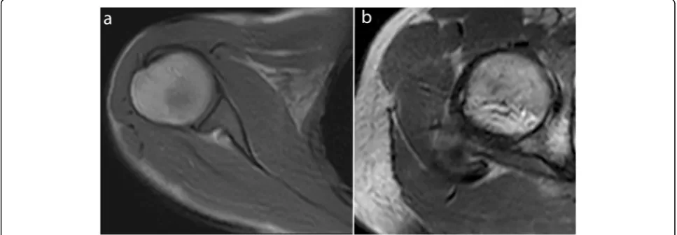
Fig. 2Real-time dynamic MR images obtained with balanced SSFP sequence at 1.5 T. Elbow in the sagittal plane in flexion (a) and knee in the sagittal plane in flexion (b) (TR, 4.6; TE, 2.3; flip angle, 40°; pixel size, 1.09 × 1.46 mm)
Table 2
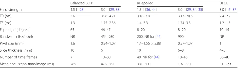
Sequence parameters reported in the recent literature for real-time joint dynamic MRI sequences at 1.5 T and 3.0 T
Balanced SSFP RF-spoiled UFGE
Field strength 1.5 T [28] 3.0 T [29,33] 1.5 T [36,44] 3.0 T [29,34,35] 3.0 T [5,37]
TR (ms) 3.6 3.98–4.71 3.18–7.8 3.13–20.6 2.4–2.7
TE (ms) 1.3 1.75–2.36 1.4–3.3 1.74–3.3 1.2–1.3
Flip angle (degree) 65 46–47 8–20 8–20 10–15
Bandwidth (Hz/pixel) NR 454–930 200, NR for [44] 990 NR
Pixel size (mm) 1.6 0.94–1.07 1.4–1.56 × 2.88 0.57–1.07 1
Slice thickness (mm) 10 6 10 6–8 4–5
Number of time frames 7 10–60 40, NR for [44] 10–16 30–40
Mean acquisition time/image (ms) 285 475–562 331–500 197–351 31–233
Proximal joints (shoulder, hip)
Intermediate joints (elbow, knee)
Distal joints (wrist, hand, ankle, foot)
Proximal joints
Dynamic imaging of proximal joints is challenging
be-cause these joints are hard to reach for RF coil
position-ing. The setup should be optimized for achieving
maximal ROM within the MRI scanner bore. The
pa-tient is typically positioned supine, head-first into the
bore for the shoulder examination and feet-first for the
hip examination, with the joint close to the MR bore
center, which allows more space for movement to occur
[
28
]. For the shoulder examination, the arm is positioned
along the patient
’
s side at rest [
28
,
58
]. The coils are
placed around the joint and could be maintained by a
harness to avoid displacement during motion (Fig.
5
,
Movie
5
) [
58
].
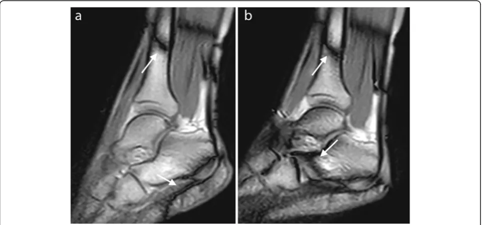
Fig. 3Real-time dynamic MR images obtained with the RF-spoiled sequence at 3.0 T. Wrist in the coronal plane in ulnar deviation (a) and ankle in the sagittal plane in plantar flexion (b) (TR, 20.6; TE, 1.8; flip angle, 15°; pixel size, 1.3 × 1.67 mm)
Fig. 4Real-time dynamic MR image obtained with the UFGE sequence at 3.0 T. Finger in the sagittal plane in flexion (TR, 4.7; TE, 2.3; flip angle, 15°; pixel size, 1.09 × 1.46 mm)
Table 3
Clinical applications of joint dynamic MRI
Shoulder Subacromial impingement [49,50]
Wrist Scapholunate instability [51]
Extensor carpi ulnaris tendon instability [32]
Finger Pulley injuries [52]
Hip Femoroacetabular impingement [53]
Knee Patellofemoral instability [16,23,43,54]
Anterior cruciate ligament deficiency [55–57]
Post-traumatic medial laxity [36]
Intermediate joints
For the study of the elbow, the patient is positioned prone,
head-first, arm above the head, with the elbow in the
cen-ter of the RF coil which is held by a support (Fig.
6
a) [
26
].
The kinematic study of the knee joint can be performed
on a subject in a supine position, feet-first, with the flex
coil held by a device around the joint (Fig.
6
b) [
59
,
60
].
The installation of a cushion under the knee increases the
flexion/extension and degrees of freedom [
5
,
43
], which
are limited by the bore diameter and the size of the lower
limb [
61
]. Unrestrained knee flexion/extension can also be
achieved with the patient in side lying position and a large
flex coil placed on the knee, and the other limb put on the
coil to prevent its displacement (Fig.
6
c) [
44
]. Some
au-thors have also proposed prone positioning of the patient,
allowing passive knee flexion [
61
] or knee flexion against
resistance [
18
,
62
].
Distal joints
All degrees of motion of distal joints can be studied
within the MRI scanner. For the wrist and hand, the
pa-tient is positioned prone, head-first, with the upper limb
raised above the head. Langner et al. studied the
abduc-tion/adduction motion of the wrist with a flex coil
positioned on the table parallel to the motion without
any support [
51
].
The distal situation of these joints makes it possible to
use a
“
rigid
”
RF coil. Bayer et al. performed dynamic
MRI sequences of the finger with a sky boot-shaped coil
[
63
], whereas Schellhammer and Vantorre used a knee
coil to study finger motion [
52
]. The hand can also be
positioned within a head coil to investigate all degrees of
freedom (Fig.
7
a) [
29
,
33
], whereas Kaiser et al. used an
extremity coil to explore wrist pronation-supination
[
32
]. Given the difference between the coil size and the
small joint volume, the use of pads or foams to hold the
hand in a central position within the coil and to avoid
undesired motion is required (Fig.
7
b) [
32
].
For the ankle, the patient is in the supine position, and
images can be obtained using a flex coil that is
posi-tioned around the joint with a device adapted to the RF
coil model (Fig.
7
c) [
22
,
24
,
26
].
Limitations, solutions, and perspectives
Real-time dynamic MRI is based on high temporal
reso-lution, but it also requires sufficient contrast, SNR, and
spatial resolution for joint motion evaluations through
image post-processing. The image quality also depends
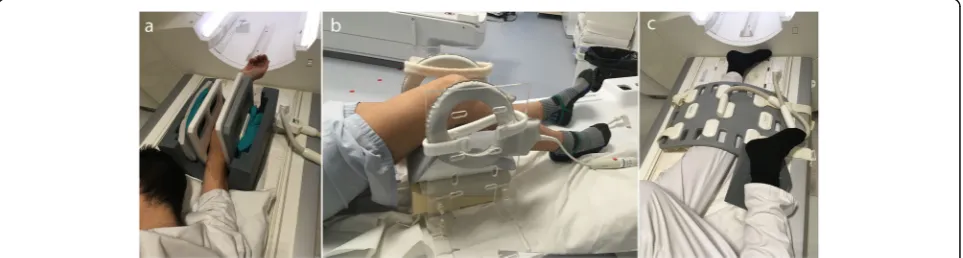
Fig. 5Patient and coil positioning for shoulder (a) and hip (b) motion evaluation in an Achieva dStream 3.0 T Philips MRI scanner
on the MRI scanner, magnetic field strength, and RF coil
type [
10
]. Acquiring MRI at 3.0 T using flex coils with
16 channels can improve the acquisition time, SNR, and
spatial resolution (Table
2
).
However, these gradient echo sequences can be affected
by some artifacts that worsen with higher magnetic field
strength. Moreover, these sequences are susceptible to
magnetic field inhomogeneities, resulting in signal loss
and deformation. For example, a chemical shift artifact
can appear as a black border at the fat/water interface.
These artifacts can be reduced with a larger bandwidth,
even if it decreases the SNR [
33
]. Chemical shift artifacts
can also be reduced with in-phase TE [
64
].
Susceptibility and motion artifacts can be reduced with
acceleration techniques such as parallel imaging with
phased-array RF coils and partial or radial sampling of
the
k
-space with iterative reconstructions [
29
,
65
–
68
],
which allow improvements in image quality and
tem-poral resolution.
The balanced SSFP sequence is also deteriorated by
band artifacts due to off-resonance effects from B
0non-uniformity, which are not present in RF-spoiled and
UFGE sequences. These band artifacts can appear over
the joint and disturb image analysis, in particular at 3.0
T (Fig.
8
) [
29
,
45
]. They can be reduced by minimizing
TR or by using a 3D shim at the beginning of the
exam-ination [
26
,
64
]. Other methods to reduce these band
ar-tifacts include the use of alternating TR, which widens
the space between these bands [
69
]; multiple-offset
method; frequency modulation [
70
]; or the use of
Fig. 7Patient and coil positioning for wrist (a), finger (b), and ankle motion evaluation (c). The wrist is positioned within a head coil in an Achieva dStream 3.0 T Philips MRI scanner (a). The hand is positioned within an extremity coil (which is opened to show positioning inside) for finger motion evaluation in an Achieva dStream 3.0 T Philips MRI scanner (b). The flex coil is positioned in the support of ankle motion evaluation in an Optima 1.5 T GE (c). Some cushions and sandbags are added to maintain the joint in the selected plane
specific algorithms [
71
]. Band artifacts can also be reduced
by positioning the joint in an intermediate position during
calibration, considering its ROM [
26
]. Field
inhomogen-eity is increased in the case of large difference in size
be-tween the joint and the RF coil, as for finger examination.
Adding dielectric pads around the joint can improve the
homogeneity of the field and reduce artifacts [
33
].
New post-processing methods are being developed to
register images obtained during motion with
high-resolution 3D static images. These methods make it
pos-sible to track bone motion in real-time [
31
,
37
], to
ob-tain 3D reconstructions of the bone structures during
motion [
34
], and, therefore, to provide accurate
quanti-tative biomechanical data.
Conclusion
Three gradient echo sequences (balanced SSFP, RF-spoiled,
and UFGE sequences) are available for real-time dynamic
MRI of joints of the upper and lower limbs during
continu-ous motion, considering their excellent temporal resolution,
good SNR, and contrast. However, real-time dynamic MRI
requires the adaptation of the sequence parameters,
rigor-ous patient and coil positioning to allow an evaluation with
a sufficient level of quality. These real-time sequences can
be incorporated within a daily protocol for joint MR
ana-lysis due to their short acquisition time and would allow to
better understand dynamic outcomes from specific joint
disorders or to diagnose conditions not otherwise detected
with static imaging. Future work may be focused on
post-processing method integration in current exams to improve
the SNR and to obtain 3D reconstruction while maintaining
a short acquisition time.
Supplementary information
Supplementary informationaccompanies this paper athttps://doi.org/10. 1186/s13244-020-00868-5.
Additional file 1: Movie 1.Balanced SSFP sequence of the shoulder in the axial plane during rotation at 3.0 T (same parameters as Fig.1). This sequence shows glenohumeral rotation, subscapular tendon excursion and the contraction of the subscapularis and infraspinatus muscles during internal and external rotation, respectively.
Additional file 2: Movie 2.Balanced SSFP sequence of the wrist in the coronal plane during radial/ulnar abduction at 3.0 T (same parameters as Fig.1). This sequence shows wrist bones relationship during motion.
Additional file 3: Movie 3.RF-spoiled sequence of the ankle in the sa-gittal plane during flexion at 3.0 T (same parameters as Fig.3). This se-quence allows us to investigate tibiotalar motion and Achilles tendon excursion.
Additional file 4: Movie 4.UFGE sequence of the finger in the sagittal plane during flexion at 3.0 T (same parameters as Fig.4). This sequence allows the evaluation of bones motion and flexor tendon excursion.
Additional file 5: Movie 5.Shoulder motion in the three degrees of freedom after coil positioning.
Abbreviations
3D:Three-dimensional; CT: Computed tomography; MRI: Magnetic resonance imaging; RF: Radio frequency; ROM: Range of motion; SNR: Signal-to-noise
ratio; SSFP: Steady-state free precession; TE: Echo time; TR: Repetition time; UFGE: Ultra-fast gradient echo
Acknowledgements
The authors thank the application engineers from GE Healthcare, Philips Healthcare, and Siemens Healthineers for their valuable help, and the radiographers from Brest University and Military Teaching hospitals.
Authors’contributions
MG was the major contributor in writing and organizing the manuscript. MG and BB conducted the literature search. BB and DBS supervised the work and made substantial contributions to the design of the study. KM, SB, and FR read, reviewed, and contributed with their expertise in their different fields to the final manuscript. All authors read and approved the final manuscript.
Funding
The authors declare that this work has not received any funding.
Availability of data and materials Not applicable
Ethics approval and consent to participate Not applicable
Consent for publication Not applicable
Competing interests
The authors declare that they have no competing interests.
Author details
1
Department of Radiology, Military Teaching Hospital Clermont-Tonnerre, Rue du colonel Fonferrier, 29240 Brest, Cedex 9, France.2Department of
Radiology, University Hospital Morvan, Brest, France.3Laboratory of Medical
Information Processing (LATIM), INSERM-UMR 1101, Brest, France.4University
of Western Brittany (UBO), Brest, France.5University Hospital, Brest, France.
6IMT Atlantique, UBL, Brest, France.7Department of Physical and Medical
Rehabilitation, University Hospital Morvan, Brest, France.8Department of
Paediatric Physical and Medical Rehabilitation, Fondation Ildys, Brest, France.
9
Department of Radiology, University Hospital La Cavale Blanche, Brest, France.
Received: 23 November 2019 Accepted: 2 April 2020
References
1. Tashman S, Collon D, Anderson K, Kolowich P, Anderst W (2004) Abnormal rotational knee motion during running after anterior cruciate ligament reconstruction. Am J Sports Med 32:975–983
2. Draper CE, Besier TF, Fredericson M et al (2011) Differences in
patellofemoral kinematics between weight-bearing and non-weight-bearing conditions in patients with patellofemoral pain. J Orthop Res 29:312–317 3. Powers CM, Ward SR, Fredericson M, Guillet M, Shellock FG (2003)
Patellofemoral kinematics during weight-bearing and non-weight-bearing knee extension in persons with lateral subluxation of the patella: a preliminary study. J Orthop Sports Phys Ther 33:677–685
4. McWalter EJ, O’Kane CM, FitzPatrick DP, Wilson DR (2014) Validation of an MRI-based method to assess patellofemoral joint contact areas in loaded knee flexion in vivo: patellofemoral contact area validation. J Magn Reson Imaging 39:978–987
5. d’Entremont AG, Nordmeyer-Massner JA, Bos C, Wilson DR, Pruessmann KP (2013) Do dynamic-based MR knee kinematics methods produce the same results as static methods? Magn Reson Med 69:1634–1644
6. Carr R, MacLean S, Slavotinek J, Bain GI (2019) Four-dimensional computed tomography scanning for dynamic wrist disorders: prospective analysis and recommendations for clinical utility. J Wrist Surg 08:161–167
8. Li G, Van de Velde SK, Bingham JT (2008) Validation of a non-invasive fluoroscopic imaging technique for the measurement of dynamic knee joint motion. J Biomech 41:1616–1622
9. Teixeira P, Gervaise A, Louis M et al (2015) Musculoskeletal wide-detector CT kinematic evaluation: from motion to image. Semin Musculoskelet Radiol 19:456–462
10. Shapiro LM, Gold GE (2012) MRI of weight bearing and movement. Osteoarthritis Cartilage 20:69–78
11. Sechtem U, Pflugfelder PW, White RD et al (1987) Cine MR imaging: potential for the evaluation of cardiovascular function. AJR Am J Roentgenol 148:239–246
12. van Dijk P (1984) ECG-triggered NMR imaging of the heart. Diagn Imaging Clin Med 53:29–37
13. Melchert UH, Schröder C, Brossmann J, Muhle C (1992) Motion-triggered cine MR imaging of active joint movement. Magn Reson Imaging 10:457– 460
14. Burnett KR, Davis CL, Read J (1987) Dynamic display of the
temporomandibular joint meniscus by using“fast-scan”MR imaging. AJR Am J Roentgenol 149:959–962
15. Zhu Y, Pelc NJ (1999) Three-dimensional motion tracking with volumetric phase contrast MR velocity imaging. J Magn Reson Imaging 9:111–118 16. Muhle C, Brossmann J, Melchert UH et al (1995) Functional MRI of the
patellofemoral joint: comparison of ultrafast MRI, motion-triggered cine MRI and static MRI. Eur Radiol 5:371–378
17. Borotikar B, Lempereur M, Lelievre M, Burdin V, Ben Salem D, Brochard S (2017) Dynamic MRI to quantify musculoskeletal motion: a systematic review of concurrent validity and reliability, and perspectives for evaluation of musculoskeletal disorders. PLoS One 12:e0189587
18. Shellock FG (2003) Functional assessment of the joints using kinematic magnetic resonance imaging. Semin Musculoskelet Radiol 7:249–276 19. Shellock FG, Mink JH, Deutsch AL, Fox JM (1989) Patellar tracking
abnormalities: clinical experience with kinematic MR imaging in 130 patients. Radiology 172:799–804
20. Schmid MR, Hodler J, Cathrein P, Duewell S, Jacob HAC, Romero J (2002) Is impingement the cause of jumper’s knee? Dynamic and static magnetic resonance imaging of patellar tendinitis in an open-configuration system. Am J Sports Med 30:388–395
21. Scarvell JM, Smith PN, Refshauge KM, Galloway HR, Woods KR (2005) Association between abnormal kinematics and degenerative change in knees of people with chronic anterior cruciate ligament deficiency: a magnetic resonance imaging study. Aust J Physiother 51:233–240 22. Tokuda O, Awaya H, Taguchi K, Matsunga N (2006) Kinematic MRI of the
normal ankle ligaments using a specially designed passive device. Foot Ankle Int 27:935–942
23. Brossmann J, Muhle C, Büll CC et al (1994) Evaluation of patellar tracking in patients with suspected patellar malalignment: cine MR imaging vs arthroscopy. AJR Am J Roentgenol 162:361–367
24. Sheehan FT, Seisler AR, Siegel KL (2007) In vivo talocrural and subtalar kinematics: a non-invasive 3D dynamic MRI study. Foot Ankle Int 28:323–335 25. Shellock FG, Foo TK, Deutsch AL, Mink JH (1991) Patellofemoral joint:
evaluation during active flexion with ultrafast spoiled GRASS MR imaging. Radiology 180:581–585
26. Quick HH, Ladd ME, Hoevel M et al (2002) Real-time MRI of joint movement with trueFISP. J Magn Reson Imaging 15:710–715
27. Draper CE, Santos JM, Kourtis LC et al (2008) Feasibility of using real-time MRI to measure joint kinematics in 1.5 T and open-bore 0.5 T systems. J Magn Reson Imaging 28:158–166
28. Pierrart J, Lefèvre-Colau MM, Skalli W et al (2014) New dynamic three-dimensional MRI technique for shoulder kinematic analysis. J Magn Reson Imaging 39:729–734
29. Shaw CB, Foster BH, Borgese M et al (2019) Real-time three-dimensional MRI for the assessment of dynamic carpal instability. PLoS One 14:e0222704 30. Yen P, Katzberg RW, Buonocore MH, Sonico J (2013) Dynamic MR imaging
of the temporomandibular joint using a balanced steady-state free precession sequence at 3 T. AJNR Am J Neuroradiol 34:E24–E26 31. Gilles B, Perrin R, Magnenat-Thalmann N, Vallee JP (2005) Bone motion
analysis from dynamic MRI: acquisition and tracking. Acad Radiol 12:1285– 1292
32. Kaiser P, Kellermann F, Arora R, Henninger B, Rudisch A (2018) Diagnosing extensor carpi ulnaris tendon dislocation with dynamic rotation MRI of the wrist. Clin Imaging 51:323–326
33. Boutin RD, Buonocore MH, Immerman I et al (2013) Real-time magnetic resonance imaging (MRI) during active wrist motion—initial observations. PLoS One 8:e84004
34. Makki K, Borotikar B, Garetier M, Brochard S, Ben Salem D, Rousseau F (2019) In vivo ankle joint kinematics from dynamic magnetic resonance imaging using a registration-based framework. J Biomech 86:193–203
35. Henrichon SS, Foster BH, Shaw C et al (2020) Dynamic MRI of the wrist in less than 20 seconds: normal midcarpal motion and reader reliability. Skeletal Radiol 49:241–248
36. Studler U, White LM, Deslandes M, Geddes C, Sussman MS, Theodoropoulos J (2011) Feasibility study of simultaneous physical examination and dynamic MR imaging of medial collateral ligament knee injuries in a 1.5-T large-bore magnet. Skeletal Radiol 40:335–343
37. Clarke EC, Martin JH, d’Entremont AG, Pandy MG, Wilson DR, Herbert RD (2015) A non-invasive, 3D, dynamic MRI method for measuring muscle moment arms in vivo: demonstration in the human ankle joint and Achilles tendon. Med Eng Phys 37:93–99
38. Chavhan GB, Babyn PS, Jankharia BG, Cheng H-LM, Shroff MM (2008) Steady-state MR imaging sequences: physics, classification, and clinical applications. Radiographics 28:1147–1160
39. Bieri O, Scheffler K (2013) Fundamentals of balanced steady state free precession MRI. J Magn Reson Imaging 38:2–11
40. Hargreaves B (2012) Rapid gradient-echo imaging. J Magn Reson Imaging 36:1300–1313
41. Fuchs F, Laub G, Othomo K (2003) TrueFISP—technical considerations and cardiovascular applications. Eur J Radiol 46:28–32
42. Elster AD (1993) Gradient-echo MR imaging: techniques and acronyms. Radiology 186:1–8
43. Burke CJ, Kaplan D, Block T et al (2018) Clinical utility of continuous radial magnetic resonance imaging acquisition at 3 T in real-time patellofemoral kinematic assessment: a feasibility study. Arthroscopy 34:726–733 44. Fiorentino NM, Lin JS, Ridder KB, Guttman MA, McVeigh ER, Blemker SS
(2013) Rectus femoris knee muscle moment arms measuredin vivoduring dynamic motion with real-time magnetic resonance imaging. J Biomech Eng 135:044501
45. Lingala SG, Sutton BP, Miquel ME, Nayak KS (2016) Recommendations for real-time speech MRI. J Magn Reson Imaging JMRI 43:28–44
46. Frahm J, Voit D, Uecker M (2019) Real-time magnetic resonance imaging: radial gradient-echo sequences with nonlinear inverse reconstruction. Invest Radiol 54:757–766
47. VanPelt MD, Landrum MR, Igbinigie M, Wadhwa V, Chhabra A (2017) Kinematic magnetic resonance imaging of peroneal tendon subluxation with intraoperative correlation. J Foot Ankle Surg 56:395–397 48. Sheehan FT (2012) The 3D in vivo Achilles’tendon moment arm, quantified
during active muscle control and compared across sexes. J Biomech 45:225–230 49. Tempelaere C, Pierrart J, Lefèvre-Colau MM et al (2016) Dynamic
three-dimensional shoulder MRI during active motion for investigation of rotator cuff diseases. PLoS One 11:e0158563
50. Tasaki A, Nimura A, Nozaki T et al (2015) Quantitative and qualitative analyses of subacromial impingement by kinematic open MRI. Knee Surg Sports Traumatol Arthrosc 23:1489–1497
51. Langner I, Fischer S, Eisenschenk A, Langner S (2015) Cine MRI: a new approach to the diagnosis of scapholunate dissociation. Skeletal Radiol 44:1103–1110 52. Schellhammer F, Vantorre A (2019) Semi-dynamic MRI of
climbing-associated injuries of the finger. Skeletal Radiol 48:1435–1437 53. Burke CJ, Walter WR, Gyftopoulos S et al (2019) Real-time assessment of
femoroacetabular motion using radial gradient echo magnetic resonance arthrography at 3 Tesla in routine clinical practice: a pilot study. Arthroscopy 35:2366–2374
54. Muhle C, Brossmann J, Heller M (1999) Kinematic CT and MR imaging of the patellofemoral joint. Eur Radiol 9:508–518
55. Guenoun D, Vaccaro J, Le Corroller T et al (2017) A dynamic study of the anterior cruciate ligament of the knee using an open MRI. Surg Radiol Anat 39:307–314
56. Haughom BD, Souza R, Schairer WW, Li X, Benjamin Ma C (2012) Evaluating rotational kinematics of the knee in ACL-ruptured and healthy patients using 3.0 Tesla magnetic resonance imaging. Knee Surg Sports Traumatol Arthrosc 20:663–670
58. Matsui K, Tachibana T, Nobuhara K, Uchiyama Y (2018) Translational movement within the glenohumeral joint at different rotation velocities as seen by cine MRI. J Exp Orthop 5:7
59. Borotikar BS, Sipprell WH 3rd, Wible EE, Sheehan FT (2012) A methodology to accurately quantify patellofemoral cartilage contact kinematics by combining 3D image shape registration and cine-PC MRI velocity data. J Biomech 45:1117–1122
60. Westphal CJ, Schmitz A, Reeder SB, Thelen DG (2013) Load-dependent variations in knee kinematics measured with dynamic MRI. J Biomech 46: 2045–2052
61. Lin CC, Zhang S, Frahm J, Lu TW, Hsu CY, Shih TF (2013) A slice-to-volume registration method based on real-time magnetic resonance imaging for measuring three-dimensional kinematics of the knee. Med Phys 40:102302 62. Powers CM, Shellock FG, Pfaff M (1998) Quantification of patellar tracking
using kinematic MRI. J Magn Reson Imaging 8:724–732
63. Bayer T, Adler W, Janka R, Uder M, Roemer F (2017) Magnetic resonance cinematography of the fingers: a 3.0 Tesla feasibility study with comparison of incremental and continuous dynamic protocols. Skeletal Radiol 46:1721– 1728
64. Huang SY, Seethamraju RT, Patel P, Hahn PF, Kirsch JE, Guimaraes AR (2015) Body MR imaging: artifacts, k-space, and solutions. Radiographics 35:1439– 1460
65. Zaitsev M, Maclaren J, Herbst M (2015) Motion artifacts in MRI: a complex problem with many partial solutions. J Magn Reson Imaging 42:887–901 66. Deshmane A, Gulani V, Griswold MA, Seiberlich N (2012) Parallel MR
imaging. J Magn Reson Imaging 36:55–72
67. Tsao J, Kozerke S (2012) MRI temporal acceleration techniques. J Magn Reson Imaging 36:543–560
68. Uecker M, Zhang S, Voit D, Merboldt KD, Frahm J (2012) Real-time MRI: recent advances using radial FLASH. Imaging Med 4:461–476
69. Nayak KS, Lee HL, Hargreaves BA, Hu BS (2007) Wideband SSFP: alternating repetition time balanced steady state free precession with increased band spacing. Magn Reson Med 58:931–938
70. Foxall DL (2002) Frequency-modulated steady-state free precession imaging. Magn Reson Med 48:502–508
71. Björk M, Ingle RR, Gudmundson E, Stoica P, Nishimura DG, Barral JK (2014) Parameter estimation approach to banding artifact reduction in balanced steady-state free precession. Magn Reson Med 72:880–892
Publisher
’
s Note