Open Access
Research article
Receptor oligomerization and beyond: a case study in bone
morphogenetic proteins
Kai Heinecke
1,2, Axel Seher
3, Werner Schmitz
1, Thomas D Mueller
4,
Walter Sebald
1and Joachim Nickel*
1Address: 1Physiologische Chemie II, Biozentrum, Universität Würzburg, Würzburg, Germany, 2Institut für Humangenetik, Biozentrum, Universität
Würzburg, Würzburg, Germany, 3Universitätsklinikum Würzburg, Abteilung für Molekulare Innere Medizin, Würzburg, Germany and 4Molekulare Pflanzenphysiologie und Biophysik, Julius von Sachs Institut, Universität Würzburg, Würzburg, Germany
Email: Kai Heinecke - [email protected]; Axel Seher - [email protected]; Werner Schmitz - [email protected]; Thomas D Mueller - [email protected]; Walter Sebald - [email protected]; Joachim Nickel* - [email protected] * Corresponding author
Abstract
Background: Transforming growth factor (TGF)β superfamily members transduce signals by oligomerizing two classes of serine/threonine kinase receptors, termed type I and type II. In contrast to the large number of ligands only seven type I and five type II receptors have been identified in mammals, implicating a prominent promiscuity in ligand-receptor interaction. Since a given ligand can usually interact with more than one receptor of either subtype, differences in binding affinities and specificities are likely important for the generation of distinct ligand-receptor complexes with different signaling properties.
Results: In vitro interaction analyses showed two different prototypes of binding kinetics, 'slow on/slow off' and 'fast on/fast off'. Surprisingly, the binding specificity of ligands to the receptors of one subtype is only moderate. As suggested from the dimeric nature of the ligands, binding to immobilized receptors shows avidity due to cooperative binding caused by bivalent ligand-receptor interactions. To compare
these in vitro observations to the situation in vivo, binding studies on whole cells employing homodimeric
as well as heterodimeric bone morphogenetic protein 2 (BMP2) mutants were performed. Interestingly, low and high affinity binding sites were identified, as defined by the presence of either one or two BMP receptor (BMPR)-IA receptor chains, respectively. Both sites contribute to different cellular responses in that the high affinity sites allow a rapid transient response at low ligand concentrations whereas the low affinity sites facilitate sustained signaling but higher ligand concentrations are required.
Conclusion: Binding of a ligand to a single high affinity receptor chain functioning as anchoring molecule and providing sufficient complex stability allows the subsequent formation of signaling competent complexes. Another receptor of the same subtype, and up to two receptors of the other subtype, can then be recruited. Thus, the resulting receptor arrangement can principally consist of four different receptors, which is consistent with our interaction analysis showing low ligand-receptor specificity within one subtype class. For BMP2, further complexity is added by the fact that heterooligomeric signaling complexes containing only one type I receptor chain can also be found. This indicates that despite prominent ligand receptor promiscuity a manifold of diverse signals might be generated in this receptor limited system.
Published: 7 September 2009
BMC Biology 2009, 7:59 doi:10.1186/1741-7007-7-59
Received: 30 April 2009 Accepted: 7 September 2009
This article is available from: http://www.biomedcentral.com/1741-7007/7/59
© 2009 Heinecke et al; licensee BioMed Central Ltd.
Background
The bone morphogenic proteins (BMPs), growth and dif-ferentiation factors (GDFs) and activins belong to the large transforming growth factor (TGF)β superfamily of secreted signaling molecules [1,2]. The more than 30 TGFβ-like proteins identified in vertebrates to date [3,4] play important roles in all stages of embryogenesis [5]. In the adult organism these factors exhibit a broad range of biological effects and control various processes during regeneration and tissue repair such as growth, growth inhibition, differentiation, apoptosis, and secretion [6,7]. Based on their functional and sequence similarities TGFβ members can be divided into several subfamilies: the TGFβs (TGFβ1, β2, and β3), activins (activin A, B, C, E), BMP2s (BMP2, 4), BMP7s (BMP5, 6, 7), GDF5s (GDF5, 6, 7) and others [1,8]. Signal transduction of TGFβ members is mediated by oligomerizing two different types of trans-membrane serine/threonine kinase receptor chains termed type I and type II. Five type II receptors and seven type I receptors have been identified in mammals and the broad range of TGFβ ligands suggests a high degree of pro-miscuity in ligand-receptor interactions [1,9]. On one hand most receptors can bind several different ligands, and on the other hand most ligands can interact with more than one receptor chain of each subtype. Since members of the TGFβ superfamily transduce signals via a heterooligomeric receptor system, differences in binding affinities and specificities might generate a multiplicity of ligand-receptor complexes with different signaling prop-erties, allowing cellular responses that differ in quality and quantity.
Binding specificities and affinities between ligands and receptors have been analyzed on a semiquantitative basis by crosslinking radioactively labeled ligands with recep-tors that were overexpressed in cells. Two general binding modes have been observed via this technique. One mode, called 'sequential', is characteristic for TGFβs and activins and involves high affinity binding of the ligand to a type II receptor and subsequent low affinity interaction of this complex with a type I receptor [10,11]. Ligands following this binding mode can be directly crosslinked to a type II receptor but crosslinking to a type I receptor is dependent on the type II receptor presence. The second binding mode, called 'cooperative', is characterized by crosslink-ing to either the type I or type II receptors and has been proposed for BMPs. However, crosslinking efficiency is enhanced if both receptor types are coexpressed [1].
To better understand receptor activation and the mecha-nism underlying receptor specificity for TGFβ ligands, we determined binding affinities of different BMPs and GDFs to their cognate receptor ectodomains by surface plasmon resonance. One representative member from each of three BMP/GDF subfamilies was chosen in this study. Binding
parameters were evaluated in two ways, (1) by immobiliz-ing the receptor ectodomains of the type I and type II receptors activin receptor (ActR)-I, ActR-IB, BMP receptor (BMPR)-IA, BMPR-IB, ActR-II, ActR-IIB, and BMPR-II, and (2) by immobilizing the ligands. These two setups allow us to obtain data on the individual binding affinity as well as the avidity that is inherently linked to the dimeric nature of the ligands. To compare the binding properties of BMP/GDF receptor interaction with related receptor systems, activin A was included in this study. Possible cooperative interactions between the two receptor types were investigated by studying the formation of ternary complexes consisting of the ligand and the ectodomains of both receptor types on the biosensor chip.
The dimeric nature of the ligands suggests that coopera-tive binding via multiple interactions between ligand and receptors (avidity) should also exist in vivo. Furthermore, since certain ligands such as BMP2, BMP4 or GDF5 can interact independently with type I as well as type II recep-tors [12,13] an inherent complexity of individual ligand-receptor interactions can be expected on cell surfaces. In addition, since the ligands can bind to other cell surface components such as coreceptors (for example, DRAGON, BAMBI) [14,15] or the extracellular matrix (for example, heparin) [16] the analysis of receptor recruitment and activation is further complicated.
To analyze receptor compositions on cell surfaces and their relation to biological function, BMP2 variants were created lacking the heparin binding sites in order to reduce binding to the extracellular matrix (ECM). Addi-tional amino acid exchanges were introduced resulting in homodimeric or heterodimeric ligands with interrupted receptor binding epitopes. Binding of these variants to receptors expressed on whole cells was analyzed by radio-ligand binding assays and correlated to their biological activities.
Results
Expression and purification of receptor ectodomain and ligand proteins
Since the association rate kon as well as the binding con-stant KD determined from the sensorgrams directly
β-mercap-toethanol each receptor protein appears as a single band with an apparent molecular weight between 15 and 30 kDa, the bands of higher molecular weight most likely represent incorrectly folded multimers linked by disulfide bridges (data not shown). Purification of only mono-meric receptor proteins could be achieved since only monomeric ECDs bound to and were recovered from BMP2 affinity columns. The ECDs ActR-I and ActR-IB derived from insect cells could not be purified by affinity chromatography due to their lack of binding to BMP2. Hence, these receptors were purified to homogeneity by trimethylaminoethyl (TMAE) anion exchange chromatog-raphy followed by reverse-phase high performance liquid chromatography (RP-HPLC). All isolated proteins exhibit purities >95% (data not shown).
Biosensor experiments
As shown by the structures of several ligand-receptor com-plexes, the dimeric ligands are capable of interacting simultaneously with two receptor molecules of either sub-type. Based on this property, the ligands can interact as analyte either with one, or simultaneously with two, immobilized receptors when those are present at suffi-cient density on the biosensor (Figure 1a). Using the inverse setup, with the ligands immobilized and the receptor as analytes, individual binding of single receptor molecules to the ligands can be determined (Figure 1b). Simultaneous binding of both receptor subtypes to the ligand, as is seen in ternary complex formation, can be recorded using the experimental setup shown in Figure 1c.
Influence of ionic strength and pH value on binding affinities
The solubility of the BMP ligands strongly depends on pH and ionic strength. In order to find optimal conditions, a series of measurements with varying pH and salt
concen-tration were performed. BMP2 was perfused over biosen-sor surfaces with the ECDs of BMPR-IA, BMPR-IB or ActR-IIB immobilized and employing different buffers as indi-cated (Additional file 1).
Similar binding affinities and specificities were observed over a wide range of salt concentrations (150 to 900 mM) and pH conditions (pH 5.0 to 9.5). Thus, the binding of BMP2 to immobilized receptors is unaffected by ionic strength up to 500 mM NaCl. Above 500 mM NaCl the affinities of BMP2 for immobilized receptors decrease up to 10-fold (Additional file 1). As expected, strongest bind-ing is observed at physiological pH. More acidic or basic conditions result in a decrease (3-fold to 10-fold) of the affinities of BMP2 to all tested receptors (Additional file 1).
The observed robustness of binding, independent of pH or ionic strength, can be explained by the nature of the binding interfaces [17-24]. For the interaction of receptors of either subtype with the ligand, the binding is domi-nated by hydrophobic interactions. Since the association rates are far below the diffusion-controlled limit (<107 M -1 s-1) electrostatic steering seems not to be involved in
lig-and-receptor interaction.
Based on our results we used 4-(2-hydroxyethyl)-1-piper-azineethanesulfonic acid (HEPES) buffer containing 500 mM NaCl at a pH value of 7.4 for all biosensor measure-ments. Use of this buffer in the interaction analysis yielded binding data which do not differ from those obtained using physiological salt concentrations, but greatly reduced non-specific binding of the ligand to the carboxymethyl cellulose (CM) matrix on the chip surface.
Binding of ligands to immobilized receptors
Of the ligands tested, the highest binding affinities were observed for BMP2 with preferred binding to the type I receptors BMPR-IA (apparent KD: 0.8 nM) and BMPR-IB
(2.7 nM) and for the GDF5:BMPR-IB interaction (1.3 nM), whereas activin A showed preferential binding to the type II receptor ActR-IIB with similarly high binding affin-ities (2.1 nM) (see Table 1). In contrast, for BMP7 such a preference in binding to a receptor of either subtype was not detected: comparable affinities were observed instead for the interaction of BMP7 with the type I receptor BMPR-IB (9 nM) and the type II receptors ActR-II (8 nM) and ActR-IIB (9.2 nM). Of the four prototypic ligands tested only BMP7 bound ActR-I, and then very weakly (the sen-sorgrams could not be evaluated). Since ligand concentra-tions up to 120 nM were used the apparent KD value of this interaction is probably larger than 500 nM. The ECD of ActR-IB was not bound by any of the tested ligands.
Experimental layout Figure 1
Experimental layout. Model of biosensor experiments with ligands as analyte passed over immobilized receptor
ectodomains (ECDs) (a), receptor ECDs passed over
immo-bilized ligands (b) and ternary complexes formed by
perfus-ing an immobilized type I receptor with the ligand plus the ECD of a type II receptor (c).
B
C
The specificity of interactions between the studied recep-tors and BMP2, BMP7, and GDF5 is only moderate. The receptor BMPR-IA revealed the highest ligand specificity; it binds BMP2 with ≥20-fold higher affinity than GDF5 or BMP7. The interactions of other receptors with these lig-ands show only discrimination with a 10-fold difference in binding affinity. Among the ligands, GDF5 exhibits the highest receptor specificity, binding preferentially to BMPR-IB and ActR-IIB. The type I receptor specificity of GDF5 is defined by a single residue (Arg57), which is located in the pre-helix loop in the center of the type I receptor binding epitope [25].
For some of our data similar results have been published by other groups [18,26]. However, affinities of other lig-and-receptor interactions differ by more than two orders of magnitude. Of note, the affinities of activin A for bind-ing to the immobilized type II receptors II and ActR-IIB are reported as 10-fold to 100-fold higher compared to our data. The discrepancy is mainly due to lower dissocia-tion rates (koff) that are reported by Greenwald et al. [18,27]. In addition, the affinity of BMP7 for BMPR-IA according to our measurements is 20-fold higher than reported by Allendorph et al. [26]. One explanation might be differences in the chip surface density of the immobi-lized receptor. At low immobilization levels the distances between individual receptors might be too large to allow
Table 1: Binding parameters of interactions of soluble ligands with immobilized receptor ectodomains (ECDs)
Ligand (analyte) Type I receptor (immobilized) Type II receptor (immobilized)
ActR-I ActR-IB BMPR-IA BMPR-IB ActR-II ActR-IIB BMPR-II
Mean Mean Mean SD Mean SD Mean SD Mean SD Mean SD
BMP-2
kon × 10-4 [M-1s-1] NB NB 50 ± 12.1 25 ± 4.37 370 ± 66.6 280 ± 33.6 150 ± 25.5
koff × 103 [s-1] NB NB 0.4 ± 0.09 0.7 ± 0.09 88 ± 23.8 18 ± 4.68 70 ± 19.6
KD (kin) [nM] NB NB 0.8 ± 0.37 2.7 ± 0.82 14 ± 6.31 6.3 ± 2.39 45 ± 20.3
KD (eq) [nM] NB NB NE NE 24 ± 1.92 9.0 ± 1.17 59 ± 10.0
GDF-5
kon × 10-4 [M-1s-1] NB NB 23 ± 5.98 39 ± 4.68 140 ± 15.4 110 ± 19.8 110 ± 20.9
koff × 103 [s-1] NB NB 4.3 ± 0.77 0.5 ± 0.16 28 ± 4.48 4.5 ± 0.59 38 ± 8.74
KD (kin) [nM] NB NB 19 ± 8.36 1.3 ± 0.56 20 ± 5.40 4.0 ± 1.24 36 ± 15.1
KD (eq) [nM] NB NB NE NE 32 ± 3.84 5.6 ± 0.90 46 ± 8.28
BMP-7
kon × 10-4 [M-1s-1] NE NB 14 ± 2.24 11 ± 2.42 120 ± 20.4 140 ± 30.8 96 ± 14.4
koff × 103 [s-1] NE NB 7.9 ± 1.19 1.0 ± 0.18 6.2 ± 0.69 9.0 ± 1.26 24 ± 6.24
KD (kin) [nM] > 500* NB 58 ± 18.0 9.0 ± 3.87 5.1 ± 1.43 6.5 ± 2.34 25 ± 10.3
KD (eq) [nM] > 500* NB NE NE 8.0 ± 0.48 9.2 ± 1.28 40 ± 5.20
Activin-A
kon × 10-4 [M-1s-1] NB NB NB NB 130 ± 23.4 160 ± 22.4 53 ± 7.95
koff × 103 [s-1] NB NB NB NB 7.5 ± 1.65 1.7 ± 0.14 29 ± 6.96
KD (kin) [nM] NB NB NB NB 5.7 ± 2.28 1.1 ± 0.24 59 ± 24.2
KD (eq) [nM] NB NB NB NB 6.0 ± 0.54 2.1 ± 0.15 24 ± 4.08
The data obtained from measurements with immobilized type I receptor ECDs were fitted to a kinetic model (1:1 Langmuir binding) from which KD
(kin) (bold) is calculated as koff (× 103 s-1)/k
on (× 10-4 M-1 s-1). Due to low but significant binding of BMP7 to AR-I, affinities could not be evaluated
exactly but are estimated to be higher than 500 nM (bold, asterisks). The data obtained from ligand binding to immobilized type II receptors were best fitted by equilibrium dose response KD (eq) (bold). For this interaction the calculation of KD (kin) (bold, italic) revealed minor differences
(<twofold). All data represent mean values of at least three repeated measurements using six different ligand concentrations.
for simultaneous interaction of the dimeric ligand with two immobilized receptors. By contrast, at very high den-sities steric hindrances could occur. An investigation into this has been reported for the interaction of activin A with the type II receptor ActR-IIB [27].
However, another explanation for the latter discrepancy might be due to the usage of the detergent 3-[(3-cholami-dopropyl)dimethylammonio]-1-propanesulfonate (CHAPS). The results obtained from measurements with 0.36% CHAPS added to HBS500 buffer (see Methods) dif-fer in most of the cases, some dramatically, from those obtained without CHAPS (Figure 2). Only the interaction of activin A with ActR-II and ActR-IIB is unaffected, whereas all other ligand-receptor interactions show a reduced affinity. The binding affinity of BMP7 to BMPR-IA was reduced 20-fold. The sensorgrams for the BMP7:BMPR-IA (Additional file 2) interaction could not be directly evaluated, but a correlation of the resonance units obtained with an 80 nM ligand solution and the known Rmax value of the sensor chip yields an estimation of the apparent KD value of approximately 2 μM.
How-ever, not only was the interaction between ligands and type I receptors changed in the presence of the detergent CHAPS, but binding specificity to the type II receptors was also altered. Whereas binding of GDF5 to ActR-II showed only a 30-fold decrease, binding of the ligands to BMPR-II was completely abolished in the presence of CHAPS. Thus, the presence of CHAPS not only alters the binding affinities but also influences ligand-receptor specificities in the majority of the interactions investigated here.
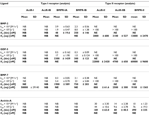
Binding of receptors to immobilized ligands
Due to the measurement of the 1:1 interaction and hence the lack of avidity, apparent affinities are much lower when the setup is based on immobilized ligands and using the soluble receptor ectodomains as analytes (Table 2). Binding constants range from 48 nM for the interac-tion of BMPR-IA with BMP2 up to 60 μM for the binding of BMPR-II to immobilized GDF5. The very weak affini-ties of the 1:1 interaction of BMPR-II to BMP2, BMP7, and GDF5 have been recently reported by Yin et al. [28]. Under this setup again BMPR-IA shows the strongest overall binding (among the type I receptors) to BMP2 (KD: 48
nM). A similar value was reported by Sachse et al. [29] for this interaction and for the binding of BMPR-IA to BMP4 [30,31], which is plausible considering that the type I receptor binding epitope (wrist epitope) of BMP2 and BMP4 share 100% amino acid identity [32]. Interestingly, using this setup with immobilized ligands, the type I receptor ActR-I measurably interacts only with BMP7.
Regarding the ligand specificities of the receptors, the results are similar to those observed with the reciprocal setup using immobilized receptors. Owing to the lack of
avidity all affinities are 'scaled' down by a factor of 50 to 1,000. However, for BMP2 and GDF5 the binding to the type II receptors benefits much more from avidity effects compared to type I receptor binding. For BMP7, which binds type I and type II receptors with similar affinities, no such significant receptor subtype specific effect on the avidity is observed. In the case of activin A simultaneous binding of the ligand to 2 type II receptors also leads to an increased affinity by a factor of 30 to 40, direct binding of activin A to type I receptors is not observed independent of the biosensor setup. The lack of type I receptor binding of activin A can be possibly explained by the known struc-tures of activin A:ActR-II complexes, which show that the type I receptor epitope in activin A might be structurally disrupted in the absence of the type II receptors [23,27].
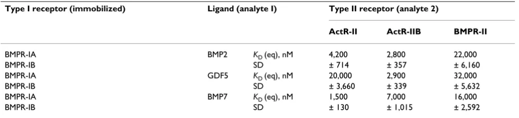
Binding affinities in ternary complexes
The crystal structures of the BMP2:BMPR-IA:ActR-II [17] and BMP2:BMPR-IA:ActR-IIB [24] ternary complexes clearly demonstrate the lack of any receptor:receptor con-tacts. Furthermore, no gross conformational changes are observed in the ligand dimer architecture of BMP2 upon complex formation, in contrast to activin A and TGFβ3. Consequently, a cooperative recruitment of the type II receptor ectodomains could be excluded from Biacore measurements [24]. To determine whether all type II receptor ectodomains bind to BMP2, BMP7 and GDF5 with identical affinities independent of the presence of a type I receptor, ternary complexes were generated on the biosensor matrix as described in the Methods section (Fig-ure 1c, Table 3). The results of the 'ternary' interactions reveal only marginal differences compared to those obtained for individual receptor-ligand interactions (see Tables 2 and 3). All differences, except for the interaction of ActR-IIB with the BMP7:BMPR-IBimmobilized complex, are within a factor of two and thus not significant consid-ering the standard deviations of regular biosensor meas-urements. An increase in affinities due to cooperativity, as shown for the binding of BMP7 to ActR-I in the presence of ActR-II [18], could not be detected in our experiments. The detection of ternary complex formation via the immobilized type I receptor ActR-I was not possible due to its low ligand binding capabilities. The reverse detec-tion to measure the binding of soluble type I receptor ECDs to a preformed ligand:type II receptor complex with the type II receptor serving as the anchor to the biosensor could not be performed, since the fast dissociation rates
koff for ligand type II receptor interaction impeded a coin-jection setup, which is the experimental basis for these measurements.
iso-Influence of 3-[(3-cholamidopropyl)dimethylammonio]-1-propanesulfonate (CHAPS) for ligand-receptor interaction Figure 2
Influence of 3-[(3-cholamidopropyl)dimethylammonio]-1-propanesulfonate (CHAPS) for ligand-receptor interaction. Binding affinities of the ligands bone morphogenic protein (BMP)2, BMP7 and growth and differentiation factor
(GDF)5 to the immobilized type I receptors BMP receptor (BMPR)-IA and BMPR-IB (a) and those of the same ligands plus
activin A to the type II receptors activin receptor (ActR)-II, ActR-IIB, and BMPR-II (b) are depicted as bar diagrams. The data
represent mean values of two individual experiments using six different ligand concentrations. Standard deviations are indicated by error bars.
immob. receptor analyte
A
1.0 E-10
1.0 E-09
1.0 E-08
1.0 E-07
1.0 E-06
BMPR-IA BMPR-IB
BMP-2 GDF-5 BMP-7 BMP-2 GDF-5 BMP-7
K
D(kin)
(M)
+ 0.36 % CHAPS - CHAPS
n.e.*
immob. receptor analyte
B
1.0 E-09
1.0 E-08
1.0 E-07
1.0 E-06
1.0 E-05
+ 0.36 % CHAPS - CHAPS
K
D(eq)
(M)
BMPR-II ActR-IIB
ActR-II
n.b.* n.b.* n.b.*
lated extracellular domains of the receptors are used, the cooperative recruitment of the type II receptor chains that are observed in crosslinking experiments on cells must therefore be generated by an alternative mechanism, such as the interaction of transmembrane or intracellular domains of the receptors.
Different types of binding kinetics
Generally, two types of binding kinetics could be observed in our experiments. The first type, which is observed for the interaction of BMP2, BMP7, and GDF5 with the immobilized type I receptors BMPR-IA and BMPR-IB, can be considered 'slow' being characterized by relatively slow association kon (1 to 5 × 10⁵ M-1 s-1) and
dissociation rates koff (0.4 to 8 × 10⁻³ s-1) (see Additional
file 3). The second type, which is seen for the majority of BMP2, BMP7 and GDF5 type II receptor interactions, is 'fast' exhibiting fast association kon (>106 M-1 s-1) and
dis-sociation rates koff (>10-2 s-1) (see Additional file 3). The
sensorgrams measuring ternary complex formation clearly display both types of binding kinetics, the slow associa-tion and dissociaassocia-tion of the ligand to/from the immobi-lized type I receptor ectodomain and the fast binding kinetics for the interaction of the soluble type II receptor ectodomain with the preformed complex (Additional file 3).
Table 2: Binding parameters of interactions of soluble receptors with immobilized ligands.
Ligand Type I receptor (analyte) Type II receptor (analyte)
ActR-I ActR-IB BMPR-IA BMPR-IB ActR-II ActR-IIB BMPR-II
Mean SD Mean Mean SD Mean SD Mean SD Mean SD mean SD
BMP-2
kon × 10-4 [M-1s-1] NB NB 3.9 ± 0.63 2.3 ± 0.56 NE NE NE
koff × 103 [s-1] NB NB 1.9 ± 0.47 8.0 ± 1.38 > 100 > 100 > 100
KD (kin) [nM] NB NB 48 ± 19.6 350 ± 146 NE NE NE
KD (eq) [nM] NB NB NE NE 3800 ± 608 3100 ± 527 13000 ± 2470
GDF-5
kon × 10-4 [M-1s-1] NB NB 0.5 ± 0.162 0.3 ± 0.09 NE NE NE
koff × 103 [s-1] NB NB 17 ± 1.90 1.0 ± 0.124 > 100 > 100 > 100
KD (kin) [nM] NB NB 3300 ± 1439 300 ± 123 NE NE NE
KD (eq) [nM] NB NB NE NE 22000 ± 2420 4700 ± 658 60000 ± 9600
BMP-7
kon × 10-4 [M-1s-1] NE NB 0.3 ± 0.05 3.1 ± 0.38 NE NE NE
koff × 103 [s-1] NE NB 5.4 ± 0.70 23 ± 5.84 > 100 > 100 > 100
KD (kin) [nM] NE NB 1900 ± 589 750 ± 283 NE NE NE
KD (eq) [nM] 58000 ± 29140 NB NE NE 880 ± 61.6 2500 ± 300 9100 ± 1365
Activin-A
kon × 10-4 [M-1s-1] NB NB NB NB 30 ± 3.30 14 ± 2.38 10 ± 1.23
koff × 103 [s-1] NB NB NB NB 44 ± 10.6 9.6 ± 2.78 76 ± 19.3
KD (kin) [nM] NB NB NB NB 180 ± 63.0 88 ± 40.5 890 ± 335
KD (eq) [nM] NB NB NB NB NE NE NE
The data obtained from the interaction soluble type I receptor ectodomains (ECDs) with the immobilized ligands were fitted to the 1:1 Langmuir binding model and the KD (kin) (bold) calculated as koff (× 103 s-1)/k
on (× 10-4 M-1 s-1). Due to the fast kinetics of the interaction between type II
receptors as analyte and the immobilized ligands BMP2, BMP7 and GDF5, the data could only be fitted by equilibrium dose response KD (eq). The dissociation rate constants (koff) of these interactions are >100 (× 103 s-1). All data represent mean values of three repeated measurements using at
least six different analyte concentrations.
The 1:1 interactions of the soluble type I and type II recep-tor ectodomains to the immobilized ligands show princi-pally comparable characteristics (in terms of fast and slow) to those of the 1:2 interactions, which are observed in the inverse situation (compare figures in Additional file 3). Binding kinetics of the 1:1 interaction are generally characterized by faster dissociation rates koff. This is expected since on the biosensor with the ligand being immobilized, the binding epitopes of the ligand act inde-pendently, thus a dissociation of the receptor analyte is irrevocable. In the 1:2 interaction dissociation of the lig-and analyte from one receptor does not automatically cause the release of the ligand from the biosensor. Since the ligand is still coupled via the second receptor, fast rebinding can occur and hence the dissociation is dramat-ically decreased. Noteworthy is the very fast dissociation of the type II receptor analytes from the immobilized BMP2, BMP7, and GDF5 resulting in sensorgrams with an almost rectangular shape (Figure 3b). Since data acquisi-tion can only proceed with a limited sampling frequency (2.5 Hz) an evaluation of the kinetic rate constants is not feasible. Thus, the dissociation rates koff can be estimated to be certainly >10-1 s-1 but more precise analysis cannot
be provided here. Hence, no predictions with regard to the association rates can be made.
The lifetimes of individual ligand-receptor complexes can be deduced from the dissociation rates. For the 1:2 inter-action of BMP2, BMP7 and GDF5 with the type I receptors BMPR-IA and BMPR-IB rather long complex lifetimes (t1/
2 = (ln2)/koff) on the order of 2 to 30 min can be
calcu-lated, whereas ligand:type II receptor complexes with the type II receptors anchored to the sensor surface exhibit half-lives of the order of a few seconds (1 to 15 s). For the 1:1 interaction, which can be considered the initial bind-ing event in the case of a sequential bindbind-ing mechanism, complex lifetimes are significantly reduced. However, the lifetimes of almost all BMP2, BMP7, and GDF5 type I receptor (1:1) complexes are still longer than those deter-mined for the 1:2 interactions of these ligands with the
type II receptors. Only activin A can form complexes with type II receptors that exhibit half-lives longer than 1 min.
Our data strongly suggest that, in all ligand-receptor sys-tems tested here, one defined receptor subtype serves as an anchor for the recruitment of the ligand from the superna-tant to the membrane surface. The other receptor subtype either does not interact with the ligand (that is, activin A with ActR-IB) or binds with a fast binding kinetic as observed for the BMP2 or GDF5 type II receptor com-plexes and thus cannot efficiently act as a membrane anchor. These data consequently suggest a sequential binding mode for BMP2 and GDF5, with an initial recruit-ment via type I receptors and a subsequent binding of the type II receptors to this intermediate ligand:type I receptor complex.
Ligand binding on whole cells
The presence of four receptor binding epitopes in the dimeric ligand creates the possibility of a whole set of individual ligand-receptor interactions on cell surfaces. In addition the ligands can interact with other cell surface components such as coreceptors (for example, DRAGON, BAMBI) [14,15] or the extracellular matrix (for example, heparin) [16]. In order to lower interactions with the extracellular matrix, we created BMP2 ligands lacking the heparin binding sites (so-called coreBMP2 variants, see Methods section). In biosensor analyses the variant core BMP2 wild type (coreBMP2wt) exhibits receptor binding characteristics identical to those of wtBMP2 indicating that the N-terminal sequences are not involved in receptor interaction. For the homodimeric coreBMP2L51P variant no binding to type I receptors is detected (KD > 1 μM), in
agreement with published data [33]. Binding to type II receptors is identical to that of wtBMP2, confirming that the mutation L51P solely destroys type I receptor binding. In the case of the heterodimeric coreBMP2wt/BMP2L51P variant a binding constant of 50 nM was determined for the interaction with BMPR-IA and of 350 nM for the bind-ing to BMPR-IB. Interestbind-ingly, the same bindbind-ing constants
Table 3: Binding affinities of soluble type II receptors in ternary complexes
Type I receptor (immobilized) Ligand (analyte I) Type II receptor (analyte 2)
ActR-II ActR-IIB BMPR-II
BMPR-IA BMP2 KD (eq), nM 4,200 2,800 22,000
BMPR-IB SD ± 714 ± 357 ± 6,160
BMPR-IA GDF5 KD (eq), nM 20,000 2,900 32,000
BMPR-IB SD ± 3,660 ± 339 ± 5,632
BMPR-IA BMP7 KD (eq), nM 1,500 7,000 16,000
BMPR-IB SD ± 130 ± 1,015 ± 2,592
The data obtained from binding of type II receptor ectodomains (ECDs) as analyte to the preformed complexes were fitted by equilibrium dose response KD (eq). The data represent mean values of two measurements using at least six different type II receptor concentrations.
could be determined when either the ligand or the recep-tors were immobilized. Furthermore, these values resem-ble the 1:1 interactions of BMPR-IA or BMPR-IB with wtBMP2 (see Table 2). So far no mutations in BMP2 have been found that are able to completely abolish type II receptor binding. The heterodimeric coreBMP2wt/A34D variant binds type II receptor ectodomains (immobilized on the biosensor) with only 3-fold lower binding affinity, and the homodimeric coreBMP2A34D variant with 10-fold lower binding affinity, compared to wtBMP2. Since a real 1:1 ligand:type II receptor interaction cannot be sim-ulated with these ligands they were not suitable for radio-ligand binding assays.
We analyzed the binding of iodinated coreBMP2wt to C2C12 cells (Figure 3a). When ligand concentrations up to 500 pM were used a binding constant of about 180 pM was determined with roughly 12,000 binding sites calcu-lated per cell. Both values agree with previously published binding data employing other BMP responsive cells [34,35].
Of note, 30% of total binding to C2C12 cells was non-specific even when coreBMP2wt was used. Since nothing is known about the detailed receptor composition for the binding sites detected in these cells similar experiments were carried out using transiently transfected COS-7 cells
Binding of radiolabeled proteins on cell surfaces Figure 3
Binding of radiolabeled proteins on cell surfaces. (a) Dose-dependent binding of iodinated core bone morphogenic pro-tein (wild type) (coreBMP2wt) to C2C12 cells (total binding, black squares). Unspecific binding as determined by addition of a
1,000-fold excess of cold ligand (blue diamonds) was subtracted resulting in specific binding of the ligand (red stars). (b)
Com-parison of specific binding of coreBMP2wt to COS-7 cells transfected with either BMP receptor (BMPR)-IA or activin receptor
(ActR)-IIB or cotransfected with both receptors chains. (c) Specific binding of the iodinated heterodimeric coreBMP2/L51P
mutant to BMPR-IA transfected COS-7 cells using ligand concentration up to 4 nM. (d) Specific binding of a radiolabeled
anti-BMPR-IA Fab fragment to either untransfected (black squares) or anti-BMPR-IA transfected (red asterisks) COS-7 cells. At all cases specific binding was fitted to a one-site binding model resulting in the indicated values for KD and Bmax.
ligand
bound
(cpm
)
ligand concentration (pM)
± 187 20
0 100 200 300 400 500
0 1000 2000 3000 4000 5000 6000
totally bound
unspecifically bound
specifically bound
Bmax:3877
KD: 183pM ±
cpm
A
0 100 200 300 400 500
0 500 1000 1500 2000 2500
3000 B (cpm)
max
4875
5294
6143
KD(pM)
5139
1389
877
ligand concentration (pM) BMPR-IA
ActR-IIB BMPR-IA / ActR-IIB
B
specifically
bound
(cpm)
0 1000 2000 3000 4000
0 2000 4000 6000 8000 10000 12000
ligand concentration (pM)
BMPR-IAempty vector
Bmax: KD:
48276 cpm 13422 pM
Bmax:
KD:
2193 cpm 12936pM
transfected receptor:
anti BMPR-IA (Fab) ligand:
D
specifically
bound
(cpm)
0 1000 2000 3000 4000
0 1000 2000 3000 4000 5000 6000 7000
Bmax: 54024 cpm
KD: 30295 pM
ligand concentration (pM)
transfected receptor:
heterodimeric coreBMP-2wt/L51P ligand:
BMPR-IA
C
specifically
bound
(Figure 3b). The conditions were chosen to keep the number of binding sites similar to those observed in non-transfected C2C12 cells, however the affinities for the lig-ands were at least fourfold lower. Importantly, the values observed for the binding of BMP2 to cells transfected with BMPR-IA or ActR-IIB were basically identical to those of the 1:2 interactions determined from Biacore measure-ments (see Table 1). Cotransfection of both receptor sub-types resulted in a marginal increase in binding affinities (<twofold), similar to what was observed from Biacore measurements when the ectodomains of both receptor subtypes were immobilized simultaneously on the bio-sensor (data not shown). These data clearly show that also on whole cells only very weak cooperativity, if any, exists in BMP2-mediated receptor recruitment.
Using up to 500 pM concentrations of the heterodimeric coreBMP2wt/L51P variant the resulting binding curves did not enter the plateau phase and thus could not be fit-ted to a one-site binding model. With higher ligand con-centrations a binding constant KD of approximately 30 nM was obtained resembling the binding affinity for the 1:1 BMP2:BMPR-IA interaction as determined from Biacore measurements (Figure 3c). Interestingly, the number of binding sites seems about 10-fold higher (approximately 150,000 per cell) compared to the meas-urements obtained with homodimeric wild-type coreBMP2 (see Figure 3a). Since we cannot exclude that other sites beside the transfected receptor are bound at higher ligand concentrations, the BMPR-IA binding sites were directly determined using a radiolabeled Fab frag-ment (AbyD, Morphosys, Martinsried, Germany), which binds specifically to the ectodomain of BMPR-IA (Figure 3d). For mock-transfected and BMPR-IA transfected cells an identical binding constant of KD approximately 13 nM was obtained for the Fab fragment, which is again consist-ent with Biacore measuremconsist-ents (data not shown). Fur-thermore, the number of BMPR-IA-derived binding sites as determined from the Fab-fragment binding is basically identical to those found in the measurements using the heterodimeric coreBMP2wt/L51P variant. In mock-trans-fected cells the number of BMPR-IA-derived binding sites is approximately 25-fold lower. Thus COS-7 cells express only minor amounts of BMPR-IA endogenously and the majority of the signal in the transfected cells is generated from the interaction with the ectopically expressed BMPR-IA. Due to the monovalent nature of our Fab fragment the number of binding sites most likely accounts for individ-ual BMPR-IA molecules on the cell surface. Consequently, the interaction of coreBMP2wt with BMPR-IA should result in similar values for maximal ligand binding (Bmax) at higher concentrations. However, when we used higher concentrations of coreBMP2wt we obtained a biphasic binding curve indicating the presence of two different kinds of binding sites (Figure 4a). Separate evaluation of
the binding affinities for the lower (0 to 500 pM) and higher (1,000 to 4,000 pM) concentrations yields KD val-ues of 1.4 and 25 nM resembling the affinities obtained from Biacore experiments for the 1:2 (high affinity) and the 1:1 (low affinity) interaction. Importantly, the major-ity (90%) of the total binding sites are low affinmajor-ity sites, which most likely reflect receptor monomers, whereas only 10% of the binding sites exhibit high binding affin-ity. These sites most likely represent receptors that are arranged as preformed dimers or even in higher ordered structures thereby allowing a simultaneous 1:2 interac-tion.
Repeating the experiment using ActR-IIB transfected cells to measure the binding of coreBMP2wt at higher concen-trations did not produce a biphasic binding curve (Figure 4b). Fitting analysis of the binding data at higher or lower ligand concentration resulted in identical values for KD
and Bmax. To determine whether the rather small Bmax val-ues are due to weaker expression of ActR-IIB, expression levels were independently tested using fluorophore tagged receptors and western blot analysis of whole cell lysates. Since no significant differences were detected between BMPR-IA and ActR-IIB transfected cells, this suggests that the majority of the ActR-IIB receptors on the cell surface are not occupied by the ligand even at concentrations of 4 nM (data not shown).
To determine, whether non-transfected BMP2 responsive cells exhibit the same distribution of monomeric or dimeric receptor assemblies C2C12 cells were incubated with iodinated coreBMP2wt (Figure 4c). Similar to BMPR-IA transfected COS-7 cells a biphasic binding curve was observed. The number of binding sites at lower and higher concentrations suggest a similar distribution of high and low affinity receptor sites, but binding affinities were four times higher for both 1:1 and 1:2 interactions compared to BMPR-IA transfected COS-7 cells. It remains unclear if the very tight binding in untransfected BMP2 responsive cells is due to the interaction of the ligand with both endogenously expressed type I and type II receptor chains resulting in a heterohexameric complex. The high affinity might likewise due to involvement of affinity-enhancing coreceptors such as DRAGON, a member of the repulsive guidance molecule (RGM) family, which might facilitate ligand binding to dimeric as well as to monomeric recep-tors. Expression of all three RGM family members could be detected in C2C12 cells by real-time RT-PCR experi-ments. The highest expression levels found for DRAGON (RGMb) were approximately 20-fold lower compared to those of BMPR-IA (data not shown).
Biological activity
Binding of radiolabeled ligands using higher ligand concentrations Figure 4
Binding of radiolabeled ligands using higher ligand concentrations. (a) Specific binding of core bone morphogenic protein (wild type) (coreBMP2wt) to BMP receptor (BMPR)-IA transfected results in a biphasic binding curve with a pro-nounced break (marked by arrow). Fitting the curve separately (0 to 500 pM and 1,000 to 4,000 pM) to a one-site binding
model the indicated values for KD and Bmax were achieved. (b) Specific binding of coreBMP2wt to COS-7 cells transfected with
activin receptor (ActR)-IIB and (c) to untransfected C2C12 cells.
0 1000 2000 3000 4000 0
1000 2000 3000 4000 5000 6000 7000 8000
Bmax:
KD:
48515 cpm 24243 pM
ligand concentration (pM)
Bmax:
KD:
5294 cpm 1389 pM
transfected receptor:
coreBMP-2(wt)
ligand: BMPR-IA
A
specifically
bound
(cpm)
B
transfected receptor:
coreBMP-2wt ligand: ActR-IIB
0 1000 2000 3000 4000
0 500 1000 1500 2000 2500 3000
ligand concentration (pM)
Bmax: 4875 cpmKD: 5139 pM
specifically
bound
(cpm)
transfected receptor:
coreBMP-2wt ligand: none
0 1000 2000 3000 4000
0 2000 4000 6000 8000 10000 12000 14000
Bmax: 3877 cpm KD: 183 pM
Bmax:
KD:
36487 cpm 8382 pM
ligand concentration (pM)
C
specifically
bound
BMP2 responsive cells and in cells transfected with BMP receptors suggesting consequences for downstream signal-ing events. We therefore used induction of alkaline phos-phatase (ALP) expression to monitor the effect of different receptor complex arrangements. In C2C12 cells BMP2 induces ALP activity in a dose-dependent manner requir-ing about 20 nM BMP2 for half-maximal response [32]. The presence and functional importance of BMPR-IA for this ALP activation has been reported previously [36,37]. Other receptors present in our C2C12 cells are ActR-I, ActR-II and BMPR-II, whereas BMPR-IB and ActR-IIB seem to be expressed at very low levels (data not shown). Since BMP2 cannot efficiently activate cells expressing ActR-I as the only type I receptor, signal transduction in C2C12 cells is most likely mediated via BMPR-IA [38,39]. Inter-estingly, the concentration for half maximal response for ALP induction in these cells correlates well with the KD
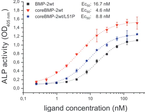
value of the 1:1 BMP2:BMPR-IA interaction, and thus sug-gests that ALP induction might be controlled via an iso-lated (not dimeric) type I receptor architecture. Therefore, ALP assays were performed employing the homodimeric coreBMP2L51P and the heterodimeric coreBMP2wt/L51P variants. The homodimeric coreBMP2L51P variant fails to induce ALP expression, which is in agreement with results published earlier using similar BMP variants that have both type I receptor sites destroyed [33,40]. However, the heterodimeric coreBMP2wt/L51P variant shows induc-tion of ALP expression similarly (difference <twofold) as coreBMP2wt (Figure 5). For comparison, wild-type BMP2 and the N-terminal truncated coreBMP2wt variant differ about fourfold in ALP induction, indicating that the pres-ence of heparin binding sites influpres-ences the induction of ALP expression more so than does the complete ablation of one type I receptor binding epitope.
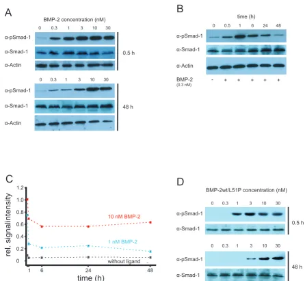
SMAD phosphorylation
In addition to ALP induction we also studied SMAD-1 phosphorylation in C2C12 cells. We performed an initial timecourse analysis of SMAD-1 phosphorylation from 30 min to 2 h after ligand addition [41] and also monitored the influence of the ligand concentration.
After incubating the cells with coreBMP2wt for 30 min half-maximal phosphorylated SMAD (pSMAD) levels were obtained using approximately 300 pM of ligand (Figure 6a). We then extended the time course analysis to examine the kinetics of long-term stimulation. Interest-ingly, the ligand concentration required for SMAD phos-phorylation increases significantly with the time of ligand exposure (Figure 6b). After 48-h incubation 10-fold to 30-fold higher ligand concentrations compared to short-term incubation (a 1-h period) were necessary to induce half-maximal pSMAD levels (see Figure 6a, c). At ligand con-centrations >3 nM high pSMAD-1 levels could also be observed after 6 h and 24 h of ligand exposure indicating
a permanent activation of the SMAD pathway (data not shown). Interestingly, the total SMAD-1 protein levels also marginally increased over time, but ligand independ-ently. Dose-dependent phosphorylation of SMAD-1 could be also observed upon stimulation with the het-erodimeric coreBMP2wt/L51P variant. Similarly, sensitiv-ity to ligand exposure decreased over time although not to the extent observed with wild-type ligand (Figure 6d). Importantly, after 48 h of ligand exposure, half-maximal pSMAD-1 levels were achieved using the same concentra-tion of the heterodimeric BMP2 variant (approximately 3 nM) as observed for wtBMP2. This is consistent with the comparable ability of wtBMP2 and heteromeric wtBMP2/ L51P variant to induce ALP, in that at variant concentra-tions required for half-maximal ALP induction (10 to 20 nM) pSMAD-1 levels are similarly high.
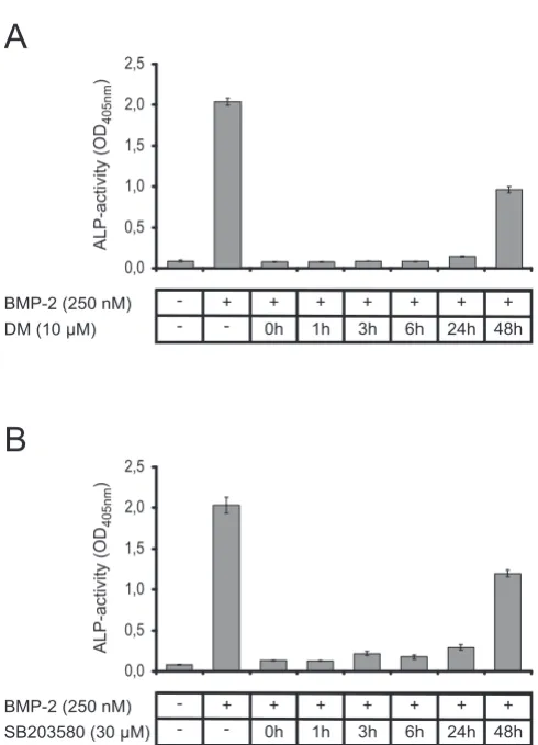
Inhibition of the SMAD and mitogen-activated protein (MAP) kinase pathway
The important points to consider are whether SMAD phosphorylation and induction of ALP expression are coupled through a common signaling cascade. It is sup-posed that SMAD phosphorylation leads to differentia-tion of C2C12 cells into the osteoblastic lineage, but it is unclear whether SMAD phosphorylation is also required for the induction of ALP gene expression at later time-points. Recently, a small-molecule inhibitor of BMP sign-aling, called dorsomorphin, was demonstrated to perturb
Biological activity of bone morphogenic protein (BMP)2 vari-ants
Figure 5
Biological activity of bone morphogenic protein (BMP)2 variants. The dose-dependent induction of alkaline phosphatase (ALP) activity in serum starved C2C12 cells is shown for the indicated ligands BMP2 wild type (BMP2wt) (black squares), coreBMP2wt (red triangles) and the het-erodimeric coreBMP2wt/L51P variant (blue asterisks). The background absorption at 405 nm of 0.09 ± 0.0075 was not subtracted to indicate the signal to noise ratio.
0,1 1 10 100
0,0 0,2 0,4 0,6 0,8 1,0 1,2 1,4 1,6 1,8 2,0
ALP
a
ctivity
(O
D
405n
m
)
ligand concentration (nM)
BMP-2wt Ec :50 16.7 nM
coreBMP-2wt Ec :50 4.6 nM
dorsoventral axis formation in Zebrafish [42]. This sub-stance selectively inhibits BMP type I receptors ActR-I, BMPR-IA and BMPR-IB and thereby prevents phosphor-ylation of SMAD1/5/8 proteins. SMAD2/3 phosphoryla-tion as well as phosphorylaphosphoryla-tion of the p38 MAP kinase is not affected by dorsomorphin. The p38 MAP kinase path-way contributes to chondrogenesis induced by GDF5 in ATDC-5 cells as well as to osteogenic differentiation of C2C12 cells mediated by BMP2 [43,44]. Several small molecule inhibitors of p38 MAP kinase activation are
cur-rently available, however not all (for example, SB202190) reduce or inhibit the induction of ALP expression [44].
To investigate if the activated SMAD1/5/8 and/or p38 MAP kinase pathways are required for the ALP induction, dorsomorphin and SB203580 were added separately to C2C12 cells at different timepoints using concentrations of 10 and 30 μM, respectively. ALP activity was analyzed 72 h after ligand addition (Figure 7). The results clearly show that the simultaneous administration of either
dor-SMAD phosphorylation Figure 6
SMAD phosphorylation. (a) A concentration-dependent phosphorylation of SMAD-1 mediated by bone morphogenic pro-tein (BMP)2 is shown at the indicated timepoints in C2C12 cells by western blotting. Analysis of SMAD-1 and actin levels acts
as loading control. (b) Time-dependent phosphorylation of SMAD-1 by BMP2 at a concentration of 0.3 nM. (c) Diagram of the
time-dependent phosphorylation levels of SMAD-1 without ligand or induced by BMP2 at the indicated concentrations. The data were obtained by scans of western blot exposures. pSMAD signals were quantified and normalized to total SMAD-1 levels
using the software ImageJ. (d) Concentration-dependent phosphorylation of SMAD-1 mediated by heterodimeric core BMP
wild type (coreBMP2wt)/L51P. α-pSmad-1
α-Actin
0 0.3 1 3 10 30
0.5 h
α-pSmad-1
α-Actin
48 h BMP-2 concentration (nM)
0 0.3 1 3 10 30
A
α-Smad-1
α-Smad-1
α-pSmad-1
α-Actin
BMP-2
0 0.5 1 6 24 48
- + + + + +
time (h)
(0.3 nM)
B
α-Smad-1
C
time (h)
rel.
signalintensity
10 nM BMP-2
1 nM BMP-2
without ligand
1 6 24 48
0 0.2 0.4 0.6 0.8 1.0 1.2
α-pSmad-1
0 0.3 1 3 10 30
0.5 h
α-pSmad-1
48 h BMP-2wt/L51P concentration (nM)
0 0.3 1 3 10 30
D
α-Smad-1
somorphin (Figure 7a) or SB203580 (Figure 7b) with lig-and (that is, at t = 0 h) completely abolishes ALP induction. Even if the inhibitors are added 24 h after the ligand only a marginal increase in ALP activity is observed. Addition of the inhibitors 48 h after ligand administration still results in a significantly reduced ALP activity com-pared to induction by 250 nM BMP2wt in the absence of these inhibitors. Similar results were obtained from anal-ogous experiments using ATDC-5 cells, thus the observed inhibition of ALP activity is not cell type specific (data not shown). These results clearly demonstrate that the induc-tion of ALP gene expression requires a permanent activa-tion of both, MAP kinase and SMAD pathways.
Discussion
In this study we investigated the binding properties of dif-ferent BMP ligands, in vitro and on whole cells, and corre-lated these properties with immediate downstream signaling events such as SMAD phosphorylation and induction of ALP expression. By performing in vitro inter-action analyses in an identical manner for three proto-typic members of the BMP subfamilies (BMP2/4, BMP5/ 6/7 and GDF5/6/7) we could compare and analyze differ-ences in detail, enabling us to deduce consequdiffer-ences for the initial steps of receptor binding and activation. Due to the dimeric nature of the BMP/TGFβ ligands their receptor binding mechanism is inherently complex, which compli-cates data acquisition and analysis. The binding of the lig-and to membrane-anchored receptors is affected by avidity as the two receptor sites lead to a strong increase in the apparent binding affinity. The increase in affinity should mainly result from slower dissociation because, based on statistical thermodynamics, it is highly unlikely that a molecule attached to two receptors can leave both sites simultaneously. If re-binding is fast the dissociation is slowed down dramatically. By contrast, statistical ther-modynamics also predicts that binding of a dimeric lig-and to membrane-anchored receptors will occur via a stepwise process, since the direct binding of ligand to two receptors simultaneously (with respect to the timing of the binding events) involves a trimolecular reaction, which is a very rare event. Thus, mechanistically, the lig-ands will most likely bind initially to a single receptor chain (unless the receptors exist as preformed dimers on the cell surface) and a second receptor chain will subse-quently be recruited into this membrane-bound complex.
Our experimental setup utilizing immobilization of either the ligands or the receptors allows the determination of the rate constants for each of these association and disso-ciation steps. With immobilized ligands our in vitro inter-action analysis delivers binding constants and kinetics for the so-called 1:1 interaction, where the receptors bind independently and no cooperativity is observed, due to the absence of allosteric mechanisms or direct contacts between the extracellular domains of the receptors. This setup provides parameters that likely resemble the situa-tion when ligand first encounters the cell surface, thereby binding to a single receptor. Our results show that for the interaction of BMP2 and GDF5, with type I receptors, as well as the binding of activin A to the type II receptors ActR-II and ActR-IIB, the 1:1 interaction occurs with 30-fold to 100-30-fold lower affinity than the 1:2 interaction, under the conditions tested. Binding of BMP2 and GDF5 to the type II receptors is even more affected by avidity, showing a 100-fold to 1,000-fold increase in affinity when going from a 1:1 to a 1:2 interaction scheme. As expected the increased binding affinities in the 1:2 interactions result from reduced dissociation rates but an increase in
Inhibition of the SMAD and p38 mitogen-activated protein (MAP) kinase pathway
Figure 7
Inhibition of the SMAD and p38 mitogen-activated protein (MAP) kinase pathway. Alkaline phosphatase (ALP) assays were carried out using C2C12 cells in the absence or presence of 250 nM of bone morphogenic
pro-tein (wild type) (BMP2wt). (a) Dorsomorphin (DM) or (b)
SB203580 was added at the indicated timepoints. The back-ground absorption at 405 nm of 0.09 ± 0.0075 was not sub-tracted to indicate the signal to noise ratio.
A
0,0 0,5 1,0 1,5 2,0 2,5
0h 1h 3h 6h 24h 48h
DM (10 μM)
BMP-2 (250 nM) - +
-
-+ + + + + +
ALP-activity
(OD
)
405nm
0,0 0,5 1,0 1,5 2,0 2,5
0h 1h 3h 6h 24h 48h
SB203580 (30 μM)
BMP-2 (250 nM) - +
-
-+ + + + + +
ALP-activity
(OD
)
405nm
the association rates was also observed in most cases. However, in the 1:1 interaction scheme, the dissociation rates for ligand:type II receptor complexes are much faster than those observed for the ligand:type I receptor interac-tion. The deduced half-lives for complexes of BMP2 and GDF5 bound to one type II receptor are, at most, on the order of very few seconds, whereas binding of these lig-ands to one of their type I receptors results in complexes with half-lives on the order of several minutes. Thus, assuming single isolated receptors for either subtype, BMP2 and GDF5 likely bind first to a type I receptor and subsequently recruit a second receptor (which possibly could be either subtype) into the membrane-anchored binary complex. The initial complex of a ligand bound to two membrane-anchored receptors likely stabilizes the complex by lowering the dissociation rate due to avidity such that the receptor recruitment can proceed without the intermediate complex falling apart.
BMP7 seems different when compared with BMP2 or GDF5 as the 1:1 interaction scheme does not reveal a clear high affinity receptor for BMP7. The type I receptor BMPR-IB and the type II receptor ActR-II exhibit almost identical affinities for BMP7. However, the dissociation rates again show that the BMP7:BMPR-IB complex has a fivefold longer half-life than the BMP7:ActR-II complex, making a sequential mechanism with binding of BMP7 first to BMPR-IB more likely. Most importantly, these hypotheses are only valid under the assumptions that no other com-ponents affect the complex lifetime and that the receptor usage of a ligand in vivo solely depends on the receptor affinity, that is, the receptor with the highest binding affinity is the receptor to be recruited by this ligand. How-ever, it is known that signaling of BMP7 and BMP6 involves the type I receptor ActR-I [45], which binds with at least 30-fold lower affinity (in the 1:1 interaction scheme) than the other type I receptors BMPR-IA and BMPR-IB. Furthermore, coreceptors such as members of the DRAGON/RGM family or β-glycan can influence binding by enhancing recruitment to the membrane sur-face and, in the case of DRAGON, even influencing recep-tor specificity [46]. Aside from the coreceprecep-tors, heparin binding sites in some of the BMP ligands also change their membrane localization, possibly forming a ligand species that is not soluble as assumed but is rather, at least in part, localized to the membrane even in a receptor-unbound state.
To determine the receptor architecture present on cells in vivo and to correlate the observation with the in vitro bind-ing affinities, we additionally performed ligand-bindbind-ing assays to whole cells. Interestingly, if the ligand concentra-tion is sufficiently high, the resulting binding curves sug-gest the presence of two different receptor species on cell surfaces. Using BMPR-IA transfected cells, high and low
affinity binding sites whose binding affinities correlate with the respective 1:2 and 1:1 interaction in vitro could be identified for wild-type BMP2. We could estimate that about 10% of the overall receptor sites represent high affinity and about 90% the low affinity sites. By using a heterodimeric BMP2 variant with only one functional type I receptor epitope, we could confirm the presence and the number of these low affinity sites. Correlating these observations with our in vitro interaction analysis we suggest that about 10% of the type I receptors are present as preformed dimers, thereby binding BMPs with very high affinity, whereas the majority of the BMP type I receptors are present as isolated single binding sites on cells.
For the BMP type II receptors the picture is similar. Despite the exclusive detection of sites correlating in their ligand binding affinities to the 1:2 interaction scheme, the number of sites is much lower than expected from expres-sion levels being comparable to those of BMPR-IA. We suggest that due to the low binding affinities for the 1:1 BMP2:ActR-IIB interaction (approximately 3 μM, see table 2), significantly high levels of ligand specifically bound to monomeric type II receptors cannot be achieved due to increased unspecific interactions and solubility limitation of the BMP ligands. These data nevertheless clearly show that the receptor architectures on cells are heterogeneous before ligand binding.
There are reports in the literature that preformed receptors and single receptors (that are oligomerized by the ligand) can address different signaling pathways, namely that pre-formed receptors lead to induction of the SMAD pathway, whereas ligand-induced receptor oligomerization leads to activation of the p38 MAP kinase cascade [47]. We inves-tigated the consequences of binding to the two different receptor species by analyzing the induction of ALP expres-sion and measuring SMAD phosphorylation. The concen-tration for half-maximal ALP expression correlates with the affinity determined for the 1:1 ligand:type I receptor interaction. Furthermore the heterodimeric BMP2 variant BMP2wt/L51P with only one functional type I receptor epitope exhibits nearly the same half maximal effective concentration (EC50), strongly suggesting that ALP
BMP2 exposure is likely mediated by preformed type I receptor dimers, which can bind BMP2 with very high affinities in the subnanomolar range. During extended exposure these preformed receptor dimers seem to disap-pear, probably due to internalization, while monomeric receptor chains predominantly remain at the cell surface. These receptor monomers bind wtBMP2, as well as the heteromeric wt/L51P variant with only one intact type I receptor site, with a lower binding affinity, resulting in a lower EC50 value. A constitutive endocytosis via clathrin-coated pits, was reported for the type I receptor BMPR-IA and BMPR-II, and also for BMPR-II, via caveola-like inter-nalization [48]. It is, however, unknown whether the internalized preformed receptor complexes reappear at the cell surface as complexes that would thus reconstitute high affinity sites, or if additional processes inhibit such reappearance and thereby keep the ligand sensitivity low. It also remains unclear, if other mechanisms such as autoregulatory feedback loops trigger, for example, total SMAD levels throughout the time the experiments take.
Our results utilizing BMP2 heterodimers with one ablated receptor epitope also clearly suggest that only one type I receptor is needed in the ligand:receptor complex to allow signaling. In contrast, earlier studies showed that two type II receptors are required for the formation of a signaling competent complex. It remains questionable whether this finding is the result of reduced type II receptor binding affinities (that is, thermodynamically controlled) or shorter half-lives of individual ternary complexes (that is, kinetically controlled). However, the recruitment of the type II receptors seems to be the limiting step in BMP2-mediated signaling.
The remaining single type I receptor sites are still capable of transducing signals via the SMAD pathway, but due to their lower ligand affinities higher concentrations are likely required. As shown by addition of dorsomorphin and SB203580, a sustained activation of the type I recep-tor resulting in both activated SMAD and MAP kinase pathways is required for the induction of BMP-responsive ALP gene expression. It is important to note that ALP is not directly activated by either SMAD and/or the p38 MAP kinase pathways, since cycloheximide abolishes BMP2 induced ALP mRNA synthesis [49]. It was demonstrated that BMP2 controls ALP expression and osteoblast miner-alization by a Wnt autocrine loop. Consequently, BMP2-mediated ALP gene expression seems to depend only on the quantity of type I receptors being activated by the lig-and. Subsequent processes seem not to be limiting.
Conclusion
A comparison of our results obtained from in vitro interac-tion analyses with binding studies performed on intact cells provides new insights into the complexity of BMP/
GDF receptor activation and its relevance for subsequent signaling events. Our data clearly demonstrate the pres-ence of distinct receptor arrangements on the cell surface, contributing to distinct cellular responses. A minor subset of receptors seems to be preformed and contains at least two receptors of each subtype, allowing the assembly of active signaling complexes at low ligand concentrations. Whether this heterohexameric ligand:receptor arrange-ment acts as a functional unit or is part of even higher ordered cell surface structures remains undetermined. However, the majority of activated BMP receptors on cell surfaces mediating long-term signal transduction by BMP2 most likely consist of the ligand, two type II recep-tors but only one type I receptor chain. Such an assembly also best explains the signaling capabilities of het-erodimeric ligands such as BMP2/7. If only one anchoring receptor can provide sufficient complex stability the recruitment of a variety of low affinity receptor chains into signaling complexes might be possible. Heterodimeric receptor arrangements (that is, ActR-I and BMPR-IA) were recently reported that were shown to be important for the signaling of homodimeric ligands such as BMP2 and BMP4 [50]. Thus, further analyses of ligand receptor inter-actions, and the identification of residues determining binding affinity and specificity of individual ligands to their receptors, might allow the construction of new homodimeric or heterodimeric ligand proteins with unique signaling capabilities. Moreover, the data pre-sented here indicate a much greater complexity in receptor recruitment and activation, as well as in resulting down-stream signaling events, than is typically appreciated for the BMP receptor system. The presumed discrepancy resulting from the disparity between ligand number and available receptor molecules, compared to highly specific biological functions addressed by the individual TGFβ family members, suggests that receptor complexes of identical composition but formed by different ligands can activate distinct signal cascades. This raises questions how parameters such as the order of receptor recruitment, complex lifetime, receptor stoichiometry, binding kinet-ics, and subtle differences in ligand receptor architectures can alter ligand specific signaling in quantity and/or qual-ity. The data presented here provide a first glimpse of how some of the aforementioned parameters influence signal-ing by BMPs.
Methods
Expression and purification of receptor ECDs