www.ijper.org
DOI: 10.5530/ijper.53.4s.154 Correspondence: Dr. A. P. Gadad,
Professor and Head Department of Pharmaceu-tics,
KLE College of Pharmacy, KLE Academy of Higher Education and Research (Deemed-to-be-University), Nehru Nagar, Belgagavi- 590010, Karnataka,INDIA. Phone: +91 9448309472 E-mail: gadadap@rediffmail. com
Submission Date: 23-06-2015;
Revision Date: 29-02-2016;
Accepted Date: 28-10-2016
ABSTRACT
Ramipril, potent anti-hypertensive agent has been used in the treatment of hypertensive disorders. It has low bioavailability of 28-35% and short biological half-life (i.e. 2-4 hr). Thus this study attempts to evaluate chitosan-alginate nanoparticles as a novel drug delivery for Ramipril to sustain drug release and improve oral bioavailability. Nanoparticles were prepared by ionotropic pre-gelation technique using chitosan and sodium alginate polymers. Total nine formulations (F1 to F9) were prepared by varying the polymer concentrations. The nanoparticles were characterized for particle size, drug content, drug entrapment efficiency, zeta potential, surface morphology (TEM), percentage yield, in-vitro diffusion study, in-vivo bioavailability studies and stability studies. All prepared formulations were in the nanosize range of 190.5±6.15nm to 361.76±3.32 and with spherical morphology. The drug content and entrapment efficiency was found to maximum for F5 formulation, percentage yield was in the range of 53.72±2.04 to 77.91±0.565% which mainly depends upon polymer concentration. The zeta potential of optimised formulation F5 was found to be -34.2mV, showed good stability of nanoparticles during storage. The in-vitro drug release profile showed the suitability of nanoparticles for pH dependent and sustained release of Ramipril for prolonged time. Kinetic modelling revealed that the in-vitro drug release followed peppas model and non-fickian diffusion. From the in vivo studies it was predicted that oral bioavailability of Ramipril nanoparticles improved 2.17 times more than the pure drug. Stability studies carried out for optimized formulation F5 showed that the nanoparticles are more stable at 5±3°C.
Key words: Ramipril, Chitosan, Sodium alginate, Ionotropic pre-gelation technique, Chitosan-alginate polymeric complex, pH sensitive, Oral bioavailability.
Development and Bioavailability Assessment of
Ramipril Nanoparticle Formulation
Annie Pereira, Anand Panchakshari Gadad*, Archana Sidagouda Patil, Panchakshari Mallappa Dandagi
Department of Pharmaceutics, KLE College of Pharmacy, KLE Academy of Higher Education and Research (Deemed-to-be-University), Nehru Nagar, Belgagavi-590010, Karnataka, INDIA.
INTRODUCTION
Ramipril, potent anti-hypertensive agent has been used in the treatment of hypertensive disorders. It belongs to Angiotensin-Con-verting Enzyme (ACE) inhibitor class. It act by selectively suppressing renin-angio-tensin–aldosterone system, inhibit ACE, prevents conversion of angiotensin I to angiotensin II, resulting in dilation of arte-rial and venous vessel. It is highly lipophilic (log p octanol/water, 3.32), poorly water soluble drug with absolute bioavailability of 28-35%. Ramipril has a low bioavailability and also short biological half-life (i.e. 2-4
hr) with fluctuations in plasma concentra
-tions and its significant first-pass metabo -lism when taken orally.1 Conventional drug
delivery system has been characterized by immediate release and repeated dosing of the drug which might lead to the risk of
dose fluctuation.2 Nanoparticles exhibit
polysaccharide obtained by the N-deacetylation of chi-tin, a product found in the shells of crustaceans the pri-mary amine groups provide special properties that make CS very useful in pharmaceutical applications. The addi-tion of CS can not only endow nanoparticles positive surface charge, but also prolong the time for which active ingredients are in contact with the epithelium and thus enhance absorption via the para-cellular transport pathway through the tight junctions.5-7 Alginate (ALG)
is a water soluble linear polysaccharide extracted from brown sea weed and is composed of alternating blocks
of 1-4 linked α-L-guluronic and β-D-mannuronic acid
residues. Alginates which are a group of hemocompat-ible polymers have not been found to accumulate in any major organs and have shown evidence of in vivo degradation. The guluronic acid residues of alginate in the presence of calcium ion undergo ionic interaction to form gels. The properties of calcium–alginate gel beads make them one of the most widely used carriers for controlled release systems.6,8 Nanoparticles prepared
from alginate alone show low stability and
encapsula-tion efficiency, but these problems can be overcome
using cationic polymers such as chitosan. Chitosan-algi-nate polymeric complex are formed due to ionic interac-tion between the carboxylate groups of alginate and the ammonium groups of chitosan. The formed nanopar-ticles are biocompatible, biodegradable, non-toxic and capable to sustain the release of encapsulated materials
more efficiently than either alginate or chitosan alone.9
This necessitated the development of novel chitosan-alginate nanoparticles as novel drug delivery system for Ramipril in order to provide pH dependent, sustained drug release and increase oral bioavailability.
MATERIALS AND METHODS
Materials
Ramipril was procured as gift sample from Unichem Laboratories, Goa. Sodium alginate was obtained as
a gift sample from Hariharathmaja Chemical Works, Kochi. Chitosan (high viscosity) was purchased from Central Institute of Fisheries Cochin, Kerala. All other reagents used were of analytical grade.
Experimental MethodsPreparation of Chitosan-Alginate nanoparticles
Chitosan-Alginate nanoparticles were prepared by
iono-tropic pregelation technique. Briefly specified quantity
of sodium alginate and calcium chloride are dissolved in distilled water separately. The pH of the sodium alginate solution was adjusted to 5.1 using 1M
hydro-chloric acid. Briefly, a known amount of chitosan was
dissolved in 1% acetic acid solution and pH was
modi-fied to 5.4 using 0.1N NaOH. Calcium chloride, a cross
linking agent, was added dropwise to 15 ml of sodium
alginate solution (0.06-0.1%w/v) containing Ramipril
[initially dissolved in 2ml of ethanol: water (1:1)
mix-ture], under stirring condition. To this 3ml of 0.5% w/v
of Pluronic F-68 was added to above solution. Then
chitosan solution 3ml of (0.03-0.07%w/v) was added
dropwise to resultant solution (Table 1) followed by
probe sonication for 30 min and the mixture was kept
at room temperature overnight for uniform particle size distribution.5,10
Evaluation of Ramipril chitosan alginate
nanoparticles Determination of particle size and Polydispersity index
The size distribution and polydispersity index (PDI) of the formulations was measured by Dynamic Light Scat
-tering Particle Size Analyzer (Nano-flex, Microtrac Inc., USA). The range of the analyzer is 0.02 nm to 2.8 μm. The average diameter and a measure of the distribution width (polydispersity) were determined from the par-ticle size distribution data. Polydispersity index varies
from 0.0 to 1.0. The closer to zero the polydispersity
value, the more homogenous are the particles. The usual
range of PDI values: 0-0.05 (monodisperse standard),
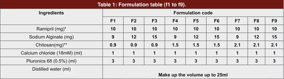
Table 1: Formulation table (f1 to f9).
Ingredients Formulation code
F1 F2 F3 F4 F5 F6 F7 F8 F9
Ramipril (mg)* 10 10 10 10 10 10 10 10 10
Sodium Alginate (mg) 9 12 15 9 12 15 9 12 15
Chitosan(mg)** 0.9 0.9 0.9 1.5 1.5 1.5 2.1 2.1 2.1
Calcium chloride (18mM) (ml) 1 1 1 1 1 1 1 1 1
Pluronics 68 (0.5%) (ml) 3 3 3 3 3 3 3 3 3
Distilled water (ml)
Make up the volume up to 25ml
0.05-0.08 (nearly-monodisperse), 0.08-0.7 (midrange polydispersity), > 0.7 (very polydisperse).11,12
Determinations of drug content
A quantity of drug loaded nanoparticles equivalent to
1mg was added to 10ml mixture of methanol and phos
-phate buffer pH 6.8 (1:10) and stirred continuously for 2 hr and then the final colloidal suspensions were ultra‐ centrifuged at 10000 rpm for half an hour. The super -natant was analysed for drug content by measuring the
absorbance at 207 nm using UV spectrophotometer.13
Entrapment efficiency
The encapsulation efficiency of nanoparticles was
determined by the separation of drug-loaded
Nanoparticles from the aqueous medium containing
non-associated Ramipril by ultracentrifugation at 12,000
rpm at 4°C for 1hr. The amount of Ramipril loaded into the nanoparticles was calculated as the difference between the total amount used to prepare the nanopar-ticles and the amount that was found in the superna-tant. The amount of free Ramipril in the supernatant
was measured by UV Spectrophotometer.14 Entrapment efficiency was then calculated as follows:
Entrapment efficiency was calculated by Eq.1
Percentage yield
Fixed volumes of Ramipril nanosuspension were cen-trifuged at 9000 rpm for 30 min at 15°C. The obtained sediment was dried and weighed.11,15 The percentage
yield was calculated by Eq.2
Shape and Surface Morphology
Transmission electron microscopy (TEM)
External morphology of Nanoparticles was determined using Transmission electron microscopy (TEM) at Pun-jab University, Chandigarh. Samples of Nanoparticles were prepared by placing one drop on a copper grid, without being stained before being examined using TEM.16
Zeta potential
The zeta potential value of optimized Ramipril loaded chitosan- alginate (CS-ALG) nanoparticle formulation was measured with the Zetasizer. To determine the zeta potential, optimized formulation was diluted with dou-ble-distilled water and placed in an electrophoretic cell.17
In vitro drug release
The release of Ramipril from nanoparticles was evalu-ated using USP type II paddle apparatus over 24 hr, dialysis membrane was loaded with nanoparticle
for-mulation containing 10 mg equivalent of drug, which was suspended initially for 2hrs in 900 ml of 0.1NHCl
buffer of pH 1.2 and then in pH 6.8 phosphate
buf-fer upto 24 hr maintained at 37±0.5°C and 50 rpm. At
regular intervals aliquots of 1 ml of the sample were withdrawn and replaced with the same volume of the respected fresh buffer solution. The amount of released
drug was assessed by UV-1700 analysis at 207 nm (Shi
-madzu UV-1700, Japan) after dilution.
In vivo studies
Healthy male wistar rats weighing 180-200 gm were
housed in polypropylene cages and maintained at room temperature under 12 hr dark/light cycles. They were fed with standard pelleted diet and water. The animals were acclimatized for one week under laboratory condi-tions before experiments on the animals. Ethical clear-ance was obtained from the Institutional Animal Ethics
Committee (Resolution No: KLECOP/IAEC/Res.20-09/08/2014) prior to the beginning of research work.
This study was aimed mainly to estimate the amount of drug in the blood withdrawn from rats at various time intervals.18 Dose for animal
Dose conversion formula from human dose to animal
dose (Rat)
The animals were divided into 2 groups
Group 1: Received pure drug Ramipril 1.08mg for 6 ani -mals in normal saline through oral route.
Group 2: Received nanoparticles containing equivalent
to 1.08 mg of Ramipril for 6 animals in normal saline
through oral route.
After 0.5, 1, 2, 4, 8, 12 and 24 hr 0.5-1ml blood was
collected from eye by retro-orbital puncture. Blood was
taken in Eppendorf containing 0.1ml of 4%w/v of sodium citrate solution and centrifuged at 5000rpm for 10 min supernatant plasma was collected and acetoni -trile was added for precipitating the plasma proteins and
vortexed for 1 min and then centrifuged at 13,000rpm
for 15 min and supernatant solution was collected and analyzed in HPLC.
HPLC method development for analysis of Ramipril in plasma samples
Instrument
A Shimadzu prominenece HPLC system (Shimedzu,
pump, DGU-20A5 online prominence degasser, SPD-M20A photodiode array detector, SIL-20AC HT Autos
-ampler, Rheodyne injection valve with 20μl loop and CTO-10ASVP-column oven. Software-LC solution V.1.25.
Chromatographic Conditions
Sheseido column C18 column 250 x 4.6mm, 5 μ diameter
was used for separation. The mobile phase containing
0.2% orthophosphoric acid: acetonitrile in the ratio of 50:50(v/v) was delivered at a flow rate 0.6 ml/min and the elution was monitored at 205 nm. Injection volume was 20μl. Pressure of 65kg/cm2 and temperature 28°C.
Preparation of 0.2% orthophosphoric acid
Measure 2000 μl of orthophosphoric acid in 1000 ml clean dry beaker and make up volume to 1000ml with
HPLC water.
Preparation of mobile phase
Mix a mixture of above solution i.e. 0.2% orthophos
-phoric acid 500ml (50%) and 500 ml of Acetonitrile HPLC (50%) and degas in ultrasonic water bath for 5 min. Filter through 0.45 μ filter under vacuum filtration.
Standard solutions and spiked samples
Accurately weigh and transfer 10 mg of Ramipril in 10ml clean dry volumetric flask add about 5ml. Aceto -nitrile, sonicate to dissolve it completely and make
vol-ume up to the mark with 0.2% orthophosphoric acid. Further pipette out 10 μl, 20 μl,30 μl,40 μl,50 μl and 60 μl of the above stock solution into a 2ml eppendorf
tube and dilute up to the mark with mobile phase to obtain workingstock solutions of concentration 5μg/ ml, 10 μg/ml,15 μg/ml, 20 μg/ml, 25 μg/ml and 30 μg/ml. Standard solutions (1 μg/ml,2 μg/ml, 3 μg/ml,4 μg/ml,5 μg/ml and 6 μg/ml) were prepared by spiking 200 μl of working solution(5μg/ml, 10 μg/ml,15 μg/ ml, 20 μg/ml, 25 μg/ml and 30 μg/ml) into 200 μl of
blank plasma and 600 μl acetonitrile. These standards
were used to construct calibration curve in plasma.
Plasma sample preparations
100 μl of plasma containing unknown concentration
of drug (withdrawn from rats at different time interval) were pipetted using micropipette into 2 ml eppendorf
tube. To this 200 μl of acetonitrile was added to pre -cipitate protein present in the plasma. This solution was capped and mixed by vortexing at high speed for 1min.
Next all tubes were centrifuged at 13,000 rpm for 15min
in eppendorf minispin centrifuge, After centrifugation
the upper organic layer was filtered through 0.2 μm
membranes into clean vials and a volume of 20 μl was
injected into HPLC system.
Short term stability studies
Optimized formulation was chosen to perform
short term stability studies. Samples were stored in glass vials for 3 months at 5±3°C in freeze and at
30±2°C/65±5%RH. After 30, 60 and 90 days samples were observed for particle size, % entrapment efficiency
and drug release were carried out for optimized formu-lation at every one month interval.19
RESULTS AND DISCUSSION
Preparation of chitosan–sodium alginate nanoparticles
The chitosan-alginate nanoparticles are successfully prepared by ionotropic gelation technique. Preparation is simple, rapid and reliable.A number of trials were carried out inorder to obtain appropriate concentration range of polymers so as to allow the formation of
tur-bid solutions and not the aggregates. The final concen
-tration range selected 0.03- 0.07% w/v and 0.06–0.1%
w/v for CS and ALG, respectively. Chitosan–alginate (CS–ALG) polyionic complexes are formed through the ionic gelation via interactions between the carboxyl groups of alginate and the amine groups of chitosan.5,8
The effect of pH on the nanoparticles formulation was
studied by Douglas and Tabrizian. It was found that size
of nanoparticle was smaller when alginate solution of pH 5.3 was combined with chitosan solution pH 5.5. This may be explained by the fact low solubility of chitosan in water at neutral and alkaline pH hence the
chitosan solution is prepared in acidic conditions. Due
Particle size and Size distribution
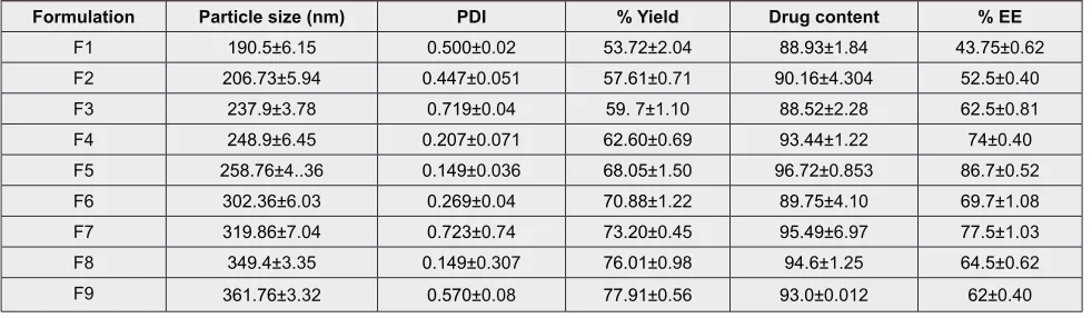
The mean particle size for formulations F1 to F9
var-ied in range of 190.5±6.15 to 361.76±3.32 (Table 2).
It was observed that mean particle size increases with the increase in the polymer concentration upto a level. Further increase in the polymer concentration above the concentration range mentioned resulted in the aggregation of the particles. The mean polydispersity index values for the Ramipril loaded chitosan alginate nanoparticle formulations F1 to F9 are in the range of
0.1499- 0.723 as shown in (Table 2). The results of PDI
can be simultaneously checked with particles size analy-sis. A monodisperse sample indicates PI value nearer to
0. However, PDI < 1 indicates polydisperse samples. Therefore, PI measurement was essential to confirm the
size distribution of the particles.8,9
Drug content
Drug content varies in the range of 88.52±2.28 to 96.72±0.853 and was determined using the UV spectro
-scopic analysis at 207 nm.
Entrapment efficiency
Encapsulation efficiency of the nanoparticles was found to vary between 43.75±0.62 and 86.7±0.52. Motwani
S K et al. suggested that at intermediate concentration of chitosan and sodium alginate the encapsulation of
drug was maximum. The percent entrapment efficiency
increases from F1 to F5 and then decreases from F6 to F9. This is because increase in the polymer concen-tration causes increase in the drug entrapment upto a level, further increase in the polymer concentration
leads to decrease in the entrapment efficiency which
can be explained on the basis of the fact that at higher concentrations of the two polymers, it is polymers that make the bulk of the nanoparticles matrix and less vol-ume is available for drug encapsulation. Formulation
F5 (86.7±0.52) show maximum entrapment efficiency.
Based on entrapment efficiency and drug content for -mulation F5 was taken as optimized for-mulation.3,9
Percentage yield
Percentage yield was found to be 53.72% to 77.91 % for formulation F1 to F9 (Table 2). Percentage practi-cal yield depends on the concentration of polymer added, as the concentration of polymer increases there is increases in the % yield. Maximum yield obtained is 77.91% for formulation F9.
TEM Analysis
Optimized formulation F5 containing chitosan (0.05%) and sodium alginate (0.08%) was subjected to TEM with different resolution. In the magnification of 10000×
particles were small, spherical (Figure 1) and discrete
while the same formulation at 30000× magnification
the surface was seen to be smooth and spherical with the size range of 245nm and 253nm.
Zeta potential
Zeta potential is an important parameter to analyze the long-term stability of the nanoparticles. It refers to the surface charge of the particles. Zeta potential of
nanoparticles is of significance on stability in suspen -sion through the electrostatic repul-sion between the particles.Zeta potential of the optimized formulation
F5 was found to be -32.2mV (Figure 2). The zeta poten
-tials of about -34.2 mV indicate good stability of for -mulation. This might be attributed to surfactant which decreases the electrostatic repulsion between the par-ticles and sterically stabilizes the nanoparpar-ticles by form-ing a coat around their surface.20
In vitro drug release
In vitro drug release from the nanoparticles was initially
carried out in 0.1N HCl buffer pH 1.2 for 2hrs fol -lowed by Phosphate buffer pH 6.8 upto 24 hr, it was Table 2: Particle size, pdi, drug content, % yield and % ee values of ramipril nanoparticles formulations F1 to
F9.
Formulation Particle size (nm) PDI % Yield Drug content % EE
F1 190.5±6.15 0.500±0.02 53.72±2.04 88.93±1.84 43.75±0.62
F2 206.73±5.94 0.447±0.051 57.61±0.71 90.16±4.304 52.5±0.40
F3 237.9±3.78 0.719±0.04 59. 7±1.10 88.52±2.28 62.5±0.81
F4 248.9±6.45 0.207±0.071 62.60±0.69 93.44±1.22 74±0.40
F5 258.76±4..36 0.149±0.036 68.05±1.50 96.72±0.853 86.7±0.52
F6 302.36±6.03 0.269±0.04 70.88±1.22 89.75±4.10 69.7±1.08
F7 319.86±7.04 0.723±0.74 73.20±0.45 95.49±6.97 77.5±1.03
F8 349.4±3.35 0.149±0.307 76.01±0.98 94.6±1.25 64.5±0.62
F9 361.76±3.32 0.570±0.08 77.91±0.56 93.0±0.012 62±0.40
Table 3: Pharmacokinetic parameter of pure ramipril and optimized formulation f5.
Formulations Cmax (mcg/
ml) Tmax (h) AUCml.h)0-t (mcg/
Pure Ramipril 7.9768 0.5hrs 114.874 Optimized
formulation F5 12.482 8hrs 249.77
Figure 1: TEM of optimized formulation F5.
Figure 2: Zetapotential of optimized formulation (F5).
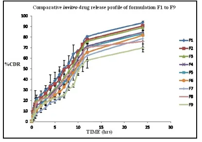
Figure 3: Comparative in vitro drug release profile of F1 to F9.
Figure 4: Comparative in vitro drug release profile of opti-mized formulation F5 and pure drug.
polymer concentration the drug will take longer time to diffuse out.
The initial release of the drug at the end of 2 hr may be due to the drug present on the surface of Nanopar-ticles. Whereas the drug which is present in the matrix inside the nanoparticles have additional barrier due to the polyelectrolyte complex which remains intact at acidic pH and when the nanoparticles are exposed to phosphate buffer pH 6.8 the polyelectrolyte complex weaken and there is slow release of drug for a prolong period of time.21
Evaluation of the release profiles of pure drug showed
that almost all the Ramipril was released
immedi-ately during first 4 hrs, suggesting that the developed
nanoparticles can be used as an important platform for sustained drug release. (Figure 4)
Among the models tested, the drug release profiles for formulations (F1 to F9) were best fitted with Kors
-meyer- Peppas model based on regression coefficients (0.9923, 0.9890, 0.9810, 0.9856, 0.9888, 0.9890, 0.9843, 0.9828, 0.9827 respectively) and n being greater 0.5 sug -gesting non-Fickian diffusion process.12
In vivo studies
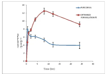
The study was carried out on male wistar albino rats to compare plasma concentration of Ramipril nanopar-ticles (optimised formulation F5) with that of pure drug given orally in normal saline.
In-vivo study revealed that formulation F5 showed greater bioavailability than that of the pure drug. The performed using dialysis bag diffusion technique. The
drug release at the end of 2 hrs in 0.1N HCl pH 1.2 for F1-25.14%, F2-22.25%, F3-20.71%, F4-17.74%, F5-16.2%, F6-14.7%, F7-10.35%, F8-20.7%, F9-14.7%
and at the end of 24 hrs the mean cumulative drug release
was F1-93.63%, F2-90.6%, F3-89.12%, F4-84.6%,
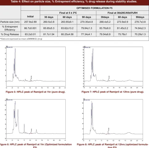
Table 4: Effect on particle size, % Entrapment efficiency, % drug release during stability studies.
OPTIMISED FORMULATION F5
Initial
Final at 5 ± 3ºC Final at 30±20C/65±5%RH
30 days 60 days 90 days 30days 60 days 90days
Particle size (nm) 257.9±2.89 260.5±3.8 263.65±9.1 270.35±4.8 268.4±5.2 273.9±0.9 278.7±3.8 % Entrapment
Efficiency 86.7±0.001 85.65±5.3 83.62±13.2 79.84±1.3 83.76±8.0 81.45±3.2 74.84±2.3
% Drug Release 83.2±0.01 81.7±1.54 80.25±4.58 77.34±4.1 79.54±6.8 75.78±1 70.29±1.3 *Data are expressed as mean ±SWWW.D. (n=3)
Figure 5: HPLC peak of Ramipril at 1hr (pure drug).
Figure 6: HPLC peak of Ramipril at 1hr (Optimized formulation F5).
Figure 7: HPLC peak of Ramipril at 12hrs (pure drug).
Figure 8: HPLC peak of Ramipril at 12hrs (optimized formula-tion F5).
HPLC peaks of pure Ramipril and optimized formu-lation F5 at 1hr and at 12 hr are shown in Figure 5, Figure 6, Figure 7 and Figure 8 respectively. AUC of 249.77 µg /ml hr, Cmax of 12.48 µg and Tmax of 8 hr was observed for formulation F5 given orally whereas pure drug showed AUC of 114.874 µg/ml hr, Cmax of 7.95 µg and Tmax 0.5hr calculated by applying trapezoidal
method. (Table 3)
AUC°-t of pure drug 114.874 µg/ml hr was increased up to 249.77µg /ml hr by Ramipril nanoparticle formula-tion through oral route. Figure 9 shows the comparative
in vivo release profile of optimized formulation F5 and
pure drug (Ramipril) after oral administration. Ramipril
nanoparticles lead to enhancement of oral bioavailabil-ity by 2.17 times than that of pure drug.22
Short term stability studies
Stability studies were carried out on the optimized
for-mulation F5 as per ICH guidelines for 90 days. By
com-paring this data with initial data it was observed that there was an slight decrease in the percentage
entrap-ment efficiency and increase in particles size due to
bet-Figure 9: Comparative in vivo release profile of optimized for-mulation F5 and pure drug (Ramipril) by oral administration.
ter stability as compared to the formulation stored at
30±2°C/65±5%RH.
Ramipril chitosan- alginate nanoparticles can be suc-cessfully prepared by ionotropic pregelation technique. In vitro release study showed that chitosan- alginate nanoparticles showed pH dependent and sustained release of drug for a prolong period of time. The oral bioavailability of Ramipril when formulated into pH sensitive chitosan alginate nanoparticles was improved by 2.17 times the pure drug.
ACKNOWLEDGEMENT
The authors would sincerely like to thank Unichem Lab-oratories, Goa for providing gift samples of Ramipril and Central Institute of Fisheries Cochin, Kerala for providing chitosan.
The authors are also grateful to
KLE University’s Dr. Prabhakar Kore Basic
Sci-ence Research Centre, Belgaum for providing the
laboratory facilities.
CONFLICT OF INTEREST
The authors declare no conflict of interest.
ABBREVIATIONS
ACE: Angiotensin-Converting Enzyme inhibitor;
TEM: Transmission electron microscopy; HPLC: High performance liquid chromatography; ALG: Algi-nate; CS: Chitosan; PDI: Polydispersity index.
REFERENCES
1. Ekambaram P, Abdul HSA. Formulation and Evaluation of Solid Lipid Nanoparticles of Ramipril. Journal of Young Pharmacists. 2011;3(3):216-20. 2. Venkateswara R, Navaneetha K. Formulation and evaluation of sustain
release tablets of Ramipril. Pharmatutor. 2014;2(7):110-9.
3. Motwani S, Chopra S, Talegaonkar S, Kohli K, Farhan JM. Chitosan–sodium alginate nanoparticles as submicroscopic reservoirs for ocular delivery:
Formulation, optimization and in vitro characterization. European Journal of Pharmaceutics and Biopharmaceutics. 2008;68(3):513-25.
4. Ranjit K, Abdul B. Nanoparticles: An overview preparation, characterization and applications. International Research Journal of Pharmacy. 2013;4(4):48-57.
5. Ping L, Ya-Ni D, Jun-Ping Z, Ai-Qin W, Qin W. Chitosan–Alginate Nanoparticles as a novel drug delivery system for Nifedipine. International Journal of Biomedical Science. 2008;4(3):221-8.
6. Tarane G, Mohammad R, Ebrahim A, Parisa Y, Alireza N, et al. Evaluation of Alginate/Chitosan nanoparticles as antisense delivery vector: Formulation, optimization and in vitro characterization. Carbohydrate Polymers. 2009;77(3):599-606.
7. Naser M, Azam B, Ali E, Heydari M. Preparation of chitosan nanoparticles containing Naja Naja Oxiana snake venom. Nanomedicine: Nanotechnology, Biology and Medicine. 2010;6(1):137-43.
8. Sangeetha S, Deepika K, Thrishala B, Chaitanya CH, Harish G, Damodharan N. Formulation and in vitro evaluation of sodium alginate nanospheres containing Ofloxacin. International Journal Applied Pharmaceutical Sciences; 2010;2(4):1-3.
9. Nagarwal RC, Kumar R, Pandit JK. Chitosan coated sodium alginate– chitosan nanoparticles loaded with 5-FU for ocular delivery: In vitro characterization and in vivo study in rabbit eye. European Journal of Pharmaceutical Sciences. 2012;47(4):678-85.
10. Kumari DC, Tharani CB, Narayanan N, Kumar S. Formulation and characterization of Methotrexate loaded sodium alginate chitosan Nanoparticles. Indian Journal of Research in Pharmacy and Biotechnology. 2015;1(6):915-21.
11. Dandagi P, Rath SP, GadadAP, Mastiholimath V. Taste masked Quinine sulphate loaded solid lipid nanoparticles for flexible pediatric dosing. Indian Journal of Pharmaceutical Education and Research. 2014;48:93-9. 12. Prajapati MD, Kumar GS, Goli D, Maulik P, Shah H. Design, development
and evaluation of nanoparticles of poorly soluble Valsartan. World Journal of Pharmacy and Pharmaceutical Sciences. 2013;2(6):5737-49.
13. Tamizhrasi S, Shukla A, Shivkumar T, Rathi V, Rathi J. Formulation And Evaluation of Lamivudine Loaded Polymethacrylic Acid Nanoparticles. International Journal of Pharmtech Research. 2009;1(3):411-5.
14. Kaur S, Rao R, Hussain A, Khatkar S. Preparation and Characterization of Rivastigmine Loaded Chitosan Nanoparticles. Journal of Pharmaceutical Sciences and Research. 2011;3(5):1227-32.
15. Jain S, Awasthi AM, Jain NK, Agrawal GP. Calcium silicate based microspheres of repaglinide for gastroretentive floating drug delivery: Preparation and in vitro characterization. Journal of Controlled Release. 2005;107(2):300-9.
16. Selvakumar K, Yadav AV. Formulation and Evaluation of Carvedilol loaded Eudragite 100 Nanoparticles. International Journal of Pharm Tech Research. 2009;1(2):179-83.
17. Sanyog J, Amit KJ, Nitin KS, Chandraiah G, Raman PS. The effect of the
oral administration of polymeric nanoparticles on the efficacy and toxicity of
tamoxifen. Biomaterials. 2011;32(2):503-15.
18. Sheikh S, Faiyaz S, Talegaonkar S, Farhan J, Mushir A. Development and bioavailability assessment of Ramipril nanoemulsion formulation. European Journal of Pharmaceutics and Biopharmaceutics. 2007;66(2):227-43. 19. Peng L, Xinyu R, Johanna L, Bert-Van V, Juha K, Jouni H, et al.
Nanosuspensions of poorly soluble drugs: Preparation and development by wet milling. International Journal of Pharmaceutics. 2014;11:215-22. 20. Fei Y, Chao Z, Yi Zheng, Lin M, Lina T. The effect of poloxamer 188
on nanoparticle morphology, size, cancer cell uptake and cytotoxicity. Nanomedicine: Nanotechnology, Biology and Medicine. 2010;6(1):170-8. 21. Saleem V, Azharuddin SK, Ali S, Patil C. Studies on different chitosan
polyelectrolyte complex hydrogels for modified release of Diltiazem Hydrochloride. International Journal of Pharmacy and Pharmaceutical Sciences. 2010;2(4):64-7.
Cite this article: Pereira A, Gadad AP, Patil AS, Dandagi PM. Development and Bioavailability Assessment of Ramipril Nanoparticle Formulation. Indian J of Pharmaceutical Education and Research. 2019;54(4s):s587-s595.
• A total of nine formulations (F1-F9) were for-mulated by varying the concentration of chitosan
(0.003%-0.07%) and sodium alginate (0.06-0.1%)
using ionotropic pregelation technique and the various parameters were evaluated.
• The size of nanoparticles were in nanosize range, spherical and discrete, particle size increase with increase in polymer concentration. Entrapment
efficiency increased with increasing the polymer
concentration to certain level and then decreased. • F5 formulation was considered as optimized
for-mulation based on higher entrapment efficiency, drug content and good Zeta potential of-32.4mV.
• In vitro release study showed initial burst effect, this may be due to drug present on the surface of nanoparticles followed by sustain release of drug. • The oral bioavailability of Ramipril when formu-lated into pH sensitive CS-ALG nanoparticles was improved by 2.17 times more than that of pure drug. From the stability studies, it can be found
that 5 ± 30 0C is the ideal temperature for storage
of nanoparticles.
SUMMARY
ABOUT AUTHORS
Anand Panchakshari Gadad: Is a Professor and Head, Department of Pharmaceutics, Col -lege of Pharmacy, K.L.E Academy of Higher Education and Research, Belagavi. He is working on areas of targeted drug delivery system viz., Gastroretentive drug delivery system, Polymeric nanoparticles, enhancing solubility of poorly soluble drugs, etc.
PICTORIAL ABSTRACT
Panchaxari Mallappa Dandagi: Is a Professor, Department of pharmaceutics, KLE Univer -sity’s College of Pharmacy, K.L.E Academy of Higher Education and Research, Belagavi. He is working on areas of targeted drug delivery system viz., Colon drug delivery system using pH
dependent polymers and also Ocular drug delivery system through Nano and microspheres.
Archana S. Patil: Is an Assistan Professor, Department of pharmaceutics, KLE University’s