RESEARCH ARTICLE
Promising upshot of silver nanoparticles
primed from
Gracilaria crassa
against bacterial
pathogens
V Lavakumar
1, K Masilamani
2, V Ravichandiran
3, N Venkateshan
4, D V R Saigopal
5, C K Ashok Kumar
1and C Sowmya
6*Abstract
Background: The study on newer antimicrobial agent from metal based nano materials has augmented in recent
years for the management of multidrug resistance microorganisms. In our present investigation, we synthesized silver
nanoparticles (AgNP’s) from red algae,
Gracilaria crassa
as beginning material which effectively condensed the silver
ions to silver nanoparticles with less price tag and no risk.
Methods: Silver nanoparticles were prepared by simple reaction of 1 mM AgNO
3with
G. crassa
extracts at room
temperature. The fabricated AgNP’s were subjected for characterization and screened against various microorganisms
for antibacterial activity.
Results: UV–Vis spectroscopy (200–800 nm), XRD, FESEM and EDAX, were performed for AgNP’s. UV–Vis spectroscopy
demonstrated the absorption edge at 443 nm and EDAX pattern is purely due to the particle size and face centered
cubic (fcc) symmetry of nanoparticles. Average size lays at 122.7 nm and zeta potential was found to be
−
34.9 mV.
The antibacterial outcome of synthesized AgNP’s (at the dose of 20 and 40 µg/ml) was evaluated against
Escherichia
coli
,
Proteus mirabilis
,
Bacillus subtilis
and
Pseudomonas aeruginosa
. The mechanism of synthesized AgNP’s bactericidal
bustle is discussed in terms of interaction with the cell membrane of bacteria. The activity was found to be
sky-scrap-ing in a dose dependent manner.
Conclusion: Thus, environmental friendly, cost effective, non hazardous stable nanoparticles were prepared by green
synthesis using red algae,
G. crassa
. Synthesized
G. crassa
AgNP’s were in acceptable size and shape. Further, it elicits
better bactericidal activity against microorganism. This will assure the out put of superior antibacterial formulation for
near future.
Keywords: Green synthesis, Red algae,
Gracilaria crassa
, Silver nanoparticles, Antibacterial activity
© 2015 Lavakumar et al. This article is distributed under the terms of the Creative Commons Attribution 4.0 International License (http://creativecommons.org/licenses/by/4.0/), which permits unrestricted use, distribution, and reproduction in any medium, provided you give appropriate credit to the original author(s) and the source, provide a link to the Creative Commons license, and indicate if changes were made. The Creative Commons Public Domain Dedication waiver (http://creativecommons.org/ publicdomain/zero/1.0/) applies to the data made available in this article, unless otherwise stated.
Background
From ancient, handling of microbial infection is an
exi-gent task for microbiologists. Countless drugs have been
found to be successful, unfortunately, it leads to mount
of drug resistance against particular pathogens with an
outlook of stern issues in concern with public wellbeing
[
1
]. Technical community is animatedly trying to expand
groundbreaking concepts in drug delivery by
challeng-ing the new microbial agent with superior mode of action
by its effectual target on the cell membrane or neither
on intracellular targets [
2
,
3
]. In 21st century,
nanotech-nology has become inevitable, not because of only claim
and also by the way of synthesis. Two way synthesis like
physical and chemical methods have several sizeable
challenges like technically protracted, expensive and
eco-logical hazardous defects [
4
]. The current art of
explo-ration in research is heading towards green synthesis of
high-yield, squat in cost, non-hazardous and eco-friendly
metallic nanoparticles by plants and microorganisms [
5
,
Open Access
*Correspondence: [email protected]
6 Department of Pharmaceutics, Raghavendra Institute of Pharmaceutical Education and Research, Anantapuram 515721, AP, India
6
]. Due to hefty surface area, high reactivity and surface
plasmon resonance, these nanoparticles were tailored
for definite application by scheming into unambiguous
size, shape and morphology leading to exhibit its broad
spectrum of activity against multi drug resistance
micro-organisms [
7
–
10
]. A quest for an environmentally
sus-tainable synthesis process has led to a few biomimetic
approaches like applying natal principles in the
forma-tion of nanoparticles. Among several, bioreducforma-tion is the
prime and widely practiced functional method in
synthe-sizing the nano materials [
11
,
12
]. Noble metal
nanopar-ticles such as silver, gold, which are geared up by plant
extracts, algae, bacteria and fungi are broadly applied in
drug delivery systems [
13
], electronics [
14
,
15
],
biosen-sors [
16
], cancer therapeutics and antimicrobial agents
[
17
–
19
]. For three decades, exploration of marine algae
has been far above the ground for the search of new and
effective natural origin medicines, because it posses
ele-vated quantity of concealed bioactive essence. Several
of such compounds, including carbohydrates, alkaloids,
steroids, phenols, saponins and flavonoids, etc. [
20
–
22
].
These multifaceted compounds exhibit a wide range of
industrial and biotechnological applications [
23
]. An
extent, these bio-molecules play decisive role in
reduc-tion of metal ions and generate the stable eco friendly
nanoparticles. Further, AgNP’s fetches superior
antibac-terial activity by interacting with thiol clusters present in
bacterial cell by cliping its replication. Literature
strap-pingly supports that these Ag
+ions unyoke the
respira-tory chains and collapse the proton motive forces across
the cytoplasmic membrane of bacteria [
24
].
Gracilaria
crassa
(
G. crassa
), a well-known red algae, having
poten-tial secondary metabolites [
25
]. In harmony to the above
information, the present study was intended to prepare
and typify silver nanoparticles from
G. crassa
, further to
explore its antimicrobial activities against highly
resist-ance microbial inhabitants.
Methods
Materials
Silver nitrate (AgNO
3; Mol. Wt: 169.87; Prod. No: 27462)
of analytical grade (AR) was purchased from Fisher
sci-entific, Mumbai, India. The nutrient agar medium was
purchased from Hi Media (Mumbai, India). All other
chemicals used were analytical grade. Microorganisms
such as
Escherichia coli
(
MTCC 443
),
Proteus mirabilis
(
MTCC 442
),
Bacillus subtilis
(
MTCC 441
) and
Pseu-domonas aeruginosa
(
MTCC 424
) were obtained from
Microbial Type Culture and Collection, Pune, India.
Seaweed collection and extraction
Gracilaria crassa
was collected along the coast of
Man-dapam (9°16
′
58.9
″
N 79°18
′
53.6
″
E), Rameswaram,
Tamilnadu, India. The freshly collected algal material was
rinsed with seawater followed by deionized water to get
rid of extra impurities. The samples were kept in shade
for 15 days drying. The algal material was ground to
pow-der and uphold stockpile by placing at
−
4°C for further
studies. The
G. crassa
extract was prepared in a conical
flask by taking 2.5 g in 100 mL of deionized water. It was
heated for 45 min at 50°C and positioned in an orbital
shaker for 24 h in order to conquer the maximum
extrac-tion of compounds. The Extracts were filtered through
whatman No. 1 filter paper and stored in refrigerator for
further studies [
26
].
Synthesis of silver nanoparticles
AgNP’s were synthesized by adopting the method
pro-posed by Sathishkumar et al. with simple modification
[
27
]. 5 ml of algal extract was added in to 95 ml of 1 mM
aqueous silver nitrate solution, in 250 ml conical flasks
and kept at 30°C in a shaker for overnight to facilitate
absolute reduction. The samples were monitored
peri-odically for its color intensity to confirm the formation of
AgNP’s.
Characterization of silver nanoparticles
UV–Vis spectral analysis
The reduction in pure silver ions was recorded by
meas-uring the UV–Vis spectrum of the synthesized AgNP’s
of
G. crassa
at room temperature with a Perkin Elmer
Lambda 25 UV–Vis spectrometer at the wavelength of
200–800 nm [
28
].
Particle size and zeta potential studies
Particle size and zeta potential experiments for
G. crassa
AgNP’s were carried out by using a HORIBA Instruments
(Singapore) Pvt Ltd, Singapore.
Powder x‑ray diffraction (XRD) analysis
The silver nanoparticles were alienated by repeated
cen-trifugation at 12,000 rpm for 10 min followed by
redis-persion of AgNP’s into deionized water for three times.
The supernatant was transferred in microwave for
dry-ing. X-ray diffraction (XRD) measurement of the AgNP’s
was carried out using Rigaku smart lab instrument
(Japan), function at voltage of 40 kV, 30 mA current with
Cu K
α
1 radiations.
FESEM‑EDAX studies
Antibacterial assay
The antibacterial evaluation of AgNP’s was conceded by
using different experimental pathogens like
Escherichia
coli
(
MTCC 443
),
Proteus mirabilis
(
MTCC 442
),
Bacillus
subtilis
(
MTCC 441
) and
Pseudomonas aeruginosa
(
MTCC
424
) maintained by department of Pharmaceutical
Biotech-nology, SVCP, Tirupati, India. Prior to experimentation,
untainted cultures was subcultured into nutrient media.
Nutrient agar plates were prepared, seeded and pierced
with 20 and 40 µg/ml of AgNP’s. Streptomycin sulphate
(20 µg/ml) was used as standard [
29
].
Results and discussion
UV–Vis spectral analysis
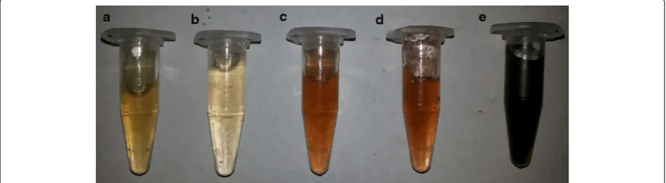
The formation of AgNP’s was defined by color
trans-formation [
30
] from pale yellow to dark brown (Fig.
1
).
The color change is due to the excitation of the surface
plasmon resonance (SPR) (Fig.
2
) which elicits
λ
max of
443 nm.
Particle size and zeta potential
Particle size determination of synthesized AgNP’s was
revealed underneath by intensity. Laser diffractions
exposed by obtained AgNP’s were in polydisperse
con-coction with average size of 122.7 nm (Fig.
3
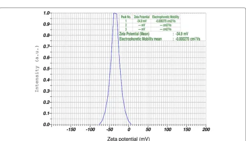
). The zeta
potential endows stability of nanoparticles and surface
charge. The zeta potential was found to be
−
34.9 mV.
Earlier reports strongly supports, when the zeta
poten-tial value positions between
−
30 and
−
50 mV, it
speci-fies good stability of nano particles [
31
]. The high
negative value (Fig.
4
) substantiates the repulsion
between the particles and thereby increases the stability
of the AgNP’s.
X‑ray diffraction (XRD) analysis
The XRD pattern of powder sample of
G. crassa
AgNP’s
exhibited peaks at 38°, 44°, 64° and 77°. Four Bragg’s
reflections corresponding to (111), (200), (220) and
(311) planes of the fcc crystal structures of metallic
sil-ver (JCPDS No. 89-3722) are interpreted from the XRD
(Fig.
5
). The orientation (111) is more predominant since
it shows high intense. Broadening of the diffraction crest
disclose the formation of pure crystalline silver [
32
].
FESEM and energy dispersive X‑ray analysis
Field emission scanning electron microscopy
investiga-tion was further confirmed the size of silver
nanoparti-cles synthesized from
G. crassa
. The size (diameter) of
the nanoparticles ranges between 60 and 200 nm and the
shapes were spherical and some are irregular (Fig.
6
). The
outcome of FESEM reports were overlapped with
ear-lier reports [
33
,
34
]. The energy dispersive X-ray
analy-sis (EDAX) depicts strong indication in the silver region,
which authenticate formation of silver nanoparticles
Fig. 1 Formation of silver nanoparticles by green synthesis. AgNP’s were formed by the reduction of silver ions by Gracilaria crassa. This figure illustrates the various stages of formation of AgNP’s; a pure algal extracts (pale yellow); b at 0 min, on immediate addition of 1 mM solution of Silver nitrate (no reaction); c after 15 min (slight reduction); d after 30 min (moderate reduction); e after 12 h of addition of 1 mM solution of Silver nitrate (Complete reduction).
Fig. 3 Particles size distribution of AgNP’s prepared from G. crassa extracts. Average particle size of synthesized AgNP’s ranges from 60 to 200 nm
(Fig.
7
). The optical absorption peak at 3 kV, which
attrib-uted to metallic silver nanocrystallites owing to surface
plasmon resonance [
35
,
36
].
Antibacterial assay
Antibacterial activity by agar well diffusion technique
was recorded after 24 h incubation of culture plates.
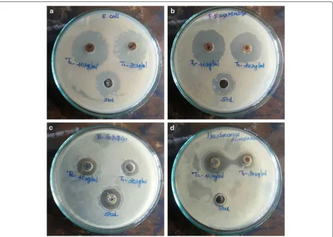
The AgNP’s demonstrate excellent antibacterial activity
against all tested microorganisms (Fig.
8
). AgNP’s showed
high spectrum of activity against
E. coli
and
P. mirabilis
at the concentrations of 20 and 40 µg/ml when compared
with standard (Fig.
9
). The significant zone of
inhibi-tion was exerted due to effect of AgNP’s on biochemical
process of the bacterial cell by interacting thiol and amino
groups of proteins and nucleic acids of cell wall [
37
–
39
].
Further, this could lead to interaction between
nanopar-ticles and microorganism which results in triggering the
discharge of highly reactive oxygen species (ROS), mostly
hydroxyl radicals and singlet oxygen [
40
–
42
]. This
aug-ments the deposition of nanoparticles on the bacterial cell
surface results large accumulation of silver nanoparticles
causing disruption of cellular functions.
Conclusion
In summary, the bio-reduction of aqueous silver ions to
silver nanoparticles (AgNP’s) was successfully done using
marine red algae,
G. crassa
in trouble-free, economy
and ecofriendly manner. The average size of silver
nano-particles was found to be 122.7 nm with high stability of
−
34.9 mV. Further characterization by UV–Vis
spectros-copy, FESEM, EDAX confirm formation nanoparticles
which are virtually spherical in shape. XRD divulges fcc
structural confirmation. The proved antibacterial
poten-tial will lend a hand to develop a powerful
antibacte-rial formulation in near future as biomedical remedies.
Hence, authors robustly propose this green synthesis
of nanoparticles can be extended to the wide range of
applications.
Fig. 5 X-ray diffraction analysis of bio synthesized silver nanoparticles using marine red algae extracts of G. crassa corresponds four Bragg’s reflections planes of the metallic silver nanoparticles.
Fig. 6 Morphological images of synthesized AgNP’s by using FESEM. This figure shows individual nanoparticles in clusters, spherical and some with irregular texture.
Abbreviations
AgNP’s: silver nanoparticles; FESEM: field emission scanning electron microscopy; EDAX: energy dispersive analysis of X-rays; XRD: X-ray diffraction; Fcc: face centered cubic; Cu: copper; mV: millivolts; SPR: surface plasmon resonance; kV: kilovolts; mM: millimolar; nm: nanometer; ROS: reactive oxygen species; MTCC: microbial type culture and collection.
Author’s contributions
LKV and SC are designed, performed and wrote the manuscript. MK contrib-uted for instrumentation. RV & VN helped in interpretation of FESEM and XRD. AK & SG were helped in screening antimicrobial activity. All authors read and approved the final manuscript.
Author details
1 Department of Pharmaceutical Biotechnology, Sree Vidhyanikethan College of Pharmacy, A.Rangampet, Tirupati 517102, AP, India. 2 Faculty of Technology, University Malaysia Pahang, Lebuhraya Tun Razak, Gambang, 26300 Kuantan, Pahang Darul Makmur, Malaysia. 3 National Institute of Pharmaceutical Educa-tion and Research, Kolkata 700032, WB, India. 4 Department of Pharmaceutical Chemistry, Sankarlingam Bhuvaneswari College of Pharmacy, Sivakasi 626130, TN, India. 5 Department of Virology, S.V. University, Tirupati 517502, AP, India. 6 Department of Pharmaceutics, Raghavendra Institute of Pharmaceutical Education and Research, Anantapuram 515721, AP, India.
Fig. 8 Antibacterial activity of AgNP’s. Zone of inhibition of silver nanoparticles against aEscherichia coli, bProteus mirabilis, cBacillus subtilis, d
Pseudomonas aeruginosa; [T1-(20 µg/ml of AgNP’s) and T2 (40 µg/ml of AgNP’s); Standard (Std-Streptomycin sulphate 20 µg/ml)].
Acknowledgements
The authors are grateful to T.N.K.V. Prasad, Institute of Frontier Technology, Regional Agricultural Research Station, Acharya N.G. Ranga Agricultural University, Tirupati for helping us to carry out particle size analysis. The authors also thankful to Center for nanotechnology, Sathyabhama University, Chennai for carrying out FESEM and XRD studies. The authors extended thanks to Sree Vidyanikethan College of Pharmacy, A.Rangampet, Tirupathi for providing facilities to carry out this work.
Compliance with ethical guidelines
Competing interests
The authors declare that they have no competing interests.
Received: 6 March 2015 Accepted: 16 July 2015
References
1. Rezai K, Weinstein RA (2010) Antimicrobial resistance-beyond the break-point: reducing antimicrobial-resistant infections in health care setting: what works. Issues Infect Dis 6:89–101
2. Sosa IO, Noguez C, Barrera RG (2003) Optical properties of metal nano-particles with arbitrary shapes. J Phys Chem B 107:6269–6275 3. Xie J, Lee JY, Wang DIC, Ting YP (2007) Silver nanoplates: from biological
to biomimetic synthesis. ACS Nano 1:429–439
4. Ahmad N, Sharma S, Md Alam K, Singh VN, Shamsi SF, Mehta BR et al (2010) Rapid synthesis of silver nanoparticles using dried medicinal plant of basil. Colloids Surf B Biointerfaces 81:81–86
5. Mandal D, Bolander ME, Mukhopadhyay D, Sarkar G, Mukherjee P (2006) The use of microorganisms for the formation of metal nanoparticles and their application. Appl Microbiol Biotechnol 69:485–492
6. Gardea-Torresdey JL, Tiemann KJ, Gomez E, Dokken K, Tehuacanero S, Jose-Yacaman M (1999) Gold nanoparticles obtained by bio -precipita-tion from gold (III) solu-precipita-tions. J Nanopart Res 1:397–404
7. Krutyakov YA, Kudrynskiy AA, Olenin AY, Lisichkin GV (2008) Synthesis and properties of silver nanoparticles: advances and prospects. Russ Chem Rev 77:233–257
8. Alivisatos AP (1996) Semiconductor clusters, nanocrystals, and quantum dots. Science 271:933–937
9. Mubarakali D, Thajuddin N, Jeganathan K, Gunasekharan M (2011) Plant extract mediated synthesis of silver and gold nanoparticles and its antibacterial activity against clinically isolated pathogens. Colloids Surf B Biointerfaces 85:360–365
10. Kelly KL, Coronado E, Zhaoand LI, Schatz GC (2003) The optical properties of metal nanoparticles: the influence of size, shape, and dielectric envi-ronment. J Phys Chem B 107:668–677
11. Song JY, Kim BS (2009) Rapid biological synthesis of silver nanoparticles using plant leaf extracts. Bioprocess Biosyst Eng 32:79–84
12. Faramarzi MA, Sadighi A (2013) Insights into biogenic and chemical production of inorganic nanomaterials and nanostructures. Adv Colloids Interface Sci 189–190:1–20
13. Perugini P, Si Simeon, Scalia S, Genta I, Modena T (2002) Effect of nano-particle encapsulation on the photostability of the sunscreen agent, 2-ethylhexyl-p-methoxycinnamate. Int J Pharm 246:37–45
14. Asharani PV, Lian Wu Yi, Gong Z, Valiyaveettil S (2008) Toxicity of silver nanoparticles in zebrafish models. Nanotechnology 19:255102 15. Jin S, Ye K (2007) Nanoparticle-mediated drug delivery and gene therapy.
Biotechnol Prog 23:32–41
16. Prow T, Grebe R, Merges C, Smith JN, McLeod DS, Leary JF et al (2006) Nanoparticle tethered antioxidant response element as a biosensor for oxygen induced toxicity in retinal endothelial cells. Mol Vision 12:616–625 17. Salata OV (2004) Applications of nanoparticles in biology and medicine. J
Nanobiotechnol 2:3
18. Husseiny MI, El-Aziz MA, Badr Y, Mahmoud MA (2007) Biosynthesis of gold nanoparticles using Pseudomonas aeruginosa. Spectrochim Acta A Mol Biomol Spectrosc 67:1003–1006
19. Gangadharan D, Harshvardan K, Gnanasekar G, Dixit D, Popat KM, Anand PS (2010) Polymeric microspheres containing silver nanoparticles as a bactericidal agent for water disinfection. J Water Res 44:5481–5487 20. Suriya J, Bharathi Raja S, Sekar V, Rajasekaran R (2012) Biosynthesis of
silver nanoparticles and its antibacterial activity using seaweed Urospora sp. Afr J Biotechnol 11:12192–12198
21. Kumar P, Senthamilselvi S, Lakshmiprabha A, Premkumar K, Ganeshkumar RS, Govindaraju M (2012) Synthesis of silver nanoparticles from Sargas-sum tenerrimum and screening phytochemicals for its antibacterial activ-ity. Nano Biomed Eng 4:12–16
22. Thangaraju N, Venkatalakshmi RP, Chinnasamy A, Kannaiyan P (2012) Synthesis of silver nanoparticles and the antibacterial and anticancer activities of the crude extract of Sargassum polycystum C. Agardh Nano Biomed Eng 4:89–94
23. Holt KB, Bard AJ (2005) Interaction of silver (I) ions with the respiratory chain of Escherichia coli: an electrochemical and scanning electrochemi-cal microscopy study of the antimicrobial mechanism of micromolar Ag+. Biochem 44:13214–13223
24. Renn D (1997) Biotechnology and the red seaweed polysaccharide indus-try: status, needs and prospects. Trends Biotechnol 15:9–14
25. De almeida CLF, Falcao DS, Lima DM, Gedson R, Montenegro DA, Lira NS et al (2011) Bioactivities from marine algae of the genus gracilaria. Int J Mol Sci 12:4550–4573
26. Das J, Das MP, Velusamy P (2014) Sesbania grandiflora leaf extract mediated green synthesis of antibacterial silver nanoaprticles against selected human pathogens. Spectrochim Acta A Mol Biomol Spectrosc 104:265–270
27. Sathishkumar G, Gobinath C, Karpagam K, Hemamalini V, Premkumar K, Sivaramakrishnan S (2012) Phyto-synthesis of silver nanoscale particles using Morinda citrifolia L. and its inhibitory activity against human patho-gens. Colloids Surf B Biointerface 15:235–240
28. Mallikarjuna K, Narasimha G, Dillip GR, Praveen B, Shreedhar B, Sree lak-shmi C et al (2011) Green synthesis of silver nanoparticles using Ocimum leaf extract and their characterization. Dig J Nanometer Bios 6:81–186 29. Shahverdi AR, Fakhimi A, Shahverdi HR, Minaian S (2007) Synthesis and
effect of silver nanoparticles on the anti bacterial activity of different antibiotics against Staphylococcus aureus and Escherichia coli. Nanomed 3:168–171
30. Sastry M, Mayyaa KS, Bandyopadhyay K (1997) pH dependent changes in the optical properties of carboxylic acid derivatized silver colloidal particles. Colloids Surf A Phy Che Eng Aspects 127:221–228
31. White B, Banerjee S, O Brien S, Turro NJ, Herman IP (2007) Zeta-potential measurements of surfactant-wrapped individual single-walled carbon nanotubes. J Phy Chem C 111:13684–13690
32. Baker C, Pradhan A, Pakstis L, Pochan DJ, Shah SI (2005) Synthesis and antibacterial properties of silver nanoparticles. J Nanosci Nanotech 5:244–249
33. Ahmad N, Sharma S, Rai R (2012) Rapid green synthesis of silver and gold nanoparticles using peels of Punica granatum. Int J Nanopar 5:185–195 34. Phanjom P, Zoremi E, Mazumder J, Saha M, Baruah SB (2012) Green
synthesis of silver nanoparticles using leaf extract of Myrica esculenta. Int J NanoSci Nanotechnol 3:73–79
35. Kaviya S, Santhanalakshmi J, Viswanathan B, Muthumar J, Srinivasan K (2011) Biosynthesis of silver nanoparticles using Citrus sinensis peel extract and its antibacterial activity. Spectrochim Acta Part A Mol Biomol Spectrosc 79:594–598
36. Rathod V, Banu A, Ranganath E (2012) Biosynthesis of highly stabilized silver nanoparticles by Rhizopus stolonifer and their anti-fungal efficacy. Int J Cur Biomed Phar Res 2:241–245
37. Choi Deng KK, Kim NJ, Ross L, Surampalli RY, Hu Z (2008) The inhibitory effects of silver nanoparticles, silver ions and silver chloride colloids on microbial growth. Water Res 42:3066–3074
38. Brett DW (2006) A discussion of silver as an antimicrobial agent: alleviat-ing the confusion. Ostomy Wound Manag 52:34–41
40. Xia T, Kovochich M, Liong M, Mädler L, Gilbert B, Shi H et al (2008) Comparison of the mechanism of toxicity of zinc oxide and cerium oxide nanoparticles based on dissolution and oxidative stress properties. ACS Nano 2:2121–2134
41. Yang H, Liu C, Yang D, Zhang H, Xi Z (2009) Comparative study of cytotox-icity, oxidative stress and genotoxicity induced by four typical nanoma-terials: the role of particle size, shape and composition. J Appl Toxicol 29:69–78
42. Franklin NM, Rogers NJ, Apte SC, Batley GE, Gadd GE, Casey PS (2007) Comparative toxicity of nanoparticulate ZnO, bulk ZnO, and ZnCl2 to a fresh water microalga (Pseudokirchneriella subcapitata): the importance of particle solubility. Environ Sci Technol 41:8484–8490
Open access provides opportunities to our
colleagues in other parts of the globe, by allowing
anyone to view the content free of charge.
Publish with
Chemistry
Central and every
scientist can read your work free of charge
W. Jeffery Hurst, The Hershey Company. available free of charge to the entire scientific community peer reviewed and published immediately upon acceptance cited in PubMed and archived on PubMed Central yours you keep the copyright
Submit your manuscript here: