Eukaryon
Eukaryon
Volume 1
Article 9
1-2005
Two to Tangle: The Story of Tau and a-Synuclein
Two to Tangle: The Story of Tau and a-Synuclein
Nijee Sharma
Lake Forest College
Follow this and additional works at:
https://publications.lakeforest.edu/eukaryon
Part of the
Molecular and Cellular Neuroscience Commons
Disclaimer:
Eukaryon is published by students at Lake Forest College, who are solely responsible for its
content. The views expressed in Eukaryon do not necessarily reflect those of the College.
Articles published within Eukaryon should not be cited in bibliographies. Material contained
herein should be treated as personal communication and should be cited as such only with the
consent of the author.
Eukaryon, Vol. 1, 28-32, January 2005, Lake Forest College
Review Article
Two to Tangle: The Story of Tau and
α
-Synuclein
Nijee Sharma*
Department of Biology Lake Forest College Lake Forest, Illinois 60045
[Role Playing Virginia M.Y. Lee
Department of Pathology and Laboratory Medicine, University of Pennsylvania,
Philadelphia, Pennsylvania]
Summary
Recent research of neurodegenerative diseases has called attention to abnormal protein aggregates found in brain lesions. Alzheimer’s disease (AD) and frontotemporal dementia with parkinsonism linked to chromosome 17 (FTDP-17) are part of a collective group of neurodegenerative diseases termed taupathies and are characterized by abnormal filamentous tau inclusions. A_ amyloid plaques and neurofibrillary tangles containing hyperphosphorylated tau are invariant features of AD. The role of tau in AD pathogenesis is controversial. However, multiple tau mutations discovered in FTDP-17 add significance to tau’s role in neurodegeneration. To study AD, our lab has identified different sites that become abnormally phosphorylated in adult tau and the potential role for dysfunctional phosphatases. To model FTDP-17, we and other labs have created transgenic mice that either overexpress wild type tau or express mutant tau. These mice develop clear neurodegenerative phenotypes. Analogous to tau aggregates, cytoplasmic inclusions known as Lewy bodies are characteristic of Parkinson’s disease (PD). _- synuclein has been linked to both familial and sporadic PD. Following this discovery, our lab has demonstrated that aggregated _-synuclein appears in Lewy bodies of human patients. Transgenic mice and Drosophila models expressing _-synuclein are being used to identify suppressors and enhancers of disease. Investigating the role of tau and _-synuclein in neurodegenerative diseases can aid in the development of effective treatments.
Introduction
A common theme among many neurodegenerative disorders is the accumulation of abnormal proteins in the brain. Taupathies and synucleinopathies are collectively grouped diseases involving protein aggregates of abnormal tau or _-synuclein, respectively. Frontotemporal dementia with parkinsonism linked to chromosome 17 (FTDP-17), Alzheimer’s disease (AD), and Parkinson’s disease (PD) belong to the above groups of diseases, though they display diverse phenotypes.
*
This paper was written for BIO346 Molecular Neuroscience. In this assignment, Nijee Sharma role-played a noted biologist, Virginia M.Y. Lee, and wrote a state–of-the-art review article on Dr. Lee’s research field, as is she were Dr. Lee herself. She then presented a PowerPoint seminar as Dr. Lee in an annual public student research conference “NeuroFrontiers” held at Lake Forest College.
FTDP-17 and AD are adult-onset neurodegenerative dementias characterized by memory deficits and the progressive loss of cognitive abilities1.
The clinical symptoms of FTDP-17 also include behavioral disturbances, muscle rigidity, body tremor, bradykinesia, and amyotrophy (muscle weakness). AD symptoms relate more to deficits in problem solving, language, judgment, and behavior1. Whereas FTDP-17
causes atrophy in the frontal neocortex, AD primarily affects neurons in the temporal lobe, hippocampus, and the parietal association cortices1. Neurofibrillary
tangles containing hyperphosphorylated tau, a microtubule-associated protein, are distinct features of both FTDP-17 and sporadic and familial AD1, 2 (Figure
1).
Along with AD, PD is also a common neurodegenerative disorder in the aging population. Symptoms of PD include resting tremor, rigidity, bradykinesia, gait disturbance, and postural instability3.
Pathologically, PD is accompanied by a loss of dopaminergic neurons in the substantia nigra and by the presence of Lewy bodies (Figure 1). Both sporadic and familial PD involve misfolding of the _-synuclein protein (Figure 1). Sporadic PD, though less understood, is more widespread than familial PD. Oxidative stress and mitochondrial dysfunction are thought to play important roles in sporadic PD3, 4. The
familial form, however, allows the development of transgenic models and offers insights into the role of α -synuclein and the pathogenesis of PD3.
Twists and Tangles
The prominent features of AD are extracellular amyloid plaques and intracellular neurofibrillary tangles containing paired helical filaments (PHF)1, 5. Amyloid
plaques are primarily composed of amyloid-_-peptides (A_), which are produced by cleavage of an amyloid precursor protein (APP)1. Mutations in APP lead to
overproduction of insoluble A_ peptide and its deposition in the neuritic plaques1, 2. Although
hyperphosphorylated tau was identified in neurofibrillary tangles (Figure 1), amyloid plaques were believed to be the dominant cause of neurodegeneration in AD. However, since tau inclusions were also seen in FTDP-17 patients, tau became a candidate gene for FTDP-FTDP-17 pathology6. The discovery of several tau gene
mutations added further significance to tau, indicating that abnormal tau might be a primary cause of neurodegeneration7.
The protein tau itself is found abundantly in axons of nerve cells and to some extent in glial cells. It binds to microtubules, stabilizes them, and facilitates transport of vesicles1. Six isoforms of the tau protein
are present in the adult human brain1, 2. All six isoforms
are produced by alternative splicing of the tau gene, and they differ by amino acid length and the number of tandem repeats (3R or 4R) of 31 or 32 amino acids1.
The microtubule binding domains are located on the tandem repeats2. Whereas the adult brain contains all
six isoforms of tau, the fetal brain only expresses the shortest tau isoform2. M o r e o v e r , t h e
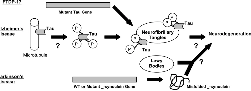
Figure 1. Role of tau and _-synuclein in neurodegenerative diseases
In FTDP-17,mutations in tau have been linked to the presence of neurofibrillary tangles containing hyperphosphorylated tau. The role of tau in pathogenesis of Alzheimer’s disease is uncertain, but hyperphosphorylated tau has been identified in neurofibrillary tangles. In Parkinson’s disease, mutations in _-synuclein have been identified, but how misfolded _-synuclein or Lewy bodies lead to neurodegeneration is unclear.
Mutations of tau in FTDP-17 are either missense, silent, and deletion mutations in the exons or intronic mutations7, 8, 9. Intronic mutations near exon 10 (E10) of
the tau gene increase the ratio of 4R-tau to 3R-tau, which is ~1 in the normal adult brain 7, 8. Studies
suggest that mutations in tau destabilize an RNA stem-loop structure that follows E10, leading to an increased encoding of E10 and an increased proportion of 4R-tau7, 8. In FTDP-17, hyperphosphorylated tau forms into
twisted filaments that are different from PHF tau of AD6.
Following Mutant Tau
Soon after tau gene mutations were reported, our lab pursued investigation of the pathological mechanisms that cause FTDP-17. One of our studies demonstrates that splicing of E10 involves three different regulatory elements10. Missense tau mutations can act on an
exon splicing enhancer within E10, and thereby increase the inclusion or exclusion of E10 in tau transcripts. Silent tau mutations can also increase inclusion of E10 by destroying the exon splicing silencer element within E10. Lastly, intronic mutations in intronic sequences flanking the 5’ splice site of E10 enhance encoding of E1010. Since the tau gene is
regulated by several elements, tau mutations can cause FTDP-17 through multiple mechanisms.
Tau Tangles Shy Away from Microtubules
Our lab has also analyzed the interaction between mutant tau and microtubules. Bacterial cells expressing recombinant tau protein with missense mutations show reduced binding of tau to microtubules10. Furthermore,
the ability of tau to promote microtubule assembly is also reduced10.
A similar study has also been performed on Chinese hamster ovary (CHO) cells. The resulting pathological phenotypes included formation of insoluble tau aggregates and decreased microtubule binding11.
Both of these studies find that decreased levels of microtubule binding correlate with increased amounts of insoluble, unbound tau in the cytoplasm, which could eventually lead to hyperphosphorylation and aggregation of tau10, 11.
Of Mice and Tau
In order to model FTDP-17, we and other labs have created transgenic mouse models that either
overexpress wild type tau or express mutant tau. Transgenic mice overexpressing the shortest tau isoform or the longest tau isoform developed accumulations of insoluble, hyperphosphorylated tau. Furthermore, these mice showed reduced fast axonal transport, axonal degeneration, diminished microtubules, and motor weakness12, 13. Since these
mice developed neuropathological symptoms by overexpression of normal tau, these studies also support the role of tau in AD pathogenesis.
Other labs have created transgenic mice expressing the common FTDP-17 mutation P301L. Similar to human pathology, these mice showed formation of neurofibrillary tangles and neuronal lesions14, 15. Behavioral and motor deficits characteristic
of FTDP-17 were also prominent in these transgenic mice14.
Tau Tangles in the AD Arena
Since PHFs in AD are composed of hyperphosphorylated forms of tau16, our lab set out to
identify the different sites on normal adult tau that become abnormally phosphorylated. Most of these sites are serine (Ser) or theronine (Thr) residues. In addition, some of these sites, i.e. Ser262 and Ser396, are located near microtubule-binding domains17, 18, 19.
Our lab proceeded to study whether these abnormally phosphorylated sites might be the result of inactivated protein phosphatases20-22. Since the
phosphorylaion levels of tau isoforms decrease with age in normal adult brain, activation of protein phosphatases is one mechanism that might contribute to dephosphorylation. Protein phosphatases 2A (PP2A) and 2B (PP2B) were shown to dephosphorylate tau in developing rat brains20. We have also
demonstrated that in human postmitotic neuron-like cells (NT2N), disrupting the microtubule network results in site-specific dephosphorylation of tau by PP2A21.
When NT2N cells are treated with inhibitors of PP2A, results indicate increased tau phosphorylation, decreased microtubule binding, destruction of selective microtubules, and axon degeneration22.
Following this series of studies, we next proceeded to compare mRNA distribution of PP2A in control and AD hippocampus. The level of PP2A mRNA was significantly reduced in AD hippocampus23.
Our data indicates that protein phosphatases may play P
Microtubule
?
TauTau
Mutant Tau Gene
WT or Mutant _-synuclein Gene
FTDP-17
Lewy Bodies Tau
Neurofibrillary Tangles
Tau
Neurodegeneration
?
Alzheimer’sDisease
Parkinson’s Disease
P
P
?
Misfolded _-synuclein
P P
P
an important role in regulating the phosphorylation state of tau, and their activity is decreased in AD neurons.
Along with inactivation of phosphatases, activation of kinases can also be responsible for abnormal tau phosphorylation. One lab has shown that increased activity of cyclin-dependent kinase 5 (cdk5) also increases levels of tau phosphorylation in several cell lines24. Brain proline-directed kinases (BPDK) are
microtubule-associated proteins that have also been shown to phosphorylate tau. Furthermore, BPDK phosphorylates sites that are found abnormally phosphorylated in PHF tau25.
Enhanced Tau Tangles
As previously described, transgenic mouse models overexpressing the shortest tau isoforms developed neurofibrillary tangles12. Moreover, similar to AD
patients, these tangles contained neurofilament (NF) subunits12. Our lab extended these findings by
designing transgenic mice that lacked neurofilaments26.
Thus, transgenic mice overexpressing the shortest tau isoform were crossbred with knock-out mice lacking neurofilaments. As a result, tau aggregation was delayed, weight loss was reduced, viability was increased, and behavioral phenotypes were improved when compared to transgenic mice with neurofilaments26. Therefore, we found that
neurofilaments may play a malevolent role in AD pathogenesis by facilitating aggregation of tau.
Plaques and Tangles: Together at Last?
Whether it is amyloid plaques or tau tangles that primarily cause neurodegeneration is highly controversial. Recently, however, two groups of scientists present evidence that links tau aggregation to amyloid plaques. In one case, transgenic mice expressing mutant tau protein were crossbred with transgenic mice expressing mutant _-amyloid precursor protein. Although these mice developed amyloid plaques at the same age, the levels of neurofibrillary tangles increased significantly27.
Similarly, to link amyloid plaques to tau tangles, another group injected A_ fibrils into brains of transgenic mice expressing mutant tau28. Findings
revealed that there is a fivefold increase in the number of neurofibrillary tangles, and they begin to appear shortly after injection of A_ fibrils28. Thus, there is a
connection between amyloid plaques and tau tangles, but how overproduction of A_ causes tau dysfunction is unclear. Also, it needs to be assessed whether these mice develop AD symptoms such as dementia. Nevertheless, these models provide a means to develop therapies that can target amyloid plaques and tau tangles27, 28.
Like Tau, Like _-Synuclein
Like tau, α-synuclein is part of the abnormal protein aggregates found in PD. The familial form of PD, with an autosomal-dominant inheritance pattern, was first linked to a missense mutation (Ala30_Thr) in
α
-synuclein by Polymeropoulos et al29. Next, our lab
discovered a link between _-synuclein and Lewy bodies, which are characteristic of PD. Substantia nigra neurons from PD patients stained positive for Lewy bodies containing _-synuclein30.
We then moved on to demonstrate that wild type and mutant _-synucleins (Ala30_Pro and Ala53_Thr)
self-aggregate in vitro31. The solubility of wild type and
mutant _-synucleins decreased, and they formed into elongated filaments. _-Synuclein polymerization was concentration and time dependent, suggesting that
initial conformational changes in _-synuclein might serve as seeds for advanced polymerization31. Also, the
A53T mutant _-synuclein was most prone to aggregate, and it may be a more pathogenic species than wild type or A30P _-synuclein31. When immunogold labeling was
done, the N- and C- terminal regions of _-synuclein were less accessible and weakly labeled, thus implying that these regions are involved in polymer formation and are buried within filaments31.
Synaptic Role of _-Synuclein
Although the functions of _-synuclein are not clearly understood, our lab has attempted to characterize the role of this protein. Using neuronal cultures, we found that expression of _-synuclein was localized primarily in presynaptic vesicles32. Furthermore, antisense
inhibition of _-synuclein resulted in decreased pools of synaptic vesicles at the presynaptic terminal. Also, we observed a decrease in levels of synapsin I and synatophysin, proteins found in synaptic terminals32.
Our results suggest that one function of _-synuclein might include maintenance of synaptic vesicle pools.
Modeling PD in Mice and Flies
To prove that _-synuclein aggregates are pathogenic, our lab created transgenic mice that expressed A53T mutant and wild type _-synuclein. Transgenic mice expressing the A53T mutant developed neuropathological symptoms similar to PD patients33.
These included accumulations of _-synuclein inclusions and severe movement disorders. Thus, formation of filamentous _-synuclein inclusions correlates with disease onset33.
Other labs have used Drosophila models to recapitulate features of PD34. Transgenic flies
overexpressing wild type and mutant _-synuclein displayed degeneration of dopamine neurons, formation of _-synuclein inclusions, and motor impairments34.
Our lab has also used Drosophila to study factors that enhance or suppress PD pathology35. In
these models, dopaminergic neuronal loss was prevented by expression of the molecular chaperone Hsp70. Although toxicity of _-synuclein was suppressed in transgenic flies, no change in aggregate pathology was evident35. Also, interference with
endogenous chaperone activity enhanced toxicity of _-synuclein35. In addition, molecular chaperones were
found to be present in Lewy bodies of PD patients35.
Our data strongly suggests that molecular chaperones might help prevent degeneration of dopaminergic neurons by suppressing _-synuclein. On the other hand, _-synuclein toxicity might be enhanced due to the trapping of chaperones in Lewy bodies35.
Increasing the activity of molecular chaperones might prove to be an effective treatment in PD patients35. It
has yet to be determined, however, whether expression of Hsp70 reverses PD symptoms, such as motor impairments, in these transgenic flies.
_-synuclein Caught in a Triangle
Along with mutations in the _-synuclein gene, familial PD has been associated with mutations in two other genes, both of which are gaining considerable importance. Mutations in the parkin gene cause autosomal recessive PD, where the onset of PD is seen at an earlier age36. As ubiquitin is known to be present
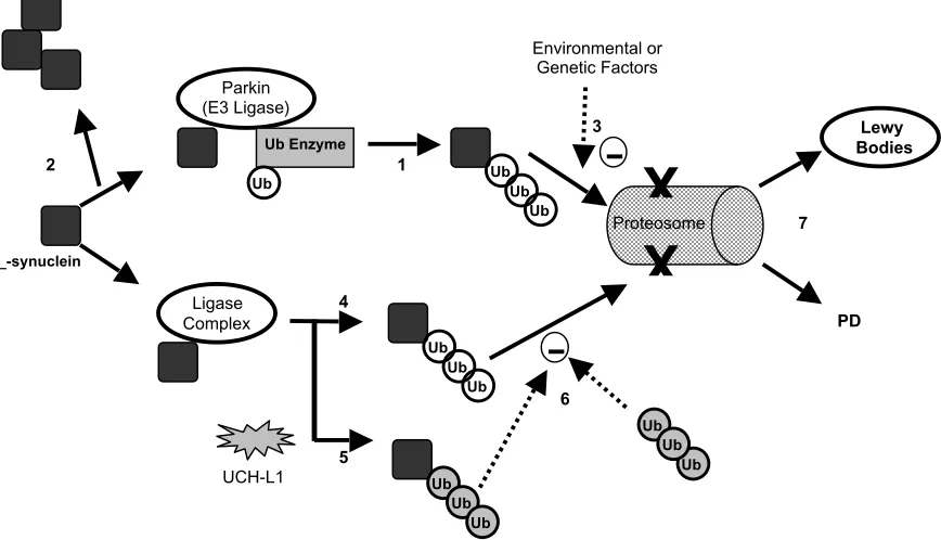
Figure 2. Proposed Models for Parkin and UCH-L1 Interaction with _-Synuclein
The parkin ubiquitin complex polyubiquinates _-synuclein and prepares it for degradation (1). Other environmental or genetic factors might inhibit proper degradation of _-synuclein, causing accumulation and fibrillization of _-synuclein in Lewy bodies (3). Mutations in parkin can cause accumulation of nonubiquinated _-synuclein (2). Loss of control of UCH-L1 ligase activity could cause extension of polyubiquityl chains on _-synuclein and inhibit its degradation (5). These ubiquitin additions require no ATP and are thus different from ubiquitin additions by ligase complexes (4) (difference in Ub is indicated by gray or white color). Polyubiquityl chains themselves can inhibit proteosomal degradation (6). Thus, in both cases, accumulation of ubiquinated _-synuclein can cause formation of Lewy bodies or PD (7).
Recently, parkin was identified as an E3 ubiquitin ligase that associates with a ubiquitin enzyme and a distinct form of _-synuclein37. Whereas normal parkin
ubiquinated synuclein, mutant parkin failed to bind _-synuclein and caused an accumulation of nonubiquinated _-synuclein37. Therefore, it is proposed
that loss of parkin function leads to pathological accumulation of _-synuclein, neuronal loss, and an absence of ubiquitin-tagged Lewy bodies, a common feature of autosomal recessive PD patients. Wild-type parkin is thought to polyubiquinate _-synuclein and prepare it for proteosome degradation; however, interference by other genetic or environmental factors might prevent degradation of these proteins and lead to formation of Lewy bodies37 (Figure 2).
A gene that has been linked to autosomal dominant PD is ubiquitin C-terminal hydolase-L1 (UCH-L1)38. UCH-L1 was recently shown to possess ligase
activity that inhibited proteosomal degradation of _-synuclein39. UCH-L1 is capable of extending the
polyubiquitin chain of _-synuclein by ATP-independent addition of ubiquitin molecules. This process differs form ubiquitylation by other ligases, such as E3, that require ATP to be present39. Furthermore, it is
hypothesized that polyubiquinated _-synuclein or polyubiquitin chains themselves inhibit proteosome degradation. Inhibition consequently promotes aggregation and fibril formation of _-synuclein pathogenic for PD39 (Figure 2).
Untangling the Problem
Although research of neurodegenerative diseases has made great advances over the past decade, the mechanisms of neurodegeneration are far from being untangled. Studying the role of tau and _-synuclein in pathogenesis of FTDP-17, AD, and PD will allow
development of effective therapies and treatments. Although it is unclear how tau and _-synuclein abnormalities lead to neurodegeneration, many studies have provided evidence for probable pathways.
Investigating tau mutations in FTDP-17 has established that hyperphosphorylated tau aggregates are sufficient to cause neurodegeneration. Events leading to hyperphosphorylation of tau in AD can include an imbalance of phosphatase or kinase activity. Emerging evidence links tau tangles to amyloid plaques; however, how amyloid plaques lead to increased tau dysfunction has yet to be established.
In PD, filamentous _-synuclein in Lewy bodies have been linked to the onset of neurodegenerative phenotypes. It is still unclear how pathogenic _-synuclein aggregates cause dopaminergic cell death. Models have also been proposed for how mutant _-synuclein, parkin and UCH-L1 genes cause PD.
Transgenic models made by our lab and others reproduce important features of FTDP-17, AD, and PD, and offer insights into pathogenesis of these diseases. Development of additional animal models will be beneficial in finding potential cures and treatments for neurodegenerative disorders.
Acknowlegements
I would like to thank Dr. DebBurman for his continuous assistance and guidance. I would also like to thank Michele McNeely and Angela Eakley for their support in helping me write and edit this paper.
References
1. George-Hyslop, P.H.; Fatter, L.A.; Goedert, M. “Alzheimer Disease and the Frontotemporal Dementias: Diseases with Cerebral Depostition of 1
Ub
Ub
3
Ub Ub
Ub
4
Ub
Parkin (E3 Ligase)
Ub Enzyme
Ub
_-synuclein
Ub Ub
Ub
Ligase Complex
UCH-L1 Ub
Ub Ub
Proteosome
Lewy Bodies
PD 2
Environmental or Genetic Factors
6
7
X
X
Fibrillar Proteins.” Scriver, C.R., Beaudet, A.L. (Ed.). The Metabolic and Molecular Basis of Inherited Disease. (8th
ed.). 2000; Vol. IV (28): 5875-5899. New York: McGraw-Hill.
2. Lee, V.M.; Goedert, M.; Trojanowski, J.Q. “Neurodegenerative Taupathies.” Annual Reviews of Neuroscience. 2001; 24:1121-159.
3. Olanow, C.W., Tatton, W.G. “Etiology and Pathogenesis of Parkinson’s Disease.” Annual Review of Neuroscience. 1999; 22: 123-44.
4. Dawson, T.M. “New Animal Models for Parkinson’s Disease.” Cell. 2000; 101: 115-18.
5. Mandelkow, E. “The tangled tale of tau.” Nature. 1999; 402: 588-9. 6. Spillantini, M.G.; Goedert, M.; Crowther, A.R., and others. “Familial multiple system taupathy with presenile dementia: A disease with abundant neuronal and glial tau filaments.” Proceedings of National Academy of Sciences. 1997; 94: 4113-118.
7. Hutton, M.; Lendon, C.L., Rizzu, P., and others. “Association of missense and 5’-splice –site mutations in tau with the inherited dementia FTDP-17.” Nature. 1998; 393: 702-5.
8. Spillantini, M.G.; Murrell, J.R.; Goedert, M., and others. “Mutation in the tau gene in familial multiple system taupathy with presenile dementia.”
Proceedings of National Academy of Sciences. 1998; 95: 7737-741.
9. Clark, L.N.; Poorkaj, P.; Wszolek, Z, and others. “Pathogenic implications of mutations in the tau gene in pallido-ponto-nigral degeneration and related neurodegenerative disorders linked to chromosome 17.” Proceedings of National Academy of Sciences. 1998. 95: 13103-107.
10. D’Souza, I.; Poorkaj, P.; Hong, M., and others. “Missense and silent tau gene mutations cause frontotemporal dementia with parkinsonism-chromosome 17 type, by affecting multiple alternative RNA splicing regulatory elements.” Proceedings of National Academy of Sciences. 1999; 96: 5598-603.
11. Vogelsberg-Ragaglia, V.; Bruce, J.; Richter-Landsberg, C., and others. “Distinct FTDP-17 missense mutations in tau produce tau aggregates and other pathological phenotypes in transfected CHO cells.” Molecular Biology of the Cell. 2000; 11: 4093-104.
12. Ishihara, T.; Hong, M.; Zhang, B., and others. “Age-dependent emergence and progression of a tauopathy in transgenic mice overexpressing the shortest human tau isoform.” Neuron. 1999; 24: 751-62.
13. Probst, A.; Gotz, J.; Wiederhold, K.H., and others. “Axonopathy and amyotrophy in mice transgenic for human four-repeat tau protein.” Acta Neuropathology. 2000; 99: 469-81.
14. Lewis, J.; McGowan, E.; Rockwood, J., and others. “Neurofibrillary tangles, amyotrophy and progressive motor disturbance in mice expressing mutant (P301L) tau protein. Nature Genetics. 2000; 25: 402-5.
15. Gotz, J.; Chen, F.; Barmettler, R., and others. “Tau filament formation in transgenic mice expressing P301L tau.” The Journal of Biological Chemistry. 2001; 276: 529-34.
16. Lee, V.M.; Balin, B.J.; Otvos, L. Jr., and others. “A68: A major subunit of paired helical filaments and derivatized forms of normal tau.” Science. 1991; 251: 675-78.
17. Seubet, P.; Dewan-Mawal, M.; Barbour, R., and others. “Detection of phosphorylated Ser262 in fetal tau, adult tau, and paired helical filament
tau.” The Journal of Biological Chemistry. 1995; 270 (32): 18917-922.
18. Otvos, L. Jr.; Feiner, L.; Lang, E., and others. “Monoclonal antibody PHF-1 recognizes tau protein phosphorylated at serine residues 396 and 404.” Journal of Neuroscience Research. 1994; 39: 669-73.
19. Goedert, M.; Jakes, R.; Crowther, R.A., and others. “The abnormal phosphorylation of tau protein at Ser-202 in Alzheimer disease recapitulates phosphorylation during development.” Proceedings of National Academy of Sciences. 1993; 90: 5066-70.
20. Mawal-Dewan, M.; Henley, J.; Van de Voorde, A., and others. “The phosphorylation state of tau in the developing rat brain is regulated by phosphorylation phosphatases.” The Journal of Biological Chemistry. 1994; 269: 30981-987.
21. Merrick, S.E.; Demoise, D.C.; Lee, M.Y. “Site-specific dephosphorylation of tau protein at Ser202
/Thr205
in response to microtubule depoylmerization in cultured human neurons involves protein phospatase 2A.” The Journal of Biological Chemistry. 1996; 271: 5589-94.
22. Merrick, S.E.; Trojanowski, J.Q.; Lee, V.M. “Selective destruction of stable microtubules and axons by inhibitors of protein
serine/threonine phosphatases in cultured human neurons (NT2N cells).”
The Journal of Neuroscience. 1997; 17(15): 5726-37.
23. Vogelsberg-Ragaglia, V.; Schuck. T.; Trojanowski, J.Q., and others. “PP2A mRNA expression is quantitatively decreased in Alzheimer’s disease hippocampus.” Experimental Neurology. 2001; 168: 402-12.
24. Patrick, G.N.; Zukerberg, L.; Nikolic, M., and others. “Conversion of p35 to p25 deregulates Cdk5 activity and promotes neurodegeneration.”
Nature. 1999; 402: 615-22.
25. Paudel, H.K.; Lew, J., Ali, Z., and others. “Brain proline-directed protein kinase phosphorylates tau on sites that are abnormally phosphorylated in tau associated with Alzheimer’s paired helical filaments.” The Journal of Biological Chemistry. 1993; 268(31): 23512-518.
26. Ishihara, T.; Higuchi, M.; Zhang, B., and others. “Attentuated neurodegenerative disease phenotype in tau transgenic mouse lacking neurofilaments.” The Journal of Neuroscience Research. 2001; 21(16): 6026-35.
27. Lewis, J.; Dickinson, D.W.; Lin, W.L., and others. “Enhanced neurofibrillary degeneration in transgenic mice expressing mutant tau and APP.” Science. 2001; 293: 1487-90.
28. Gotz, J.; Chen, F.; Dorpe, J.V., and others. “Formation of neurofibrillary tangles in P301L tau transgenic mice induced by A_42 fibrils.” Science. 2001; 293: 1491-95.
29. Polymeropoulos, M.H.; Lavedan, C., Leroy, E., and others. “Mutation in the _-synuclein gene identified in families with Parkinson’s disease.”
Science. 1997; 276:2045-47.
30. Spillantini, M.G.; Schmidt, M.L.; Lee, V.M., and others. “_-Synuclein in Lewy bodies.” Nature. 1997; 388: 839-40.
31. Giasson, B.J.; Uryu, K.; Trojanowski, J.Q., and others. “Mutant and wild type human _-synucleins assemble into elongated filaments with distinct morphologies in vitro.” The Journal of Biological Chemistry. 1999; 274(12): 7619-622.
32. Myrphy, D.D.; Rueter, S.M.; Trojanowki, J.Q., and others. “Synucleins are developmentally expressed, and _-synuclein regulates the size of the presynaptic vesicular pool in primary hippocampal neurons.” The Journal of Neuroscience. 2000; 20(9): 3214-20.
33. Giasson, B.J.; Duda, J.E.; Quinn, S.M., and others. “Neuronal _-synucleinopathy with severe movement disorder in mice expressing A53T human _-synuclein.” Neuron. 2002; 34: 521-33.
34. Feany, M.B., Bender, W.W. “A Drosophila model of Parkinson’s disease.” Nature. 2000; 404: 394-98.
35. Autuck, P.K.; Chan, H.Y.; Trojanowski, J.Q., and others. “Chaperone suppression of _-synuclein toxicity in a Drosophila model of Parkinson’s disease.” Science. 2002; 295: 865-68.
36. Kitada, T.; Asakawa, S.; Hattor, N., and others. “Mutations in the
parkin gene cause autosomal recessive juvenile parkinsonism.” Nature. 1998; 392: 605-7.
37. Shimura, H.; Schlossmacher, M.G.; Hattori, N., and others. “Ubiquination of a new form of _-synuclein by Parkin from human brain: Implications for Parkinson’s disease.” Science. 2001; 293: 263-68.
38. Leroy, E.; Boyer, R.; Auburger, G., and others. “The ubiquitin pathway in Parkinson’s disease.” Nature. 1998; 395: 451-52.