Effect of cigarette smoking on liver function test and some other
related parameters
Received: 17/10/2012 Accepted: 17/3/2013
Sangar Najat Abdul-Razaq * Bakhtiar M. Ahmed **
Background and objective: Cigarette smoking is one of the 10 greatest contributors to global death and disease. Cigarette smoke consists of many chemicals, including cytotoxic, carcinogenic and free radicals, therefore it affects many organs if not all.This
work is directed to evaluate the effects of dose response patterns of tobacco exposure on liver tissue, through assessing some serum biochemical parameters related with liver efficiency.
Methods: This study was done in Kirkuk province. It was conducted on (110) healthy male subjects, their ages ranged from 20 to 40 years. They were divided into three groups; 40
heavy smokers, 30 moderate smokers and 40 non-smokers. Blood was withdrawn for estimation of serum liver function test, lipid profile, protein electrophoresis, and lipid peroxidation test (Serum Malondialdehyde; MAD level).
Results: There were statistically significant elevations in serum alkaline phosphatase (ALP), alanine transaminase(GPT) and aspartate aminotransferase(GOT) activities in
heavy smokers while serum total bilirubin significantly was lower comparing to non smokers. Serum total protein and albumin were significantly lower in heavy smokers comparing to non smokers. The results of serum protein agarose gel-electrohporesis
showed significant changes in serum protein fractions in smoker groups. The mean level of serum total cholesterol, triglyceride, LDL, VLDL and malondialdehyde was significantly higher in heavy smoker group, while serum HDL level had a significantly lower value.
Conclusion: Cigarette smoking can affect liver efficiency and functions. These effects are dose exposure depenent.
Keywords: Cigarette smoking, Liver function test, Liver enzymes, MAD, Lipid profile.
Abstract
* Health Ministry, Health Office, Kirkuk/Iraq
**Department of Basic Science, College of Dentistry, Hawler Medical University, Erbil, Iraq
Introduction
Cigarette smoking is a major cause of preventable morbidity and mortality1.
Worldwide, more than 3 million people currently die each year from cigarette
smoking 2. The risk of death in the smokers measured by the number of cigarettes smoked daily, the duration of smoking, the
degree of inhalation and the age of initiation 3,4. Cigarette smoke contains over
4000 different chemicals, 400 of which are proven to be carcinogenic; it also contains
various oxidants such as oxygen free radicals and volatile aldehydes which are
probably the major causes of damage to biomolecules 5. Cigarette smoking yields
chemical substances with high cytotoxic potentials 6. Cigarette smoke consists of many chemicals, including nicotine, tar with its many carcinogens, and gaseous compounds including carbon monoxide
7.Cigarette smoke also contains large
numbers of free radicals that are capable of initiating or promoting oxidative injury 8.
Cigarette smokers are at greater risk for cardiovascular diseases, respiratory
disorders, cancers, peptic ulcers and gastroesophageal reflux disease,
blind-ness, bone matrix loss, and hepatotoxicity comparing with non-smokers 9. Cigarette
contact with the smoke itself such as liver. The liver is an important organ that has many tasks; such as responsibility for processing drugs, alcohol and other toxins to eliminate them from the body 10,11. Till
date, there is no previous study that concerned with the full investigation of the
effect cigarette smoke and dose exposure to tobacco smoke (the number of cigarette
smoked per a day) on liver tissue and liver functions. The present study was constructed to evaluate the link between cigarette smoking and the biochemical state of the liver throughout investigating the effect of cigarette smoking on liver function test, lipid profile and lipid peroxida-tion, and assessing the effect of cigarette
smoking on other parameters such as protein, albumin and globulins, then studying the effect of cigarette smoking on
protein electrophoresis.
Subjects: A total of (110) healthy subjects were enrolled in the present study during their attendance to outpatient department at Azadi general hospital in Kirkuk city. Their ages were ranged from (20-40) years. Obese subjects (BMI> 29) and subjects who received medications were excluded. The subjects were classified according to the number of cigarettes
smoked per day and the duration of smoking, into heavy smoker group (40
subjects), moderate smoker group (30 subjects), and non smoker group (control group)(40 subjects).
Sample collection: Five milliliter of blood was withdrawn from each subject. The blood was allowed to clot in a plain tube for 20 minute at room temperature. The serum was separated by centrifugation at 3000 rpm for 10 minutes, then each subject serum was stored in five plain tubes (5 aliquots), frozen at -20 C until the day of biochemical assay (except for the enzyme studies which were done directly).
Methods:
1. Body mass index was calculated accord-ing to Martin and Crook method 12.
2. Liver function tests, including; serum ALP, ALT and AST, and (STB) were
assed according to Kind and King method 13, Reitman and Frankel method 14,
and Walters and Gerarde method 15,
respectively.
3. Serum Protein (Gornall method) 16,
albumin (Doumas method ) 17, and globulin
(from the equation of Clarke and Dufour18).
4. Agarose gel protein electrophoresis was performed 19.
5. Lipid profile (Total cholesterol, Triglyceride, LDL, HDL, and VLDL) 16
and lipid peroxidation test (Lipid peroxidation parameter; Serum MDA level)
were performed 20,21 .
Statistical analysis: Computerized statis-tical analysis was performed using SPSS version 14 computer software. The level of statistical significance was (P< 0.05).
The results showed that serum ALP, ALT and AST levels were significantly high (P value<0.05) in heavy smoker group when compared with non smoker group,
while serum STB was significantly low (P value>0.05), Table 1. No statistically
significant differences (P value>0.05) were
observed in ALP, AST, ALT and STB levels for moderate smoker group
compar-ing with non smoker group, Table 2.
Table 1: The Mean±SE of Serum ALP, AST, ALT, STB levels for heavy smoker group compared with non smoker group.
Methods
Results
P value
Non-smoker group (N=40) Heavy
smoker group (N=40) Parameters
(unit)
0.001 [S] 71.27±2.3 78.9±0.98
ALP (IU/L)
0.001 [S] 22.7±1.2
28.53±1.3
AST (IU/L)
0.0001 [S] 18.97±0.62 28.65±0.72
ALT (IU/L)
0.069 [S] 0.95±0.04 0.82±0.02
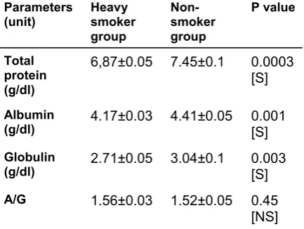
Serumtotal protein, albumin and globulin
were significantly lower (P value<0.05) in both heavy and moderate smoker groups when compared with non smoker
group ,while there was no important differences in serum albumin-globulin ratio
(A/G) between smoker groups and non
smoker group, Table 3 and Table 4. The results showed that, there was no difference in the number of bands that appear on the protein gel-electrophoretic
pattern, but there was a significant difference in the concentration of bands
among the study groups, Figure1. The results showed that the mean of albumin, α-2 and γ globulins had lower values in heavy smoker group when compared with non-smoker group but the mean of α-1 and β globulins had higher levels. In moderate smoker group the mean of albumin and α-2
globulin had lower level when compared with non-smoker group and the mean of β globulin had higher values, while there was no difference in the mean of α-1 and γ globulins between moderate smoker and non-smoker groups, Table 5 and Table 6.
Table 2: Mean±SE of Serum ALP, AST, ALT, STB levels for moderate smoker group compared with non smoker group.
Table 3: Mean±SE of Serum Total protein, albumin, globulin and (A/G) ratio for heavy smoker group compared with non smoker group.
Table 4: Mean ± SE of Serum Total protein, albumin, globulin and (A/G) ratio
for moderate smoker group compared with non smokers
Figure 1: Serum protein electrophoretic pattern for heavy, moderate and non-smoker groups.
P value
Non-smoker group (N=40) Moderate
smoker group (N=30) Parameters
(unit)
0.36 [NS] 71.27±2.3
73.5±0.78 ALP(IU/L)
0.86 [NS] 22.7±1.2
23±0.69 AST(IU/L)
0.57 [NS] 18.97±0.62 19.53±0.67
ALT(IU/L)
0.74 [NS] 0.95±0.04
1.08±0.05 STB
)mg/dl)
P value
Non-smoker group Heavy
smoker group Parameters
(unit)
0.0003 [S] 7.45±0.1 6,87±0.05
Total protein
(g/dl)
0.001 [S] 4.41±0.05 4.17±0.03
Albumin
(g/dl)
0.003 [S] 3.04±0.1 2.71±0.05
Globulin (g/dl)
0.45 [NS] 1.52±0.05 1.56±0.03
A/G
P value
Non-smoker group Moderate
smoker group Parameters
(unit)
0.007 [S] 7.45±0.1
7.11±0.08 Total
protein
(g/dl)
0.017 [S] 4.41±0.05 4.27±0.04
Albumin (g/dl)
0.104 [NS] 3.04±0.1
2.84±0.07 Globulin
(g/dl)
0.824 [NS] 1.52±0.05
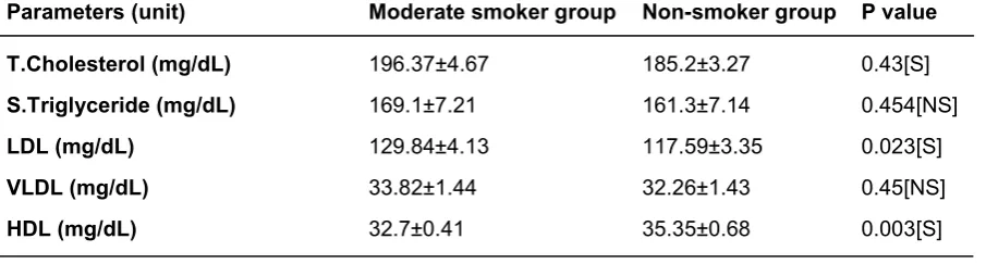
Table 7 shows that serum total cholesterol,
triglyceride, LDL and VLDL levels were significantly higher in heavy smoker group
(P<0.05) comparing with non smoker
group, while the value of serum HDL was significantly lower (P <0.05). In moderate smokers, the values of serum
γ β
α-2
α-1 Albumin
Groups
9.08 % 13.91 %
8.64 % 3.07 %
65%
Heavy smoker
10.15 % 10.88 %
6.32 % 2.06 %
70.29%
Moderate smoker
9.23 % 7.18 %
10% 1.79 %
71.28%
Non-smoker
γ β
α-2
α-1
Albumin Total protein
Groups
0.62 0.95
0.59 0.21
4.44 6.83
Heavy smoker
0.69 0.74
0.43 0.14
4.78 6.80
Moderate smoker
0.72 0.56
0.78 014
5.56 7.83
Non-smoker
P value Non-smoker group
Heavy smoker group
Parameters(unit)
0.002[S] 185.2±3.27
202.27±4.21
T.Cholesterol (mg/dL)
0.006[S] 161.3±7.14
186.05±5.06
S.Triglyceride (mg/dL)
0.003[S] 117.59±3.35
133.69±4.05
LDL (mg/dL)
0.006[S] 32.26±1.43
37.21±1.01
VLDL (mg/dL)
0.02[S] 35.35±0.68
31.37±0.3
HDL (mg/dL)
P value Non-smoker group
Moderate smoker group
Parameters(unit)
0.43[S] 185.2±3.27
196.37±4.67
T.Cholesterol (mg/dL)
0.454[NS] 161.3±7.14
169.1±7.21
S.Triglyceride (mg/dL)
0.023[S] 117.59±3.35
129.84±4.13
LDL (mg/dL)
0.45[NS] 32.26±1.43
33.82±1.44
VLDL (mg/dL)
0.003[S] 35.35±0.68
32.7±0.41
HDL (mg/dL)
total cholesterol and LDL were significantly higher (P<0.05) than that of non-smokers, while serum HDL was significantly lower (P<0.05), Table 8. There was no significant difference (P>0.05) in serum triglyceride
and VLDL levels between moderate smokers and non smokers.
Table 5: The percentage of protein fractions in heavy, moderate and non smoker groups on agrose gel-electrophoresis.
Table 6: The concentrations of protein fractions in heavy, moderate and non smoker groups on agarose gel-electrophoresis.
Table 7: The Mean ± SE of Serum total cholesterol, triglyceride, LDL, VLDL and HDL levels for heavy smoker group compared with non smoker group.
Table 9 showed that serum malondialde-hyde (MDA) level was significantly higher in heavy smoker group when compared to non smoker group, while there was no sig-nificantly change in case of moderate smokers.
Non-smoker group Moderate smoker group
Heavy smoker group
Parameters(unit)
4.79±0.28 4.99±0.3 [NS]
6.70±0.31[S]
MDA (µmole/L)
Discussion
The results showed a significant rise in se-rum ALT, AST and ALP activity in cigarette smokers when compared to control group. This may occur due to nitrosative stress which is a condition that occurs when the production of highly reactive nitrogen-containing chemicals, such as nitrous oxide, exceed the ability of the human body to neutralize and eliminate them. Nitrosative stress can lead to reactions that alter protein structure thus interfering with normal body functions 22,23. Cigarette smoke contains a large number of chemical substances with hepatotoxic potential including nicotine 6. The results show
that there was no statistically significant difference in serum ALT, AST, ALP and TB between moderate smoker group and control group. From the results of the present study, one can conclude that there is a dose response relationship between the number of cigarettes/day smoked and serum ALT, AST, ALP and total bilirubin
levels. From the results, a statistically significant reduction in serum total protein,
serum albumin and serum globulin in heavy smoker group were observed. The results
also show that there were significant reductions in the serum total protein and
serum albumin levels in moderate smoker group, while serum globulin levels didn’t change. Cigarette smoking is associated with increased oxidative stress. Albumin has antioxidant properties, through binding to copper ions and scavenging HOCl, then
the oxidized albumin may be cleared rapidly from the circulation and degraded
24,25. The present study revealed that
se-rum protein electrophoretic pattern changed in both heavy and moderate smokers when compared with non-smoker group. To the best of our knowledge and on the basis of active literature searching, other researchers did not investigate the protein fractions by protein-electrophoresis in their studies. From the results, it can be suggested that cigarette smoke contain many potential hepatotoxic substances which affect liver function through its effect on nserum protein electrophoresis frac-tions, this is due to its effect on their syn-thesis and metabolism in the liver. There is a great relationship between liver and lipid because all lipid profile parameters are synthesis and metabolized in liver 26-28. The present study revealed that serum TG, TC, LDL, and VLDL were significantly higher in heavy smokers as compared to both mod-erate and non-smokers thereby revealing a direct dose response relationship. Serum HDL level was significantly lower in heavy smokers when compared to non-smokers. Lipid alteration in cigarette smokers may be due to nicotine which stimulates the secretion of catecholamines resulting in increasing the rate of lipolysis and the increased concentration of plasma free fatty acids (FFA) which further result in increasing the releasing of hepatic FFAs and hepatic triglycerides along with VLDL to the blood stream 23,29 The results of the
present study showed that, serum TC and LDL levels were significantly higher in moderate smokers when compared with non-smokers. From the results of this work one can emphasize that lipid profile is affected by the dose response of tobacco exposure. Serummalondialdehyde (MDA)
level was significantly higher in heavy smoker group when compared with non smoker group. There are several reasons why smokers would be expected to have a higher level of lipid peroxidation compared with non-smokers. First, smokers are
prone to oxidation from the inhalation of large numbers of gas-phase and other radicals giving rise to increased oxidative damage 30-31. Second, depletion of plasma
antioxidants otherwise protecting against oxidative damage such as lipid
peroxida-tion has consistently been observed among smokers 32. Finally cigarette smoke also
induces oxidative stress by stimulating NADPH oxidase and decreasing antioxi-dant defences, leading to lipid peroxidation
33. These effects could lead to increased
hepatocellular damage 34.
Cigarette smoking can affect liver func-tions, through its effect on serum ALP, ALT and AST total protein, albumin, globulin, protein fractions in gel-electrophoresis pattern. It also affect lipid profile and lipid peroxidation.
1. Funck C., Mathilde R., Michel L., Arnould J., Verstuyft C., Martine L., et al.. Effects of type of smoking (pipe, cigars or cigarettes) on biological indices of tobacco exposure and toxicity. Lung Cancer. 2006;54: 11–8.
2. Aurelio, L.. Biochemical markers of cardiovascular damage from tobacco smoke. Curr. Pharm. Des. 2005;11: 2190–208.
3. Janson C., Chinn S., Jarvis D., Zock J., Torén K. and Burney P. Effect of passive smoking on respiratory symptoms, bronchial responsiveness,
lung function, and total serum IgE in the European Community Respiratory Health Survey: a cross-sectional study. Lancet. 2001;358
(9299):2103-9.
4. Lubin J.H., Alavanja M.C., Caporaso N, Brown L.M., Brownson R.C., Field R.W., et al. Cigarette smoking and cancer risk: modeling total exposure and intensity. Am J Epidemiol. 2007;166(4): 479-89.
5. Yeh, C., Graham B, Powell C., Mesia-Vela S., Wang Y., Hamade N., et al. Effect of cigarette smoking dose on oxidized plasma proteins. Environ. Res.2008; 106: 219–25.
6. Yuen S.T., Gogo A.R., Luk I.S., Cho C.H., Ho J.C.
and Loh T.T. The effect of nicotine and its interaction with carbon tetrachloride in the rat liver. Pharmacol. Toxicol.1995; 77: 225–30.
7. Benowitz N., Hall S., Stewart S., Wilson M., Dempsey D. and Jacob P. Nicotine and Carcino-gen Exposure with Smoking of Progressively
Re-duced Nicotine Content Cigarette. Cancer Epidemiol. Biomarkers Prev. 2007; 16: 2479–85.
8. David E.E., Shashank J.D. and Hongsheng G. Effect of cigarette Smoking on pentane excretion in alveolar breath. Clinical chemistry 1996; 42(2): 103 -308.
9. Spiro S. G. and Silvestri G. A. (2005). One hundred years of lung cancer. Am. J. Respir. Crit. Care Med.; 172: 523–9.
10. Yu M.W., Hsu F.C., Sheen I.S., Chu C.M., Lin D.Y., Chen C.J., et al. Prospective study of hepatocellular carcinoma and liver cirrhosis in as-ymptomatic chronic hepatitis B virus carriers. Am J Epidemiol. 1997; 145: 1039-47.
11. Pessione F., Ramond M.J., Njapoum C., Duchatelle V., Degott C., Erlinger S., et al. Cigarette smoking and hepatic lesions in patients with chronic hepatitis C. Hepatology. 2001; 34: 121 -5.
12. Martin A and Crook T. Normal female gonadal function in: Clinical chemistry and metabolic medi-cine, 7thed hodder Arnold. 338Eeuston road, Lon-don. 2006:148-9.
13. King P.R. and King E. J. Estimation of plasma Phosphatase by determination of hydrolysed phe-nol with amino-anti-antipyrine. J. Clin. Path.1954; 7:322-326.
14. Frankl H.D. and Merrit J.H. Enzyme activity in the serum and common bile duct. Am J Gastroenterol. 1959; 31: 166-9.
15. Walters M. and Gerarde H. Colourmeteic evalution of serum bilirubin. Microchem J. 1970; 15:231-243.
16. Tietz N.W. Text book of clinical chemistry, 3rd Ed. C.A. Burtis, E.R. Ashwood, W.B. Saunders 1999.
17. Doumas B.T. and Biggs H.G. Determination of serum albumin,– Standard methods of clinical chemistry – Acad. Press. N.Y.1972; 7:175-188.
18. Clarke W. and Dufour D.R. Contemporary Practice in Clinical Chemistry. AACC Press; Wash-ington. 2006: 200-206.
19. Hames B. D. Gel Electrophoresis of Proteins: A Practical Approach 3rd ed., Oxford University Press, New York. 1998:341-55.
20. Botsoglou, N.A., Rapid, Sensitive, and Specific Thiobarbituric Acid Method for Measuring Lipid Peroxidation in animal Tissue, Food and Freestuff Samples, J. Agric. Food Chem. 1994; 42:1931-7. 21. Jentzsch, A.M., et al., Improved Analysis of
Malondialdehyde in Human Body Fluids, Free Rad. Biol. Med. 1996; 20:251-6.
22. Chakraborty A. and Selvaraj S. Differential modulation of xenobiotic metabolizing enzymes by
Conclusion
vanadium during diethylnitrosamine induced hepa-tocarcinogenesis in Sprague-dawley rats. Neo-plasma. 2000; 47: 81– 9.
23. Pannuru P., Vaddi D. and Nallanchakravarthula V. Influence of Chronic Cigarette Smoking on
Se-rum Biochemical Profile in Male Human Volunteers. Journal of Health Science. 2009; 55(2) 265–70.
34. Halliwell B. Albumin – an important extracellular antioxidant. Biochem Pharmacol. 1988; 37: 569-71.
25. Hu M., Louie S., Cross C., Motchnik P. and Halliwell B. Antioxidant protection against hypochlorous acid in human plasma. J Lab Clin
Med. 1993; 121: 257-62.
26. Wilhelmsen L. Coronary heart disease. Epidemi-ology of smoking & intervention studies of smoking. Am Heart J. 1988; 115: 242-247.
27. Gill H.C. The cardiovascular pathology of smoking. Am Heart J. 1988; 115(1 Pt 2): 250– 7.
28. Neki N.S. Lipid Profile in Chronic Smokers – A Clinical Study. JIACM. 2002; 3(1): 51- 4.
29. Simons L.A., Simons J. and Jones A.S. The interaction of body weight, age, cigarette smoking and hormone usage with blood pressure and plasma lipids in an Australian community. Aus NZ J Med. 1984; 14: 215-21.
30. Frei B., Forte T.M., Ames B.N. and Cross C.E. Gas phase oxidants of cigarette smoke induce lipid peroxidation and changes in lipoportein properties in human blood plasma. Biochem J. 1991;277:133- 8.
31. Pryor W. and Stone K. Oxidants in cigarette smoke. Radicals, hydrogen peroxide, peroxynitrate and peroxynitrite. Ann. N. Y. Acad. Sci.1993; 686: 12–27.
32. Lykkesfeldt J., Viscovich M. and Poulsen H. Ascorbic acid recycling in human erythrocytes is induced by smoking in vivo. Free Radic Biol Med. 2003; 35: 1439– 47.
33. Agarwal, R. Smoking, oxidative stress and inflammation: impact on resting energy expenditure in diabetic nephropathy. BMC Nephrol.
2005; 6: 13–21.