VIEWS & REVIEWS
Rahul Lakshmanan, FRANZCR Matthew E. Adams,
FRCR
David S. Lynch, MRCPI Justin A. Kinsella,
MRCPI
Rahul Phadke, FRCPath Jonathan M. Schott,
FRCP
Elaine Murphy, MRCP Jonathan D. Rohrer,
MRCP
Jeremy Chataway, FRCP Henry Houlden, FRCP Nick C. Fox, FRCP Indran Davagnanam,
FRCR
Correspondence to Dr. Davagnanam: [email protected]
Redefining the phenotype of ALSP and
AARS2
mutation
–
related leukodystrophy
ABSTRACT
Objective:To provide an overview of the phenotype of 2 clinically, radiologically, and pathologi-cally similar leukodystrophies, adult-onset leukoencephalopathy with axonal spheroids and pig-mented glia (ALSP) and alanyl-transfer RNA synthetase 2 mutation–related leukodystrophy (AARS2-L), and highlight key differentiating features.
Methods:ALSP andAARS2-L cases were identified from the adult-onset leukodystrophy data-base at our institution. In addition, cases with imaging findings were identified from a literature review. The phenotypic features were determined by combining published cases with those from our database.
Results:A combined total of 74 cases of ALSP and 10 cases ofAARS2-L with neuroimaging data were identified. The mean age at onset was 42 years in ALSP and 26 years inAARS2-L. Cog-nitive and motor symptoms were the most common symptoms overall in both. Ovarian failure was exclusive to AARS2-L, present in all known female cases. Both ALSP and AARS2-L showed a confluent, asymmetric, predominantly frontoparietal, periventricular pattern of white matter disease with subcortical U-fiber sparing; pyramidal tract and corpus callosum involvement; and diffusion changes in the white matter which we have termed“deep white matter diffusion dots.”
Central atrophy and corpus callosal thinning were prominent in ALSP and disproportionately mild inAARS2-L when present. ALSP also occasionally showed ventricular abnormalities and calcifi-cations in the frontal periventricular white matter, features not seen in AARS2-L. AARS2-L demonstrates white matter rarefaction which suppresses on fluid-attenuated inversion recovery MRI sequences, a feature not seen in ALSP.
Conclusions:ALSP andAARS2-L share similar clinical, imaging, and pathologic characteristics with key differentiating features that we have highlighted. Neurol Genet 2017;3:e135; doi: 10.1212/NXG.0000000000000135
GLOSSARY
ADC5apparent diffusion coefficient;ALG5Adult-onset Leukodystrophy Group;ALSP5adult-onset leukoencephalopathy with axonal spheroids and pigmented glia;CSF1R5colony-stimulating factor receptor 1;DWI5diffusion-weighted imaging; FLAIR5fluid-attenuated inversion recovery; HDLS5 hereditary diffuse leukoencephalopathy with axonal spheroids; POLD5pigmentary orthochromatic leukodystrophy;SWI5susceptibility weighted imaging;tRNA5transfer RNA.
Adult-onset leukodystrophies are a rare (estimated prevalence of 2 in 100,000
1) and
diagnos-tically challenging group of conditions.
2MRI is pivotal in identifying the presence of a
leu-kodystrophy; however, MRI findings are commonly etiologically nonspecific.
3Two
leukodystrophies with similar clinical, imaging, and histopathologic
4phenotypes are
adult-onset leukoencephalopathy with axonal spheroids and pigmented glia (ALSP), the most
common adult-onset leukodystrophy,
5and a novel leukoencephalopathy due to autosomal
From the Lysholm Department of Neuroradiology (R.L., M.E.A., I.D.), the National Hospital for Neurology and Neurosurgery; Department of Molecular Neuroscience (D.S.L., H.H.), UCL Institute of Neurology; the Leonard Wolfson Experimental Neurology Centre (D.S.L., J.A.K.), the National Hospital for Neurology and Neurosurgery, UCL Institute of Neurology; Dementia Research Centre (J.A.K., J.M.S., J.D.R., N.C.F.), Department of Neurodegeneration, UCL Institute of Neurology, UK; Department of Neurology (J.A.K.), St Vincent’s University Hospital, University College Dublin, Ireland; Division of Neuropathology and Department of Neurodegenerative Disease (R.P.), Charles Dent Metabolic Unit (E.M.), Department of Neuroinflammation (J.C.), Neurogenetics Laboratory (H.H.), and Department of Brain Repair and Rehabilitation (I.D.), the National Hospital for Neurology and Neurosurgery, UCL Institute of Neurology, UK.
Funding information and disclosures are provided at the end of the article. Go to Neurology.org/ng for full disclosure forms. The Article Processing Charge was paid by the Medical Research Council UK.
recessive mutations in the mitochondrial
alanyl
–
transfer RNA (tRNA) synthetase 2
gene (
AARS2
-L).
6ALSP was previously
referred to as hereditary diffuse
leukoence-phalopathy with axonal spheroids (HDLS) or
pigmentary orthochromatic leukodystrophy
(POLD);
7,8however, ALSP is now preferred
as the unifying term for leukodystrophies due
to autosomal dominant mutations in the
colony-stimulating factor receptor 1 (
CSF1R
)
gene.
5Mutations in the
CSF1R
gene occur in
the tyrosine kinase domain of the
colony-stimulating factor receptor 1 which is
primar-ily expressed in microglia in the CNS and
results in microglial dysfunction in ALSP.
9,10Mutations in the
AARS2
gene cause errors in
the mitochondrial aminoacyl tRNA synthase
gene responsible for encoding alanine onto
mitochondrial tRNA during mitochondrial
translation
11and has been recently found to
be the cause of an ovario-leukodystrophy
6and an infantile cardiomyopathy.
11The aim
of this article is to summarize the
character-istic imaging appearances of ALSP and
AARS2
-L and highlight their phenotypic
sim-ilarities and discriminating clinical and
imag-ing features.
METHODS Standard protocol approvals, registrations, and patient consents. This review was performed with the approval of the University College London Hospital Trust
Institutional Review Board. All patients included in our local cohort had written consent to participate.
Local patient cohort. Eight cases of genetically confirmed ALSP and 4AARS2-L cases with imaging were identified from an adult leukodystrophy database maintained by the Adult-onset Leukodystrophy Group (ALG) at the National Hospital for Neurology and Neurosurgery, Queen Square, London. Brain histopathology was available in 2 of our patients with ALSP and in 1 patient with AARS2-L. Histopathologic specimens were reviewed by an experienced consultant neuropathologist with a special interest in white matter pathologies.
Consensus review of local imaging.A review of the imaging findings in our 12 patients was performed by a neuroradiology fellow (R.L.) and 2 experienced neuroradiologists (M.E.A. and I.D.). Images were reviewed on a PACS workstation using Ag-fa Impax 6 (AgAg-fa-Gevaert N.V., Mortsel, Belgium) and 4 diag-nostic 3mp monochrome Barco (Kortrijk, Belgium) monitors. The neuroradiologists were blinded to patient demographics and clinical information and assessed the imaging findings for each case according to the following categories: white mat-ter involvement (signal characmat-teristics, lobar predominance, symmetry, extent, involvement of subcortical U fibers, and periventricular white matter); involvement of the corpus callosum, pyramidal tracts, brainstem, basal ganglia, and cer-ebellum; atrophy; ventricular abnormalities; diffusion-weighted imaging (DWI) abnormalities; gradient echo and/ or susceptibility weighted imaging (SWI) abnormalities; progression over time; calcifications on CT, and vascular abnormalities. The findings of each case were tabulated after a consensus review.
Literature review.A review of the published literature was per-formed in the English language through a search of PubMed using each of the following search terms:“axonal spheroids,”
“leu(c)kodystrophy and spheroids,” “HDLS,” “pigmented glia,”
“leu(c)kodystrophy and pigmentary,” “pigmentary orthochro-matic leu(c)kodystrophy or POLD,” “ALSP,” “CSF1R,”
“alanyl-tRNA synthetase 2,”and“AARS2.”For both ALSP and
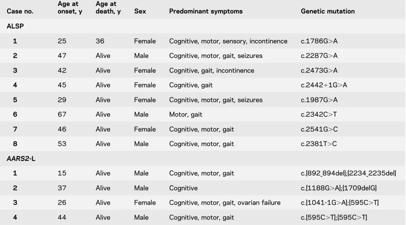
Table 1 Local patient demographics and clinical details
Case no.
Age at onset, y
Age at
death, y Sex Predominant symptoms Genetic mutation
ALSP
1 25 36 Female Cognitive, motor, sensory, incontinence c.1786G.A
2 47 Alive Male Cognitive, motor, gait, seizures c.2287G.A
3 42 Alive Female Cognitive, gait, incontinence c.2473G.A
4 45 Alive Female Cognitive, gait c.244211G.A
5 29 Alive Female Cognitive, motor, gait, seizures c.1987G.A
6 67 Alive Male Motor, gait c.2342C.T
7 46 Alive Female Cognitive, motor, gait c.2541G.C
8 53 Alive Male Cognitive, motor, gait c.2381T.C
AARS2-L
1 15 Alive Male Cognitive, motor, gait c.[892_894del];[2234_2235del]
2 37 Alive Male Cognitive c.[1188G.A];[1709delG]
3 26 Alive Female Cognitive, motor, gait, ovarian failure c.[1041-1G.A];[595C.T]
4 44 Alive Male Cognitive, motor, gait c.[595C.T];[595C.T]
AARS2-L, only genetically confirmed cases which had imaging data were included in the analysis.
Imaging phenotype.The imaging findings in the articles re-viewed were summarized and combined with the cases from our cohort to define the imaging phenotype for both ALSP and
AARS2-L.
RESULTS Local patient cohort. Of the 8 cases of ALSP, 6 (cases 1–6 in table 1) had been previously reported,12,13with the 2 remaining ALSP cases having not been previously described. All 4 cases ofAARS2-L have been published.4The demographic, clinical, and genetic information for these 12 cases is summarized in table 1. All patients with ALSP andAARS2-L in our local cohort had MRI of the brain; CT head studies were available in 3 patients with ALSP (table 1, ALSP cases 2–4) and 2 patients with AARS2-L (table 1,AARS2-L cases 1 and 2).
Histopathology. Histopathology was available for ALSP cases 1 and 2 and forAARS2-L case 3 in table 1. Both ALSP cases 1 and 2 had histopathologic findings obtained from right frontal lobe biopsies which showed findings consistent with ALSP in case 1, table 1, and HDLS in case 2, table 1. In the original description of AARS2-L, pathologic findings from a muscle biopsy were reported without description of brain histopathology.6 A right parietal lobe biopsy in AARS2-L case 3 in table 1 showed histopathologic findings consistent with ALSP with numerous axonal spheroids and pigmented glia and has been described in greater detail in the original report of the case.4 CSF1R gene testing in this patient showed no pathogenic mutation, and mutations in theAARS2 gene were instead identified.
Literature review.A total of 41 original articles were identified which contained imaging data, 40 articles for ALSP and 1 article forAARS2-L. This yielded a total of 103 cases of ALSP (including 2 previously undescribed cases from our institution) and 10 cases of AARS2-L. Twenty-nine cases of ALSP were excluded from the analysis because of the lack of a confirmed CSF1R mutation, this left at a total of 74 genetically confirmed ALSP cases. The summarized clinical, imaging, and pathologic data for all genetically confirmed ALSP and AARS2-L cases from our institution and the literature are presented in table 2.
Imaging phenotype. The imaging phenotypes were determined by combining the information available from imaging descriptions in the literature with the cases from our institution. The denominators below represent the total number of cases where information regarding each particular imaging descriptor was present.
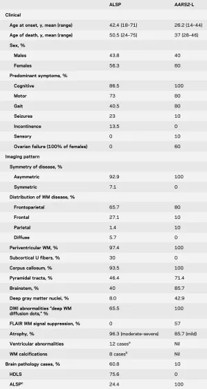
Table 2 Summarized clinical, imaging, and pathologic phenotypes of ALSP and AARS2-L
ALSP AARS2-L
Clinical
Age at onset, y, mean (range) 42.4 (18–71) 26.2 (14–44)
Age of death, y, mean (range) 50.5 (24–75) 37 (28–46)
Sex, %
Males 43.8 40
Females 56.3 60
Predominant symptoms, %
Cognitive 86.5 100
Motor 73 80
Gait 40.5 80
Seizures 23 10
Incontinence 13.5 0
Sensory 0 10
Ovarian failure (100% of females) 0 60
Imaging pattern
Symmetry of disease, %
Asymmetric 92.9 100
Symmetric 7.1 0
Distribution of WM disease, %
Frontoparietal 65.7 80
Frontal 27.1 10
Parietal 1.4 10
Diffuse 5.7 0
Periventricular WM, % 97.4 100
Subcortical U fibers, % 30 0
Corpus callosum, % 93.5 100
Pyramidal tracts, % 46.4 71.4
Brainstem, % 40 85.7
Deep gray matter nuclei, % 8.0 42.9
DWI abnormalities“deep WM diffusion dots,”%
65.5 100
FLAIR WM signal suppression, % 0 57
Atrophy, % 96.3 (moderate–severe) 85.7 (mild)
Ventricular abnormalities 12 casesa Nil
WM calcifications 8 casesb Nil
Brain pathology cases, % 60.8 10
HDLS 75.6 0
ALSPc 24.4 100
Abbreviations: ALSP5adult-onset leukoencephalopathy with axonal spheroids and pig-mented glia; DWI5diffusion-weighted imaging; FLAIR5fluid-attenuated inversion recov-ery; HDLS5hereditary diffuse leukoencephalopathy with axonal spheroids; WM5white matter.
aIdentified from our own institutional cases and through review of published images in the
literature. Only raw numbers are presented.
bReported cases in the literature and from review of our own cases. Only raw numbers
presented.
cFeatures of both pigmentary orthochromatic leukodystrophy and HDLS reported or
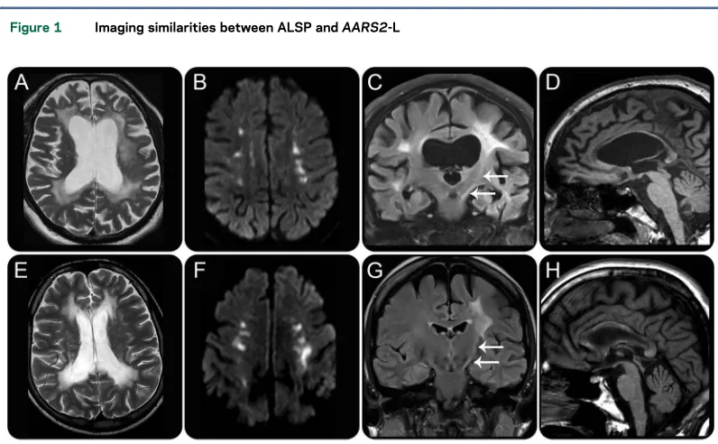
ALSP.White matter signal.White matter hyperintensity was present on T2-weighted (T2-w) sequences in all patients with data (74/74). Of those with T2-w fluid-attenuated inversion recovery (FLAIR) imaging, none showed suppression of white matter signal to suggest white matter rarefaction. Of the 16 patients with T1-weighted (T1-w) imaging, all showed hypo-intensity in affected regions (figure 1, A, D, and E). Distribution of white matter abnormality. Predomi-nance of the white matter signal abnormality was most commonly seen in a frontoparietal distribution (46/70), followed by a frontal predominant distribu-tion (19/70). Diffuse involvement was seen in 4/70, with a parietal predominant distribution present in only 1/70. Of those with reports of symmetry or asymmetry, 39/42 had asymmetric white matter sig-nal changes. A substantial majority of patients showed confluent white matter signal changes (30/33) as opposed to patchy involvement (3/33). Subcortical U fibers were involved in 9/30 patients, and it was noted in our patient cohort that the U fi-bers are generally spared until very late in the disease time course. Conversely, the periventricular white matter tends to be involved in most cases (37/38). However, in our patient cohort, it was noted that the immediate periependymal periventricular white matter tends to show a thin rim of sparing until later in the disease course.
Corpus callosum involvement.Involvement of the cor-pus callosum was present in 58/62 cases, with gener-alized callosal thinning present in 36/58 (figure 1D). The splenium was most commonly involved (12/13), followed by the genu (8/13) and the callosal body (7/13), with the location of callosal involvement spatially linked to the location of deep white matter disease.
Pyramidal tract involvement. Involvement of the pyramidal tracts was present in 13/28 (figure 1C). The pyramidal tract signal changes tended to occur later in the natural history of the disease in our patient group.
Deep gray matter nuclei and cerebellar involvement.The deep gray matter nuclei are seldom involved in ALSP (2/25). These include a case which showed basal gan-glia calcifications14and another case which described striatal volume loss.15Cerebellar involvement has on-ly been described in a single case where the cerebellar peduncles were involved,16 as a rule, however, the cerebellum tends to be spared (22/23).
DWI abnormalities.Abnormally high signal on the DWI trace images was described in 19/29 patients with diffusion data, suggesting that diffusion abnor-malities are seen in a majority of patients. On appar-ent diffusion coefficiappar-ent (ADC) maps, these areas either exhibit truly restricted diffusion or, as seen in some of our cases, diffusivity approximates that of normal white matter. The diffusion abnormalities
Figure 1 Imaging similarities between ALSP andAARS2-L
are punctate and are most commonly seen in a linear arrangement, with lesion clusters aligned parallel to the ependymal surface of the lateral ventricles with a predisposition for the corona radiata (14/19) and centrum semiovale (10/19). DWI abnormalities may also arise in the splenium of the corpus callosum, although this is seen less commonly (4/19). In our patient cohort, we have observed that in contrast to diffusion abnormalities secondary to conventional infarction, these punctate areas of punctate DWI hy-perintensity often persist over time, sometimes enlarging and coalescing. Other authors have described DWI abnormalities with a similar temporal evolution in ALSP15,17,18 which correlates with the temporal observations we have made in our patient group. To summarize the diffusion findings, we have coined the term“deep white matter diffusion dots,” and the findings are illustrated in figure 1B.
Atrophy.Almost all patients (52/54) showed atro-phy. In our cohort, the degree and location of atrophy correlated with the severity and location of white
matter signal abnormality and was progressive over time (figure 1A).
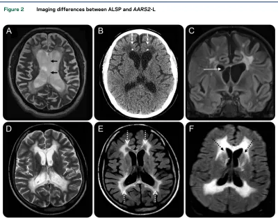
Ventricular abnormalities. An observation in our cohort of patients was the presence of ventricular abnormalities. Ventricular septations were seen in 3/8 patients (figure 2C) and were commonly located within the frontal horns or near the foramen of Monro. Other ventricular abnormalities observed were septum pellucidum fenestrations (1/8, figure 2A) and a cavum septum pellucidum and/or cavum vergae (5/8 patients). This had been previously noted in 4 patients in a large CSF1R series,16 and upon review of the published images of ALSP, a cavum septum pellucidum or cavum vergae was evident in a further 6 patients.19,20Ventricular enlargement was seen in almost all patients with data (26/27) and was proportional to the degree of volume loss.
Calcifications.In total, calcifications have been re-ported in 8 patients10,14,21,22; these were most com-monly reported to be located in the periventricular white matter adjacent to the frontal horns.10,14,22
Figure 2 Imaging differences between ALSP andAARS2-L
Three of our cases had CT data and only one of those showed calcifications which were similarly present in the periventricular white matter adjacent to the fron-tal horns (figure 2B).
Vascular.Five of 8 patients in our series had vascu-lar imaging, including digital subtraction cerebral angiography in case 1, table 1. None of our patients showed vascular abnormalities, and no vascular abnormalities have been reported in the literature in association with ALSP. Of those patients in our group who had either T2*or SWI, none showed evidence of microhemorrhages.
Enhancement. Of the 6 cases where there was enhanced imaging, no enhancement was observed.
AARS2-L.White matter signal.All patients showed T2-w hyperintense, T1-w hypointense white matter signal abnormalities (figure 1, E, G, and H). Of those pa-tients with FLAIR data, 4/7 showed suppression in the periventricular regions, presumably as a result of white matter rarefaction (figure 2E). These areas of FLAIR suppression did not show substantial volume loss, which is a feature similar to that seen in vanish-ing white matter disease.23
Distribution of white matter abnormality.Eight of ten patients showed predominant involvement of the frontoparietal white matter. The remaining 2 patients showed frontal and parieto-occipital predominant involvement. Involvement was asymmetric in all pa-tients with data (7/7) and was most commonly con-fluent (6/7) rather than patchy. Subcortical U fibers
were always spared, and the periventricular white matter was always involved (figure 1E).
Corpus callosum involvement.All patients with data showed involvement of the corpus callosum (7/7), figure 1H. The splenium was always involved with genu, and body involvement was present in 5/7 pa-tients. The corpus callosum showed only mild thin-ning (6/7), much less severe than that seen in ALSP (figure 1H).
Pyramidal tract involvement. Five of 7 patients showed pyramidal tract involvement; this was most commonly seen to involve the posterior limb of the internal capsule and the corticospinal tract in the brainstem (figure 1G). The involvement of the pyra-midal tracts was asymmetric. One of 7 patients showed specific involvement of the frontopontine tract.6
Deep gray matter nuclei and cerebellar involvement. Three of 7 patients showed T2-w hyperintensity in the caudate heads (figure 2F). One of 7 showed severe atrophy of the cerebellum.
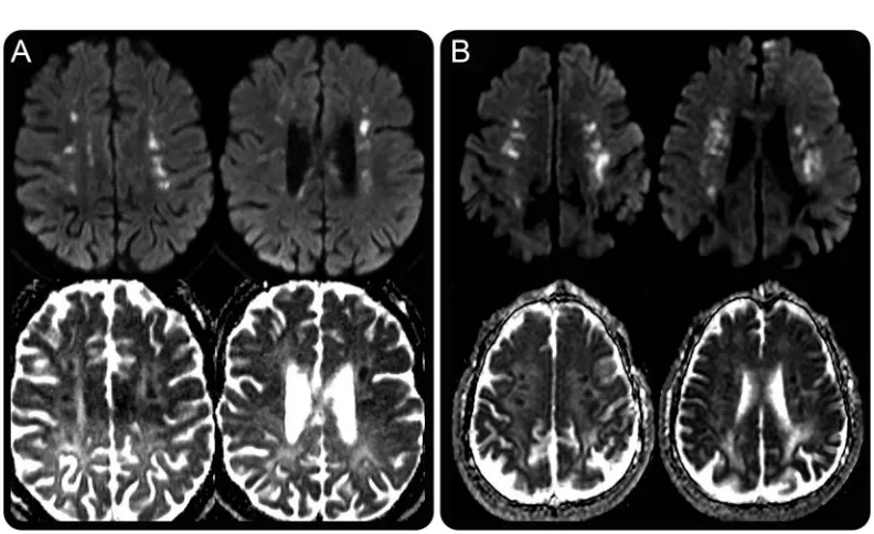
Diffusion abnormalities.All patients with diffusion data (5/5) showed restricted diffusion, with similar “deep white matter diffusion dots”as seen in ALSP, with linear, punctate, and partly confluent areas of restricted diffusion seen in the corona radiata and centrum semiovale with the lesion clusters aligned roughly parallel to the ependymal surface of the lateral ventricles (figure 3B). Restricted diffusion was seen in the corpus callosum of 3/5 patients, with the genu,
Figure 3 “Deep white matter diffusion dots”in ALSP andAARS2-L
body, and splenium all involved with equal fre-quency. Progression information was available in only one patient (case 3, table 1) in whom there was no temporal change in the diffusion during a 7-month follow-up period.
Atrophy. Atrophy was present in 6 of 7 patients with data and was disproportionately mild compared with the degree of white matter signal abnormality. This is in contrast to ALSP in which atrophy was gen-erally severe and proportional to the degree of white matter disease.
Ventricular abnormalities.No ventricular abnormali-ties were evident in any of the patients.
Calcifications.No calcifications were evident in the 2 patients who had CT data.
Vascular.Case 3 in table 1, a 26-year-old female, showed a single microhemorrhage on a susceptibility weighted study. No gradient echo or SWI data were available for the other cases. No angiographic data were available in any of the 10 cases.
Enhancement.No enhancement was detected in the 2 patients with enhanced MRIs.
DISCUSSION ALSP and AARS2-L are linked in many ways; they share common clinical features, imaging characteristics, and can share identical histopathologic appearances.4There are, however, some important differentiating features, both on clinical and imaging grounds, which can help to favor one diagnosis over the other, many of which are presented in table 2. ALSP has shown to have an older age at onset, typically occurring in the fifth decade as opposed toAARS2-L where the onset of disease tends to occur in the third decade of life. All known females withAARS2-L present as an ovario-leukodystrophy with ovarian failure, a feature which is not seen in ALSP. Cognitive impairment and motor dysfunction are the most common presenting symptoms in both ALSP and AARS2-L, with both conditions also presenting with gait abnormalities, seizures, and incontinence. Sensory symptoms have been reported in 1 patient with AARS2-L and in no patients with ALSP.
There are many imaging features that ALSP and AARS2-L have in common. Both show frontopar-ietal predominant, slightly asymmetric, confluent T2-w hyperintense, T1-w hypointense white mat-ter signal abnormalities which have a predilection for the periventricular and deep white matter and tend to spare the subcortical U fibers (figure 1, A and E). Both conditions show involvement of the corpus callosum; however, in ALSP, the callosal involvement is associated with severe thinning, which is not a feature of AARS2-L (figure 1, D and H). Both conditions demonstrate pyramidal tract involvement, typically in the posterior limb
of the internal capsule and brainstem (figure 1, C and G).
The most striking shared imaging feature of ALSP and AARS2-L is the presence of DWI “deep white matter diffusion dots” which, to our knowledge, are not described in any other leukodystrophy. These DWI lesions are an imaging mimic of internal border-zone infarction and thus can potentially mislead clini-cians into thinking that there is an underlying vascular pathology; however, the persistence and progression of these lesions over months to years differentiate them from areas of acute infarction. These areas of diffusion abnormality are multifocal, punctate, with the lesion clusters usually being roughly parallel to the ependymal surface of the lateral ventricles (figure 3, A and B). These lesions are typically located in the centrum semi-ovale and/or corona radiata but can also occur else-where especially along the corpus callosum or supratentorial pyramidal tracts. The diffusion abnor-malities in ALSP have ADC values which appear sim-ilar to or slightly lower than normal white matter, whereas the foci observed inAARS2-L tend to exhibit more marked reductions in ADC, as would be seen in acute infarcts. Diffusion abnormalities are seen in a wide range of leukodystrophies, with restricted dif-fusion due to presumed intramyelinic edema observed in metachromatic leukodystrophy, globoid cell leuko-dystrophy, X-linked adrenoleukoleuko-dystrophy, hyperho-mocystinemias, Canavan disease, maple syrup urine disease, phenylketonuria, and leukodystrophy with brainstem and spinal cord involvement and high lac-tate.24 The patterns of diffusion abnormality in the aforementioned diseases are diverse; however, none demonstrate the characteristic pattern of“deep white matter diffusion dots,”which is seen in 66% of pa-tients with ALSP and all known cases ofAARS2-L with DWI included in this review.
our cases (figure 2B) and has been reported by others.10,14,21
Ventricular abnormalities, including ventricular septations, septum pellucidum fenestrations, or a cav-um septcav-um pellucidcav-um and/or cavcav-um vergae (figure 2, A and C), were evident in 7/8 ALSP cases in our institutional database and were not described in any cases of AARS2-L. Ventricular septations are usually seen in the setting of previous ventriculitis or intraven-tricular hemorrhage,25and outside of this context is an unusual finding. Although conjecture, it is possible that the septations we have identified in patients with ALSP are secondary to an inflammatory response triggered within the ventricles. The presence of a cavum septum pellucidum or cavum vergae is of unknown signifi-cance, and when present in the general population is considered a normal variant. The frequent observation of a cavum septum pellucidum and/or cavum vergae in our case group may be incidental; however, the preva-lence in the general population is felt to be in the vicinity of 12%–20%,26which is in contrast to 63% in our local ALSP series. It is possible that damage to the white matter of the septum pellucidum allows com-munication of ventricular CSF with the potential space between the septal laminae, giving rise to a communi-cating form of a septum pellucidum, similar to that described in chronic traumatic encephalopathy.27
We have synthesized the phenotypic data for pa-tients with ALSP andAARS2-L with the intention of highlighting both the similarities and differences between the clinical, imaging, and pathologic pheno-types of the 2 conditions. Distinguishing between ALSP and AARS2-L radiologically is of particular importance, given that the histopathologic findings are near identical in both4and a brain biopsy in the absence of the correct genetic test may be misleading. Also recently, there has been a report of possible halted progression of ALSP in a CSF1Rmutation– positive patient who underwent stem cell trans-plantation,28 potentially indicating a future thera-peutic implication for correctly diagnosing these entities, although this is yet to be confirmed. As the genotypic spectrum of ALSP andAARS2-L expands, detailed genotype-phenotype correlations for each condition will be needed to determine the phenotypic significance of each mutation.
AUTHOR CONTRIBUTIONS
Rahul Lakshmanan: study concept and design, data collection and anal-ysis, radiologic analanal-ysis, and drafting and revising manuscript. Matthew E. Adams: study concept and design, data collection and analysis, radio-logic analysis, reviewing manuscript for intellectual content, and revising manuscript. David S. Lynch and Justin A. Kinsella: data collection and analysis and reviewing and revising manuscript. Rahul Phadke: reviewing manuscript for intellectual content. Jonathan M. Schott: reviewing man-uscript for intellectual content and revising manman-uscript. Elaine Murphy and Jonathan D. Rohrer: reviewing manuscript for intellectual content. Jeremy Chataway, Henry Houlden, and Nick C. Fox: reviewing
manuscript for intellectual content and revising manuscript. Indran Da-vagnanam: study concept and design, data collection and analysis, radio-logic analysis, reviewing manuscript for intellectual content, revising manuscript, and final approval of manuscript.
ACKNOWLEDGMENT
The authors acknowledge the patients for their participation in this study. They also thank doctors Günes¸ Altıokka Uzun, Merih Karbay, Zeynep Tüfekçioglu, and Has¸met Hanagasıfrom the Department of Neurology, Istanbul School of Medicine, Istanbul University, Istanbul, Turkey, for their contribution of cases to the adult leukodystrophy database at the National Hospital for Neurology and Neurosurgery, Queens Square, Lon-don, United Kingdom.
STUDY FUNDING
Supported by the Medical Research Council, Wellcome Trust, and NIHR UCLH/UCL Biomedical Research Centre (BRC). Prof. Fox is a National Institute for Health Research (NIHR) senior investigator. The Dementia Research Centre is an ARUK coordinating center and re-ceives support from the NIHR Queen Square Dementia Biomedical Research Unit, the MRC Dementias Platform UK, and the Leonard Wolfson Experimental Neurology Centre.
DISCLOSURE
Dr. Lakshmanan, Dr. Adams, and Dr. Lynch report no disclosures. Dr. Kinsella has received travel funding from Novartis and has received research support from the Stanley Thomas Johnson Foundation, Bayer Scher-ing Ireland, Pfizer Ireland, and ELItech UK. Dr. Phadke reports no disclo-sures. Dr. Schott has served on scientific advisory boards for Eli Lilly and Axon Neuroscience; has received travel funding and speaker honoraria from the US Alzheimer’s Association, the European Academy of Neurology, and the Association of British Neurologists; receives publishing royalties from Henry Stewart Talks; has been a consultant for Eli Lilly and Roche; and has received research support from Eli Lilly, Alzheimer’s Research UK, UK Alzheimer’s Society, EPSRC, Medical Research Council, EU, Horizon 20:20, Wolfson Foundation, Wellcome Trust, and Brain Research Trust. Dr. Murphy has received speaker honoraria from Vitaflo International, Shire HGT, Genzyme Inc., and Nutricia; has served on the editorial board of
Orphanet Journal of Rare Disease; and has received research support from Shire HGT, Genzyme, and Nutricia. Dr. Rohrer is supported by an MRC Clinician Scientist Fellowship (MR/M008525/1); has served on a scientific advisory board for Ionis Pharmaceuticals; and has received research support from the NIHR Rare Diseases Translational Research Collaboration. Dr. Chataway has served on scientific advisory boards for Roche and Merck and has received support from Novartis, Teva, Sanofi, and MRC-EME. Prof. Houlden has received research support from the Medical Research Council (MRC) UK, The BRT, The MDA USA, Muscular Dystrophy UK, Rosetr-ees Trust, The Wellcome Trust, the National Institute for Health (NIHR) UCL/UCLH BRC, and (NIHR) UCLH/UCL Biomedical Research Centre. Prof. Fox has served on a scientific advisory board for Biogen; has served on the editorial boards ofAlzhemier’s Disease and Associated Disorders, Neurode-generative Diseases,Alzheimer’s Research and Therapy, andLancet Neurology; holds a patent for QA Box for automated checking of MRI scans; receives publishing royalties from Springer; has been a consultant for Janssen, Roche/ Genentech, Eli Lilly, Novartis Pharma AG, Sanofi, GSK, and Biogen; and has received support from Novartis, Sanofi, Genentech/Roche, GSK, Biogen, Eli Lilly, Janssen, MRC (UK), NIA, NIHR, and the Wolfson Foundation. Dr. Davagnanam receives study support from the National Institute for Health Research University College London Hospitals Biomedical Research Centre. Go to Neurology.org/ng for full disclosure forms.
Received October 5, 2016. Accepted in final form January 4, 2017.
REFERENCES
3. Parikh S, Bernard G, Leventer RJ, et al. A clinical approach to the diagnosis of patients with leukodystro-phies and genetic leukoencephelopathies. Mol Genet Metab 2015;114:501–515.
4. Lynch D, Zhang WJ, Lakshmanan R, et al. Adult onset leukoencephalopathy with axonal spheroids and pig-mented glia: recessive mutations in AARS2 identified in a series of CSF1R negative patients. JAMA Neurol 2016; 73:1433–1439.
5. Rademakers R, Baker M, Nicholson A, et al. Mutations in the colony stimulating factor 1 receptor (CSF1R) gene cause hereditary diffuse leukoencephalopathy with sphe-roids. Nat Genet 2012;44:200–205.
6. Dallabona C, Diodato D, Kevelam S, et al. Novel (ovario) leukodystrophy related to AARS2 mutations. Neurology 2014;82:2063–2071.
7. Nicholson A, Baker M, Finch N, et al. CSF1R mutations link POLD and HDLS as a single disease entity. Neurol-ogy 2013;80:1033–1040.
8. Wider C, van Gerpen J, DeArmond S, Shuster EA, Dick-son D, Wszolek Z. Leukoencephalopathy with spheroids (HDLS) and pigmentary leukodystrophy (POLD) A single entity? Neurology 2009;72:1953–1959.
9. Ginhoux F, Greter M, Leboeuf M, et al. Fate mapping analysis reveals that adult microglia derive from primitive macrophages. Science 2010;330:841–845.
10. Konno T, Tada M, Tada M, et al. Haploinsufficiency of CSF-1R and clinicopathologic characterization in patients with HDLS. Neurology 2014;82:139–148.
11. Götz A, Tyynismaa H, Euro L, et al. Exome sequencing identifies mitochondrial alanyl-tRNA synthetase muta-tions in infantile mitochondrial cardiomyopathy. Am J Hum Genet 2011;88:635–642.
12. Lynch DS, Jaunmuktane Z, Sheerin UM, et al. Hereditary leukoencephalopathy with axonal spheroids: a spectrum of phenotypes from CNS vasculitis to parkinsonism in an adult onset leukodystrophy series. J Neurol Neurosurg Psychiatry 2016;87:512–519.
13. Ahmed R, Guerreiro R, Rohrer J, et al. A novel A781V mutation in the CSF1R gene causes hereditary diffuse leucoencephalopatahy with axonal spheroids. J Neurol Sci 2013;332:141–144.
14. Ueda S, Yamashita H, Hikiami R, Sawamoto N, Yoshida K, Takahashi R. A novel A792D mutation in the CSF1R gene causes hereditary diffuse leukoencephalopathy with axonal spheroids characterized by slow progression. eNeurologicalSci 2015;1:7–9.
15. Lee D, Yun JY, Jeong JH, Yoshida K, Nagasaki S, Ahn T-B. Clinical evolution, neuroimaging, and volumetric anal-ysis of a patient with a CSF1R mutation who presented with progressive nonfluent aphasia. Parkinsonism Relat Disord 2015;21:817–820.
16. Sundal C, Van Gerpen J, Nicholson A, et al. MRI char-acteristics and scoring in HDLS due to CSF1R gene mu-tations. Neurology 2012;79:566–574.
17. Terasawa Y, Osaki Y, Kawarai T, et al. Increasing and persistent DWI changes in a patient with hereditary dif-fuse leukoencephalopathy with spheroids. J Neurol Sci 2013;335:213–215.
18. Mateen F, Keegan BM, Krecke K, Parisi JE, Terenerry MR, Pittock SJ. Sporadic leucodystrophy with neuroaxo-nal spheroids: persistence of DWI changes and neurocog-nitive profiles: a case study. J Neurol Neurosurg Psychiatry 2010;81:619–622.
19. Kim E-J, Shin J-H, Lee JH, et al. Adult-onset leukoence-phalopathy with axonal spheroids and pigmented glia linked CSF1R mutation: report of four Korean cases. J Neurol Sci 2015;349:232–238.
20. Kleinfeld K, Mobley B, Hedera P, Wegner A, Sriram S, Pawate S. Adult-onset leukoencephalopathy with neuro-axonal spheroids and pigmented glia: report of five cases and a new mutation. J Neurol 2013;260:558–571. 21. Meyer-Ohlendorf M, Braczynski A, Al-Qaisi O, et al.
Comprehensive diagnostics in a case of hereditary dif-fuse luekodystrophy with spheroids. BMC Neurol 2015; 15:1–6.
22. Fujioka S, Broderick D, Sundal C, Baker M, Rademakers R, Wszolek Z. An adult-onset leukoencephalopathy with axonal spheroids and pigmented glia accompanied by brain calcifications. J Neurol 2013;260:2665–2668.
23. van der Knaap M, Pronk JC, Scheper GC. Vanishing white matter disease. Lancet Neurol 2006;5:413–423. 24. Patay Z. Diffusion-weighted MR imaging in
leukodystro-phies. Eur Radiol 2005;15:2284–2303.
25. Andresen M, Juhler M. Multiloculated hydrocephalus: a review of current problems in classification and treat-ment. Childs Nerv Syst 2012;28:357–362.
26. Sarwar M. The septum pellucidum: normal and abnormal. AJNR Am J Neuroradiol 1989;10:989–1005.
27. Corsellis J, Bruton C, Freeman-Browne D. The aftermath of boxing. Psychol Med 1973;3:270–303.
DOI 10.1212/NXG.0000000000000135
2017;3;
Neurol Genet
Rahul Lakshmanan, Matthew E. Adams, David S. Lynch, et al.
related leukodystrophy
−
mutation
AARS2
Redefining the phenotype of ALSP and
This information is current as of February 15, 2017
reserved. Online ISSN: 2376-7839.
Published by Wolters Kluwer Health, Inc. on behalf of the American Academy of Neurology. All rights an open-access, online-only, continuous publication journal. Copyright Copyright © 2017 The Author(s).
is an official journal of the American Academy of Neurology. Published since April 2015, it is
Services
Updated Information &
http://ng.neurology.org/content/3/2/e135.full.html
including high resolution figures, can be found at:
References
http://ng.neurology.org/content/3/2/e135.full.html##ref-list-1
This article cites 28 articles, 4 of which you can access for free at:
Citations
http://ng.neurology.org/content/3/2/e135.full.html##otherarticles
This article has been cited by 6 HighWire-hosted articles:
Subspecialty Collections
http://ng.neurology.org//cgi/collection/mri
MRI
http://ng.neurology.org//cgi/collection/dwi
DWI
http://ng.neurology.org//cgi/collection/ct
CT
http://ng.neurology.org//cgi/collection/all_genetics
All Genetics
http://ng.neurology.org//cgi/collection/all_demyelinating_disease_cns
All Demyelinating disease (CNS)
following collection(s):
This article, along with others on similar topics, appears in the
Permissions & Licensing
http://ng.neurology.org/misc/about.xhtml#permissions
its entirety can be found online at:
Information about reproducing this article in parts (figures,tables) or in
Reprints
http://ng.neurology.org/misc/addir.xhtml#reprintsus
Information about ordering reprints can be found online:
reserved. Online ISSN: 2376-7839.
Published by Wolters Kluwer Health, Inc. on behalf of the American Academy of Neurology. All rights an open-access, online-only, continuous publication journal. Copyright Copyright © 2017 The Author(s).
is an official journal of the American Academy of Neurology. Published since April 2015, it is