December 31, 2019
Isolation and Identification of E-Coli from Infected Cases from Broilers Farms In 2019
Author’s Details: Marwa. M. Khedr, May. F. Abdelaty and Mahmoud. M. Abotaleb
Central Laboratory for Evaluation of Veterinary Biologics, (CLEVB), Abbassia, Cairo, Egypt&Reference Laboratory of Quality control on Poultry Production, Animal Health Research Institute, Dokki, Giza, Egypt
Received Date: 08-Dec-2019 Accepted Date: 26-Dec-2019 Published Date: 31-Dec-2019
_________________________________________________________________________________________
Abstract
E. coli infections are responsible for great economic loss and causing a serious threat to the poultry industry. One hundred and fifty samples were collected from different poultry farms for isolation of E.coli bacteria. Different percentage rates were 6%, 8.6% and 5.3 obtained from apparently healthy birds, freshly dead and diseased birds respectively. Thirty E.coli isolates were identified as (O1, O2, O6, O78 and O157). E.Coli serotypes lethality tests in chicks showing clinical signs which appeared on the 5th day and the mortalities recorded at the 7th day post infection with percentage 7% of the challenged group. Lethality tests in mice showed mortality 100% at 3rd day post challenge in pathogenic serotypes. Molecular characterization was done for detection of iss, ombA, fimH, iroN and pabC virulence genes for E.coli serotypes. All isolates were positive for all genes except papC was negative. Antibiotic susceptibility test was applied for all for detection of the suitable antimicrobial agent in the infected poultry farms with E.coli in which all isolated serotypes were sensitive to cefatriaxone, tobramycin except O6 and were intermediate, with spectinomycin, amikacin and fosfomycin. Recently isolated serotypes may help in local vaccine production.
Key words: E.coli, pathogenicity, virulence genes, antibiotic
_________________________________________________________________________________________
1. Introduction
Escherichia coli (E.coli) is considered as a member of the normal microflora of all warm-blooded animals including poultry (Kaper et al., 2004). However, in the debilitated or in immune suppressed hosts, or when gastro-intestinal barriers are violated, even normal “non-pathogenic” strain of E.coli can cause infection to poultry, humans and animals. Moreover, there are certain E. coli strains designated as avian pathogenic E.coli
which can cause localized or systemic infection outside the avian gut, which indicates as Extraintestinal Pathogenic E. coli (ExPEC). The infection caused by ExPEC is termed colibacillosis (Nakazato et al., 2009) characterized by yolk sac infection, omphalitis, respiratory tract infection, septicemia, salpingitis and peritonitis affect broiler chickens aged 4–6 weeks as well as hazard economic impact in the infected birds’ productivity, high mortality, high cost condemnation of infected carcasses at slaughter, prophylaxis and treatment (Lutful et al., 2010).
More than 1000 serotypes are known, but only a few are considered as important in avian pathology (Sojka and Carnaghan. 1961). Serotypes O1, O2, and O78 mostly stands behind the infection and represent 15-61% of the isolates, yet other types still exist (Dho-Moulin and Fairbrother. 1999) (Rahmanet al., 2004), therefore, Serotyping of potentially pathogenic E-coli strains is necessary to improve (Stordeur and Mainil. 2002).
(Barbieri et al., 2013) (Dissanayake et al., 2014). P-fimbriae (papC) is important in the later stages of infection for the adhesion to internal organs (Dho and Fairbrother 1999).
Detection of pathogenic E. coli strains that cause septicemia becomes important for effective treatment with antimicrobial therapy and control resulting in reducing both the incidence and mortality which associated with colibacillosis (Dho et al., 1990; Susantha et al., 2001; Jeffrey et al., 2002; Geornaraset al., 2004; Zhao et al.,
2009; Qabajah and Yaqoub. 2010) which is considered as challenging during its identification as pathogenic or nonpathogenic E. coli is a regular host of the digestive tract of poultry. The effective control of E.coli
infection depended on suitable antimicrobial drugs due to the hazard use of antibiotics in humans and animals subsequently, has become the major factor for the emergence and spread of drug-resistance traits among pathogenic and commensal bacteria. The development of multi-drug resistance in E.coli is one of the major concerns worldwide (Von Baum and Marre. 2004). So, in this study, depending on the isolation of E.coli
serotypes from different infected poultry farms in Cairo and Giza governorates for the detection pathogenic types by lethality test and PCR for further local vaccine production and detect the suitable antibiotic for the infected poultry in the infected farms.
2. Materials and Methods:
2.1. Samples
:
One hundred and fifty samples were collected from apparently healthy birds (cloacal swabs), diseased birds (cloacal swabs) and freshly dead birds (liver, heart, spleen, lung and gall bladder samples) from suspected chickens with E. coli infection as shown in Table (1). The samples were transferred immediately to sterile buffered peptone water, then wrapped with ice, kept in a box and transferred directly into the lab which then isolated, characterized and serotyped in CLEVB (Central Laboratory for Evaluation of veterinary Biologics) and AHRI (Animal Health research institute).
Table 1: Sampling of examined organs samples
Source Number of samples
Apparently healthy 60
Diseased 40
Freshly dead 50
Total 150
2.2 Preparation of sample:
The carcasses were promptly necropsied according to the standard procedures described by (Lowenstin. 1986). About 25 g of each were collected from the internal portion aseptically in a sterile plastic bag (Falconpack, UAE). Samples were kept at + 4 °C for a maximum of 24 until culturing.
2.3 Reference E.coli strain
E.Coli ATCC 25922 (AHRI) was used as a positive control strain and E.coli reference antisera 295347 Lot 473096. 2.4
Isolation and identification of
E. coli
:
a) Bacteriological methods
A loopful of the culture suspension was streaked on to MacConkey agar (Lab M, Neogen Europe Lt) and incubated sample for 24 h at 37 °C aerobically. Isolation of E. coli was performed according to (Quinn et al., 2002).
b
)
Biochemical test:Suspected E. coli colonies were then transferred onto nutrient agar for further identification using biochemical tests. The serotypes were subjected to an API 20E identification system (Kwon et al., 2008).
c) The VITEK 2 automated system (bioMérieux): was used for confirmation of putative E.coli isolates (Espinar et al., 2011).
d) Serological identification
December 31, 2019
A-Lethality of E. coli isolates in day-old SPF chicks (Vidotto et al., 1990):
Unvaccinated two day-old SPF chicks (8 chicks / serotype) obtained from El- Fayuim farm were used, which challenged orally with 0.5 ml containing 9.0 × 10⁸ CFU/ml. Daily monitoring of clinical Signs, postmortem lesion of the inoculated serotypes for 7days post inoculation and monitored daily.
B-Lethality of E.coli isolatesin Mice (Wyattet al., 2003):
Female swiss mice (8th to 10th weeks of age) were housed in micro-isolator cages and provided with food and water ad libitum. Mice were lightly anesthetized with Metofane (methoxyfluorane) (Schering-Plough Animal Health Co., Union, N.J.) in a glass desiccator and challenged with 0.2 ml of E.coli culture containing 1 × 10⁹ CFU in order to measure morbidity and mortality. Control mice were inoculated with 0.2ml of the PBS diluents.
2.6 Detection of virulence genes for pathogenic E.coli isolates:
The DNA extraction was performed using the QIAamp DNA Minikit (Qiagen, Germany, GmbH) with modification from the manufacture's recommendations.
Oligonucleotide Primer: Primers used were supplied from Metabion (Germany) are listed in Table (2).
PCR amplification: Primers were utilized in a 25- µl reaction containing 12.5 µl of Emerald Amp Max PCR Master Mix (Takara, Japan), 1 µl of each primer of 20 pmol concentrations, 4.5 µl of water, and 6 µl of DNA template. The reaction was performed in an applied biosystem 2720 thermal cycler.
Table 2: Primers sequences, target genes, amplicon sizes and cycling conditions
Primer designation
Primer Denaturation
Amplification (35 cycles) Final
Extension
Primers sequences
5`---3` Product Reference Sequence
denaturation Annealing Extension
Issgene 94°C/5min 94°C /5min 54°C/30sec 72°C/30sec 72°C/7min.
F. ATGTTATTTTCTGCCGCTCTG
R. CTATTGTGAGCAATATACCC 266bp
Yaguchi, et al., (2007)
ompAgene 94°C/5min 94°C/30 sec. 58°C/1min 72°C/1min 72°C/10min F. AGCTATCGCGATTGCAGTG
R. GGTGTTGCCAGTAACCGG
919bp Ewers et al., (2007)
iroN gene 95°C/5min 94°C/30sec 50°C/40sec 72°C/50sec 72°C/10min F. ATCCTCTGGTCGCTAACT
R. CTGCACTGGAAGAACTGTTCT
847bp Ewers et al., (2007)
fimH gene 95°C/5min 94°C/30sec 50°C/40sec 72C/45sec 72°C/10min F. GCAGAACGGATAAGCCGTGG
R. GCAGTCACCTGCCCTCCGGTA
508bp Ghanbar and
Salehi. (2010)
papC gene 94°C/5min 94°C/30sec 50°C/40sec 72°C/45sec 72°C/10min F. TGATATCACGCAGTCAGTAGC
R. CCGGCCATATTCACATAA
501bp Wen-jieet al., (2008)
2.7Antimicrobial susceptibility test:
The antimicrobial susceptibility testing of E.coli isolates was conducted using the Kirby- Bauer disc diffusion method on Mueller-Hinton agar (Lab M, Neogen Europe Lt) according to the guidelines of the Clinical and Laboratory Standards Institute.(CLSI/NCCLS. 2009). the antimicrobials and their concentrations used for the susceptibility testing were listed in the table (3)
Table 3: The antimicrobials and their concentrations used for the susceptibility testing:
Test group Antimicrobial agent Disk content Zone diameter nearest whole mm Resistant Intermediate Sensitive Aminoglycosides Tobramycin (TOB10)
10 µg ≤12 12-14 ≥15
Amikacin (AK30)
30 µg ≤14 15-16 ≥17
Tetracyclines Oxytetracycline (TE30) 30μg ≤11 12-14 ≥15
Fluoroquinoiones Ciprofloxacin (CIP5) 5μg ≤15 16-20 ≥21
Levofloxacin (LEV5) 5μg ≤ 13 14-16 ≥ 17
Penicillins Penicillin 10 μg ≤10 - ≥28
Cephalosporins Ceftriaxone 30µg ≤13 14-20 ≥21 Folate pathway
inhibitors
Trimethoprim+sulfamethoxazole SXT
1.25-23.75Μg ≤10 11-15 ≥16
Clindamycin DA 2 μg ≤14 15-20 ≥21
Fosfmycins Fosfomycin 200 μg ≤12 13-15 ≥16
Quinolones Naldixic acid 30 μg ≤13 14-18 ≥19
Spectinomycin 10 μg ≤12 13-14 ≥15
Enrofloxacin 10 μg ≤12 13-16 ≥17
3. Results:
3.1Results of isolation and characterization:
As shown in table (4): A total of 30 suspected E.coli isolates were Gram negative, lactose fermenter on MacConkey, showed greenish metallic sheen on eosin methylene blue agar. On Nutrient agar medium showed Rounded, non-pigmented colonies, while on MacConkey agar medium appeared rounded, non-mucoid pink colonies (lactose fermenter) on the surface of the medium. The same isolates on Eosin Methylene blue (EMB) agar showed a distinctive yellow, green metallic.
Depending on the results of the API 20E identification system, the suspected E. coli isolates were 30 out of 150 suspected cases. The isolates gave positive reactions with ONPG, LDC, ODC, IND, GLU, MAN, SOR, RHA, SAC, MEL and ARA tests and negative reaction with ADH, CIT, H2S, URE, TDA, VP, GEL, INO, AMY and OX tests. Based on data revealed from The VITEK 2 automated system (bioMérieux). The thirty isolates were confirmed to be E.coli.
Table 4: Incidence of Ecoli in the examined organs samples
Source Number of examined organ samples
Number of positive samples
Apparently healthy 60 9(6%)
Diseased 40 13(8.6%)
Freshly dead 50 8(5.3%)
Total 150 30(20%)
The thirty suspected isolates that biochemically themorphological identified as E. coli which was classified into different serotypes using polyvalent and monovalent using standard antisera. The data revealed from the table (5), showed different serotypes (O1, O2, O6, O78 and O157) were (6, 6, 5, 9 and 4 isolates) were belonged to (O1, O2, O6, O78 and O157), respectively.
Table 5: Results of the serotyping of isolation E.coli.
Serotypes No. of serotypes percentage
O1 6 20%
O2 6 20%
O6 5 16.6%
O78 9 30%
O157 4 13.3%
3.3. Results of lethality test: A- in chicks:
Clinical signs appeared by 5-day post infection in the infected group. These signs were dullness, depression, dropping of wings, off food, ruffled feather and inability to stand and gradually developed to brown diarrhea and gasping. Mortalities recorded at 7th day post-infection in the infected chicks which were (7%). Upon necropsy of died or sacrificed, birds congestion of liver, lung, spleen and kidneys and the 2 ceca filled with yellowish to greenish or brownish contents with gas. Later on, severe pericarditis, perihepatitis and gas distended ceca were observed.
B- in mice:
Mice mortalities were 100% at day 3 post inoculation.
December 31, 2019
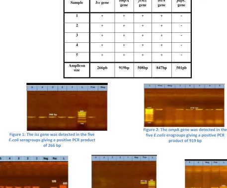
Samples were confirmed for the presence of virulence genes (iss gene, ompA gene, fimH gene, iroN gene and
papC gene) based on conventional RT-PCR assay. The conventional PCR assay was able to detect iss, ombA,
fimH, iroN except pabC virulence genes was negative for E.coli serotypes, as shown in table (6) and figures (1,2,3,4 and 5).
Table 6: PCR amplifications of E .coli serotypes:
Sample Iss gene ompA gene
fiM1 gene
iroN gene
papC gene
1 + + + + -
2 + + + + -
3 + + + + -
4 + + + + -
5 + + + + -
Amplicon
size 266pb 919bp 508bp 847bp 501pb
Figure 1: The iss gene was detected in the five
E.coli serogroups giving a positive PCR product of 266 bp
Figure 2: The ompA gene was detected in the five E.colis erogroups giving a positive PCR
product of 919 bp
Figure 4: The PapC gene was amplified in the five E.coliserogroups giving a
negative PCR product of 501 bp
1:O1 2:O2 3:O6 4:O78 5:O157 Neg: negative control Pos: positive control L: Marker
Figure 1: The fimH gene was detectedinthe five E.colis erogroups giving a positive PCR
product of 508 bp
Figure 4: The iroN gene was amplified in the five E.coliserogroups giving a
5 Results of antimicrobial susceptibility testing of E. coli isolates:
The results of antimicrobial susceptibility test in table (7) showed that there was variation in the susceptibility of E.coli serotypes to the antibiotics drugs revealed high sensitivity percent (100%) to cefatriaxone, amikacin, fosfomycin and spectinomycin followed by tobramycin, trimethoprim, sulfamethoxazole and nalidixic acid revealed susceptibility (80%) thenoxytetracyclin, ciprofloxacine, levofloxacin, ampicillin, enrofloxacine (60%) and complete resistance was observed to clindamycin and penicillin (0%).
Table 7: antimicrobial susceptibility testing of E. coli isolates
Test group Antimicrobial agent
E.coli pathogenic serotypes
Sen. %
O1 O2 O6 O78 O157
Cephems Cefatriaxone (S) (S) (S) (S) (S) 100%
Aminoglycosides Tobramycin (TOB10)
(S) (S) (I) (S) (S) 80%
Amikacin (AK30)
(S) (S) (S) (S) (S)
100%
Tetracyclines Oxytetracycline (TE30) (S) (S) (R) (R) (S) 60%
Fluoroquinoiones Ciprofloxacin (CIP5) (R) (S) (R) (S) (S) 60%
Levofloxacin (LEV 5) (R) (S) (R) (S) (S) 60%
Penicillins Penicillin (R) (R) (R) (R) (R)
0%
Ampicillin AMP10 (S) (S) (R) (R) (S)
60%
Folate pathway inhibitors
Trimethoprim+sulfamethoxazole
(S) (S) (S) (R) (S)
80%
Clindamycin DA (R) (R) (R) (R) (R)
0%
Fosfmycins Fosfomycin (S) (S) (S) (S) (S)
100%
Quinolones Naldixic acid (S) (S) (I) (S) (S)
80%
Spectinomycin (S) (S) (S) (S) (S)
100%
Enrofloxacin (R) (S) (R) (S) (S)
60%
Sen: Sensitivity percent
S: Sensitive reaction I: Intermediate reaction R: Resistant reaction
4. Discussion
E. coli infection in poultry remains of great concern due to the impact in the poultry industry and public health sectors. Therefore evaluation of virulence and antibiotic resistance determinants is essential for predicting preventive measures.
The obtained results in the current study showed that on examination of 150 chicken samples, E. coli species were isolated with an overall percentage of 20% (Table 4). The highest isolation rate of Salmonella species was in apparently healthy birds (6%) followed by diseased birds (5.3%) and freshly dead (3.6%). This finding was almost in agreement with the report of (Dashe et al., 2003). E. coli isolated from samples collected from infected hearts, lungs, air sacs, liver, spleen, ovaries, kidney, oviduct, intestine and bursa of fabracious by (Osman et al., 1989).
December 31, 2019
fifth day after infection and the mortalities recorded at the 7thday post infection within one bird with a percentage 7% in the challenged group these signs similar to the general signs of E. coli infection recorded by (Awaad, 1972). E. coli isolates lethality in Mice showed mortality 100% at day 3 post challenged and this results similar to the results recorded by (Wyatt et al., 2003).
Several virulence determinants have been reported in different E. coli serovars. The distribution of seven virulence genes that play an important role in Salmonella for invasion, enterotoxin production and pathogenesis in the host were investigated.
iss gene that is known to be associated with serum resistance was found in 100% of the examined isolates. This finding is in conformity with that recorded by the result obtained by (Ewers et al., 2004) and (Moemen et al., 2014) showed a high incidence of the iss virulence gene as 82.7%, 73.8% respectively. Also, (rochal et al.,
2008) (Wen-jie et al., 2008) and (Yaguchi et al., 2007) recorded similar results.
All of the examined isolates were positive for the OmpA gene and this result matches the result detected by (Mahmoud et al., 2018) and (Ewers et al., 2007) who isolated the OmpA gene from E.coli serotypes O2, O26,O78,O127,O1 and O91.
The iroN gene that is responsible for iron acquisition systems was found in all serotypes and this result like the result proved by (Carli et al., 2015) that demonstrated a high frequency of the bacterial genes cvaC,
iroN, iss, iutA, sitA, tsh, fyuA, irp-2, ompT and hlyF in pathogenic strains and similar to the result showed by (Ewers et al., 2007).
The fimH virulence factor is seemed to be an essential unit for protecting the APEC isolates against the host immune system (Mellata 2013). In the present study, fimH gene was present in all the examined serotypes and this result like the result proved by (Akbar et al., 2018) who said That the prevalence of type 1 fimbrial adhesion gene (fimH) in APEC isolates was 66.66%and similar to the results showed by (Ghanbar and Salehi 2010) . Another study has shown a higher prevalence of the fimH virulence gene (Ragione 2002).
P-fimbriae (pap) is important in the later stages of infection for the adhesion to internal organs was not detected in all examined serotypes. Other reports have also revealed the prevalence of
the
Papc gene, with a percentage of 24.3% (rochal 2008).Regarding antibiogram of Salmonella isolates, the obtained results showed that 100% of the isolates were susceptible to cefatriaxone, amikacin, fosfomycin and spectinomycin followed by tobramycin, trimethoprim, sulfamethoxazole and nalidixic acid revealed susceptibility (80%) then oxytetracyclin, ciprofloxacine, levofloxacin, ampicillin, enrofloxacine was (60%). while high resistance rates were observed against clindamycin and penicillin. The results of the current study are nearly similar to the results obtained by (Zehor et al., 2017) which showed high level of resistance to tetracyclines (94.12%), sulfamethoxazole-trimethoprim (88.89%), enrofloxacin (86.27%), nalidixic acid (85.62%) and all the strains were susceptible to cefotaxime, excepting three, These findings were in close agreement with the results of (Shecho et al., 2017) who reported 100 and 92.3% susceptibility of E. coli isolates to ciprofloxacin and sulfamethoxazole-trimethoprim, respectively in Ethiopia (Edilu et al., 2019)showed that E. coli isolates were completely (100%) susceptible to ciprofloxacin, norfloxacin and sulfamethoxazole-trimethoprim and majority of the isolates were also susceptible to gentamicin (93%), streptomycin (85%), nalxidic acid (83%), kanamycin (75%).
Conclusions
animals including limiting the availability of antimicrobials in the illegal market needs to be addressed. Moreover, the establishment of guidelines for prudent use of antimicrobials in farm animals with effective enforcement is required in Egypt.
6. References:
i.
Alexander DJ and Senne DA.(2008): Newcastle disease, other avian paramyxoviruses and Pneumovirus infections. Disseases of poultry.ii. Akbar, A., Taghi, Z.,Mahmoud, J and Reza, G. (2018): ECOR phylotyping and determination of virulence genes in Escherichia coli isolates from pathological conditions of broiler chickens in poultry slaughter-houses of southeast of Iran. Vet Res Forum. 9(3): 211–216.
iii.
Ammar A.M, El-Hamid M.I, Eid S.E.A, El Oksh A.S.(2015): Insight into antimicrobial resistance and virulence genes of emergent multidrug resistant avian pathogenic Escherichia coli in Egypt:How closely related are they? Rev. Med. Vet. 2015;166(9-10):304–314.iv. Awaad M.H. (1972):Studies on E. coli Infection in Chickens: M. V. Sc. Thesis, Fac. Vet. Med., Cairo Univ., Egypt.
v.
Barbieri N. L., de Oliveira A. L., Tejkowski T. M., Pavanelo D. B., Rocha D. A., Matter L. B., Callegari-Jacques S. M., de Brito B. G. and Horn F.(2013):Genotypes and pathogenicity of cellulitis isolates reveal traits that modulate APEC virulence. PLoS ONE. 8(8) doi: 10.1371/journal.pone.0072322.e72322.vi. Carli, S., Ikuta, N., Lehmann, FK., da Silveira, VP., de MeloPredebon, G., Fonseca, AS. and Lunge, VR. (2o15): Virulence gene content in Escherichia coli isolates from poultry flocks with clinical signs of colibacillosis in Brazil. Poult Sci. 94(11):2635-40.
vii.CLSI/NCClS (2009): Performance Standards for Antimicrobial Disk Susceptibility Tests; Approval Standard-Tenth Edition and Performance Standards for Antimicrobial Susceptibility Test; M02-A10 and M100-S20.Published online 2017 Jul 28. doi: 10.14202/vetworld.2017.830-835.
viii. Dashe, Y., Raji, M., Abdu,PandOladele B. (2003): Distribution of aerobic bacteria in visceral organs of sick and apparently healthy chickens in Jos, Nigeria. Int Res J Microbiol. 4:79–83.
ix. Dho-Moulin M. and Fairbrother J. M. (1999): Avian pathogenic Escherichia coli (APEC) Veterinary Research. 30(2-3):299–316.
x. Dho, M., Vandenboseh, J. F., Girardeau, J. P., Bree, A., Barat, T. and Lafont, J. P. (1990): Surface antigens from E. coli O2, and O78 strains. Infect. Immun., 58: 740-745.
xi. Dissanayake D. R., Octavia S. and Lan R. (2014): Population structure and virulence content of avian pathogenic Escherichia coli isolated from outbreaks in Sri Lanka. Veterinar Microbiology. 31(168):403– 412.
xii.Edilu, J.,Kebede, A.,Morka, D.,Endrias, Z.,Bizunesh, M. ,and Ayichew, T. (2019): Identification and antimicrobial susceptibility profile of Escherichia coli isolated from backyard chicken in and around ambo, Central Ethiopia. BMC Vet Res.15: 85.
xiii.
Edwards, R. and Ewing, H. (1972) Identification of Enterobacteriaceae. Burgess Publishing Co.,Minneapolis. p709.
December 31, 2019
xv. El-Sukhon SN, Musa A and Al-Attar M.(2002): Studies on the bacterial etiology of airsaculitis of broilers in northern and middle Jordan with special reference to Escherichia coli, Ornithobacteriumrhinotracheale, and Bordetella avium. Avian Dis. 46(3):605–12.
xvi.Ewers, C.;Li, G.; Wilking, H.; Kiebling, S.; Alt, K.; Antao, E.M.; Laturnus, C.; Diehl, I.; Glogge, S.; Homeier, T.; Bohnke, U.; Steinruck, H.; Philipp, H.c.andWieler, L.h. ( 2007): Avian pathogenic, uropathogenic, and newborn meningitis-causing Escherichia coli: how closely related are they? International Journal of Medical Microbiology .Volume 297, Issue 3, 11 June 20.
xvii. GamalYounis, AmalAwad, and Nada Mohamed (2017): Phenotypic and genotypic characterization of antimicrobial susceptibility of avian pathogenic Escherichia coli isolated from broiler chickens. Vet World. 2017 Oct; 10(10): 1167–1172. doi: 10.14202/vetworld.2017.1167-1172.
xviii. Geornaras, I., Hastings, J.W., and Holy, A. (2004): Genotypic analysis of Escherichia coli strains from poultry carcasses and their susceptibilities to antimicrobial agents. Applied Environmental Microbiol. 67: 1940-1944.07, Pages 163-176.
xix.Ghanbarpour and Salehi (2010): Determination of Adhesion Encoding Genes of Uropathogenic in Escherichia coli isolates from omphalitis of chicks. American of Animal and veterinary sciences 5(2): 91-96.
xx.Jeffrey, J.S., Nolan, K. H., Tonooka, S., Wolfe, W., Giddings, S. M., Horne, S. L ., Foley, A. M., Lynne, J. O., Ebert L. M., Elijah, G., Bjorklund, S., Pfaff- McDonough, J., Singer, R. S., and Doetkott, C. (2002): Virulence factors of Escherichia coli from Cellulitis or Colisepticemia lesions in chickens. Avian Diseases, 46: 48-52.
xxi.Kaper, JB., Nataro, JP. and Mobley HL(2004):. Pathogenic Escherichia coli. Nat Rev Microbiol. 2:123– 140.
xxii. Kwon, S. G.; Cha, S. Y.; Choi, E. J.; Kim, B.; Song, H. J. and Jang, H. K. (2008): Epidemiological prevalence of avian pathogenic E. coli differentiated by multiplex PCR from commercial chickens and hatchery in Korea. J. Bacteriology and Virology. 38(4): 179-188.
xxiii. Lowenstine, L. (1986): Necropsy procedures. In: Harrison LR, editor. Clinical avian medicine and surgery. W. B. Saunders; p. 298–309.
xxiv. Lutful, KS. (2010): Avian Colibacillosis and Salmonellosis: a closer look at epidemiology, pathogenesis, diagnosis, control and public health concerns. Int J Environ Res Publi Health. 7:89–114. xxv. Mahmoud,A., Ghada, M., Amgad, A. and Abeer, M. (2018): Serotyping and Virulence Genes Detection
in Escherichia coli Isolated from Broiler Chickens.jbs.vol.18 no.1.2018.p.46-50.
xxvi. Mellata M. (2013): Human and avian extra-intestinal pathogenic Escherichia coli: Infections, zoonotic risks, and antibiotic resistance trends. Foodborne PathogDis.10(11):916–932.
xxvii. Moemen, A. M, Mostafa, A. S. and Elshimaa, R(2014): Virulence Genes Content and Antimicrobial Resistance in Escherichia coli from Broiler Chickens. Vet Med Int. 2014; 2014: 195189.
xxviii. Mude, S., Naod, T., Jelalu, K., and Yimer, M. (2017): Cloacael Carriage and Multidrug Resistance Escherichia coli O157:H7 from Poultry Farms, Eastern Ethiopia. J Vet Med. . doi: 10.1155/2017/8264583.
xxix. Nakazato, G., Tatiana, A., Eliana, G., Marcelo, B. and Wanderley, D.(2009): Virulence factors of avian pathogenic Escherichia coli Pesq. Vet Bras. 29:479–486.
xxx. Orndorff, P. E. (1994): Escherichia coli type 1 pili, p. 91-111. In V. L. Miller, J. B. Kaper, D. A. Portnoy, and R. R. Isberg (ed.), Molecular genetics of bacterial pathogenesis. ASM, Washington, D.C. xxxi. Osman, E., Osman, K., Mehmet, C. and Ersin, I. (1989): Haem-agglutination, Hydrophobicity,
Enterotoxigenicity and Drug-Resistance characteristics of avian E. coli. Avian diseases 33: 631-635.
xxxiii. Ragione,Rand Woodward, M. (2002): Virulence factors of Escherichia coli serotypes associated with avian coli septicaemia. Res Vet Sci. 2002;73:27–35.
xxxiv. Rahman.M.,Samad, M., Rahman, J. and andKabir, S.(2004): Bacterio-pathological studies on salmonellosis, colibacillosis and pasteurellosis in natural and experimental infections in chickens. Bangladesh J Vet Med. 2:1–8.
xxxv. Rocha, A., Rocha, S ., Lima-Rosa, C., Souza, G.,Moraes, H., Salle, F.,Moraes, L. and Salle, Carlos T.P. (2008): Genes associated with pathogenicity of avian Escherichia coli (APEC) isolated from respiratory cases of poultry.Bras. vol.28 no.3 Rio de Janeiro.
xxxvi. Rodriguez-Siek K. E, Giddings C. W., Doetkott C., Johnson T. J., and Nolan L. K. (2005): Characterizing the APEC pathotype. Vet. Res.36: 214:256.
xxxvii. Salama, S. S; Afaf A. K.; Elham A. E. and Taha, M. M. (2007): Molecular Strategies for the differentiation and identification of local E. coli isolated from chicken: I. characterization of protein profile.B.S. Vet. Med. J. January, 17 (1): 25-28.
xxxviii.Shecho, M., Thomas, N., Kemal, J. and Muktar, Y. (2017): Cloacael carriage and multidrug resistance Escherichia coli O157:H7 from poultry farms, eastern Ethiopia. J Vet Med. 2017. 10.1155/2017/8264583.
xxxix. Susantha, M. G., Riddell, C., Andrew, A. P., and Allan, B. J. (2001): Phenotypic andgenotypic characterization of virulence factors of Escherichia coli isolated from broiler chickens with simuilaneous occurrence of cellulitis and other colibacillosislesions.Canadian J. Vet.Res., 65: 1 -6.
xl. Sojka, W.J. and Carnaghan, R.B.A. (1961): Escherichia coli infection in poultry. Res. Vet. Sci., 2: 340-352.
xli.Stordeur, P. and Mainil, J. (2002): Avian colibacillosis. Ann. Med. Vet., 146: 11-18.175
xlii. Vidotto, M.C. , Muller, E.E. , de Freitas, J.C. , Alfieri, A.A., Guimaraes, I.G. and Santos, D.S. (1990): Virulence factors of avian Escherichia coli. Avian Diseases, 34 (1990), pp. 531-538.
xliii. Von Baum H.andMarre, R. (2005): Antimicrobial resistance of Escherichia coli and therapeutic implications. Int J Med Microbiol. 2005;295:503–511.
xliv. Wen-jie, J.,Zhi-ming, Z., Yong-zhi, Z.,Ai-jian, Q., Hong-xia, S.,Yue-long, L.,Jiao, W.andQian-qian, W.(2008): Distribution of Virulence-Associated Genes of Avian Pathogenic Escherichia coli Isolates in China. Agricultural Sciences in China 7(12):1511-1515 ·
xlv.Wyatt, B.,Steven, R., Mog,2. and Frederick, J. (2003): Pathogenicity and Immune Response Measured in Mice following Intranasal Challenge with Enterotoxigenic Escherichia coli Strains H10407 and B7A. Infect Immun. 2003 Jan; 71(1): 13–21.
xlvi. Yaguchi, K.; Ogitani, T.; Osawa, R.; Kawano, M.; Kokumai, N.; Kaneshige, T.; Noro,T.; Masubuchi, K. and Shimizu, Y.(2007): Virulence factor of avian pathogenic Escherichia coli strains isolated from chickens with colisepticemiain japan. Avian Dis. 51(3):656-662.
xlvii. Zhao, L., GAO, S., HUNAN, H., Xu, x., Zhu, x., Yang, w., Gao, Q., and Liu, x. (2009): Comparison of virulence factors and expression of specific genes between uropathogenic Escherichia coli and avian pathogenic E.coli in a murine urinary tract infection model and chicken challenge model. Microbiology-Sgm, 155: 1634-1644.