eggs using FPT and ELISA methods
Razzagh Mahmoudi1, Reza Norian2Abstract
Due to the indiscriminate use of antibiotics in poultry industry and non-compliance withdrawal times. The aim of present study is detection of enrofloxacin residue in chicken eggs from Northwest regions of Iran (Tabriz, Urmia and Ardabil cities) using Four Plate Test (FPT) and ELISA methods. At the first time, one hundred and fifty egg chickens were collected randomly from market of Northwest regions of Iran and antibiotics residue was determined by FTP method. The results of FPT revealed that up to 60.66% of the samples (91/150) were contaminated with the antibiotics residue; the egg samples of Ardebil (42.85%) and Tabriz (27.27%) had the highest and lowest percentage of contamination. The ELISA assay showed that out of 91 positive samples in FPT, 78 samples (85.71%) were positive for enrofloxacin. ELISA analyses demonstrated that the maximum levels of (56.17 ppb) and the highest contamination rate (43.58%) were observed in Ardabil egg samples. Concluding, the findings of this study suggest that the indiscriminate use and non-compliance withdrawal times of enrofloxacin in poultry farms of northwest regions of Iran. The raising awareness of poultry breeders about the risks of antibiotic residues in food products of animal origin, along with the exact compliance withdrawal times can be very helpful.
Keywords: Antibacterial, Egg, Enzyme Linked Immunoassay, Residue
Journal of Research & Health Social Development & Health Promotion
Research Center Vol.5, No.2, Summer 2015
Pages: 159-164 Original Article
1. Correspondence to: Assistante Professor of Food Hygiene & Aquatics Department, Faculty of Veterinary Medicine, University of Tabriz, Tabriz, Iran
Tel/Fax: +98 41 36378743
Email: [email protected]
2. Laboratory of Food Quality Control
and Hygiene, Veterinary Medicine Office,
Qazvin Province, Iran
Received: 28 Aug 2014 Accepted: 04 Oct 2014
How to cite this article: Mahmoudi R, Norian R. Determination of enrofloxacin residue in chicken eggs using FPT and ELISA methods.
J Research Health2015; 5(2): 159-164.
Introduction
Antibiotics are the most important group of antimicrobial drugs, widely prescribed for human, and animals. It is estimated that 100– 200 thousand tons of antibiotic substances are annually produced in the world [1,2]. According to World Health Organization (WHO), about half of the worldwide-produced antibiotics are consumed for non-human applications [3]. Antimicrobials are used by the poultry industry to enhance growth and feed efficiency and to reduce bacterial disease [4]. In layer hens, antimicrobials are used only to treat and to
prevent bacterial infections. [5].
used in sub-therapeutic levels as feed additives for promoting and protecting normal growth of meat production livestock [9].
Enrofloxacin is one of the third generation members of the fluoroquinolone antibacterial agents. This antibiotic that is very useful against a wide variety of infections in animals and used as a prophylaxis or treatment of infectious diseases. Also, the high level usage of this drug in animals and humans, or the use of less potent quinolones, particularly in developing countries, has been attributed to the rapid development of bacterial resistance to these agents combined with less withdrawal time of antibiotics in meat producing industry. Existence of antibiotic residues in food stuff can pose hazards to human health, including sensitivity to antibiotics, allergic reactions and imbalance of intestinal micro flora, bacterial resistance to antibiotics in microorganisms, as well as in the food industry [10]. Hence, routine quality assurance of food stuff regarding antibiotic residues is warranted [6].
European Union (EU) countries establish a maximum residue level (MRL) of 30 ng/g of muscle, liver and kidney tissue for the sum of enrofloxacin and its metabolite ciprofloxacin [11]. Screening methods for FQs in foods of animal origin include microbial growth inhibition [12], enzyme linked immunosorbent assay (ELISA) [13], thin layer chromatography (TLC) and others [14]. It is extremely important to protect treated food destined for human consumption.
The purpose of this study was to analyses enrofloxacin residue in egg samples producing from Northwest regions of Iran.
Method
This was a descriptive cross-sectional study, conducted in 2014 to determine enrofloxacin antibiotic residue in egg samples (150 samples), as study population. Egg samples were collected from the main centers of distribution of eggs in all three cities in northwest of Iran (the number of 50 samples from each Ardabil, Tabriz and Urmia cities). Random cluster sampling method was used and sample size
In the present study, proportion of P (presence of antibiotic residue in egg samples) was estimated at around 30% in accordance with reports of egg samples from northwest regions of Iran [15], with approximately a 7% error, sample size was determined 150.
Samples moved to the Food hygiene laboratory of University of Tabriz and sampling of the egg (yolk and white) was performed in the sterile conditions and under biological hood. For microbiological assay (Four Plate Test) the culture medium of Muller Hinton Agar (MHA) with three different pH of 6.0, 7.2, and 8.0 (V381547, Merck, Germany) were prepared and autoclaved. Following cooling to 45–50 °C, the bacterial suspensions were added and plates were poured. Four different inoculated media were used for the antibiotic detection: medium I: MHA, pH 6.0; medium II: MHA, pH 7.2, and medium III: MHA, pH 8.0, and were seeded with Bacillus subtilis (ATCC 6633, 104–105 CFU/ml); medium IV: MHA, pH 8.0, seeded with Micrococcus luteus (PTTC ATCC 1169, 106 CFU/ml). In the following, egg yolk and white were collected aseptically and under the biological hood of sterile swabs taken. The samples were homogenized using Ultra Turrax T25. Ten microliters of each sample was applied to paper discs (Mast Diagnostics, BD0638W) and the discs, after drying at 40 °C for 10– 15 min, were put on the earlier prepared and seeded agar plates. Plates incubated for 24 hour at 37 ° C were placed. Micrococcus luteus plates containing bacteria due to the slow growth have been in oven for 48 hours. Finally, the results were read using a digital caliper. Inhibition zone was created around the raw samples in culture medium for the presence of antibiotic residues positive detected was measured by using a digital caliper [16].
In short, the mean width of the inhibition zone was calculated. The criterion for positive samples was the inhibition zone of 2 or more than 2 mm in width.
the ELISA technique was performed according to manufacture’s instruction (RIDASCREEN Enrofloxacin ELISA kit (R 1501), r-biopharm, Germany). In brief, 4 g of each sample was weighed and homogenized with mixer, then the homogenized samples were mixed with 3 ml of distilled water and 6 ml of ethyl acetate was added. The suspension was vortexed for 10 min and centrifuged at 3000g for 10 min at room temperature. A 4 ml of ethyl acetate supernatant (corresponding to 2 g of sample) was transferred into a fresh tube and dried at 60 °C under a weak stream of N2. The residue was re-dissolved in 1 ml isooctane/chloroform (2:3) mixture. A half milliliter of the enrofloxacin buffer was added to this solution and vortexed intensively for approximately 1 min. The solution was centrifuged at 3000g for 10 min at room temperature. A 50 µl of the aqueous (upper) layer was used per well in the assay. The absorbance of the samples was read at 450 nm and the amount of enrofloxacin was calculated based on calibration curve. The level of enrofloxacin was expressed as ppb of tested samples. The mean lower detection limit of the RIDSCREEN enrofloxacin test was 6ppb and the recovery rates were >80% for all samples. Statistical analysis
Statistical analysis was carried out using normal confidence intervals and analysis of variance (ANOVA) with SPSS software. Significant level was considered P<0.05.
Results
The results of FPT test showed that out of
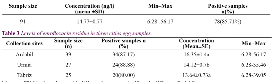
150 samples, 91 (60.66%) were positive as demonstrated the diameter of inhibition zone higher or equal than 2 mm. It came clear that the obtained results included all media with various chosen pH and both selected bacteria. The maximum and minimum diameter of inhibition zone observed from Ardabil egg samples (22.5 mm for B. subtilis) and Tabriz egg samples (5.8 mm for S. aureus) respectively. In addition, the highest contamination rate (42.85%) was observed in Ardebil egg samples. The results of FPT are presented in Table1. To determine the level of enrofloxacin in positive samples, the quantitative method of ELISA was conducted and the results showed that out of 91 positive samples from former assay (FPT), 78 samples (85.71%) were positive. The levels of contamination with enrofloxacin are depicted in Table 2. Based on ELISA findig the minimum detected level of enrofloxacin was 6.28 ppb and the maximum measured level of enrofloxacin (56.17ppb) in the Ardabil egg samples. Interestingly, we found the highest contamination rate (42.85%) was observed in Ardebil egg samples (Fig1).
Table 1 The results of FPT presented as the width of inhibition zone (mm)
Collection
site B.subtilisInhibition Zone (Mean ± SE)M.loteus Ardabil 19.7±1.2 12.8±2.2
Urmia 11.8±1.8 9.4±0.8
Tabriz 9.5±1.1 7.6±0.5
Discussion
Recently the
Table 2 Occurrence of enrofloxacin residue in all samples of egg (ELISA method).
Sample size Concentration (ng/l)
(mean ±SD) Min–Max Positive samplesn(%)
91 14.77±0.77 6.28-.56.17 78(85.71%)
Table 3 Levels of enrofloxacin residue in three cities egg samples.
Collection sites Sample size(n) Positive samples n(%) Concentration(Mean±SE) Min–Max
Ardabil 39 34(87.17) 16.35±1.4a 6.28-56.17
Urmia 27 24(88.88) 14.12±0.7b 6.28-35.46
Tabriz 25 20(80.00) 13.64±0.73a 6.28-39.05
Means ± SEM in the column with different letters are significantly different (P<0.05). occurrence of antibiotic residues in meat, egg
and edible viscera of food-producing animal due to broad use of antibiotics for the treatment of different diseases is a major concern for public health. This study reports for the first time the residue of enrofloxacin (which is mostly used by the poultry keepers for the treatment of different diseases) in egg samples in Northwest region of Iran using both FPT and ELISA methods.
One of the commonly used assays for antibacterial residues screening in food and feed materials is microbiological assay. Today microbiological testing mostly used in screened food samples at the macro level. Therefore, these tests are easy to perform and towards the cost of consumption, have an ability to detect multiple types of antibiotic residue. This method Cause to reduce the number of Samples sent for tests [17]. In microbiological assay, the broad inhibition zone formation indicating the high concentration of antibacterial in examined foodstuff. In the present study FPT assay was conducted to screen the wide range of egg samples. Results of FPT showed that 60.66% of egg samples were positive in terms of having antibacterial effects.
The maximum and minimum diameter of inhibition zone observed from Ardabil egg samples (22.5 mm for B. subtilis) and Tabriz egg samples (5.8 mm for S. aureus) respectively. Although there was different size of inhibition zone by various samples, it was not clear which antibiotic residue was detected by using this method. Additionally, there are factors influencing the results of FPT assay such as pH
of the growth medium and tissue components [14,18].
Based on the results of Monitoring of antibiotic residue in chicken eggs in Tabriz city by FPT, from total 60 samples, 18 (30%) cases are diagnosed to be contaminated to antibiotic residues and the most contamination to antibiotic residues were related to Macrolides groups [15].
The screening procedure is the first step in studying samples to prove the existence or absence of drug residues. This method must be inexpensive, Ability to perform a large number of samples and have minimal false positive and false negative results. Also, all samples contained Antibiotic residues that more than maximum residue level (MRL) should showed positive [19].
To confirm the results of former qualitative assay, all positive samples were subjected to ELISA assay as a group specific test. The results showed that 85.71%, of the samples were enrofloxacine positive. To explain the reason for this discrepancy between two methods, one should note the facts such as multiantibacterial detection capability of FPT and matrix effect [20]. Our results demonstrated that the minimum and maximum measured level of enrofloxacin was 6.28 and 56.17 ppb respectively.
in human and veterinary medicine. These antibiotics have a wide spectrum of action and high efficacy against infectious disease. They are highly effective in treating against Gram positive, Gram negative and Micoplasma in infection processes. The high level of use in animals and humans, and to some degree of misuse in the sense of unnecessary use or use of quinolones with poor activity in some developing countries, has been blamed for the rapid development of bacterial resistance to these agents which means a hazard to human health [21].
The administration of fluoroquinolones to food-producing animals without an adequate withdrawal time (WDT) may lead to violative concentrations of residues in foods destined for human consumption. These residues represent a risk to public
health, including stimulation of bacterial resistance, alterations on intestinal microflora and hypersensivity reactions [22].
Drug withdrawal (WDTs) time is the time required for drug residue to reach a safe concentration for human or animal consumption, defined as MRL. This parameter is generally based on data derived from healthy animals and established on the basis of drug residue levels in various tissues, e.g. kidney or muscle [23]. The obtained results revealed a detectable level of enrofloxacin residues which confirm widespread misuses of antibiotic especially enrofloxacin in farms and lack of application of recommended withdrawal times. To deliver safe food for human consumption, WDTs of pharmaceutical formulations of a drug must be fulfilled.
Acknowledgements
This work was financially supported by University of Tabriz, Tabriz, Iran.
Contributions
Study idea and design: MR
Data collection and analysis: MR, NR
Writing, compiling, and editing of article: MR
Conflict of interest
"The authors declare that they have no competing interests."
References
1- Kummerer K. Significance of antibiotics in the environment. J Antimicrob Chemother2003, 52(1):5– 7.
2- Wise R. Antimicrobial resistance: priorities for action. J Antimicrob Chemother2002: 49 (4): 585–86. 3- World Health Organization (WHO): Use of antimicrobials outside human medicine and resultant antimicrobial resistance in humans. 2002.
4- Donoghue DJ. Antibiotic residues in poultry tissues and eggs: human health concerns. Poultry Scie2003; 82: 618–21.
5- Stolker AA, Brinkman UA. Analytical strategies for residue analysis of veterinary drugs and growth-promoting agents in food-producing animals – a review. J Chromatogr A2005; 1067(1-2): 15–53. 6- Farahmand S, Aref S, Nordehr R, Rahim M, Fariba G. Enrofloxacin Residue in Chicken Tissues from Tehran Slaughterhouses in Iran. Pakistan J Nut2007; 6: 409-13.
7- Weihai X, Xinting W, Liping D, Gan Z. Residues of enrofloxacin, furazolidone and their metabolites in Nile tilapia (Oreochromisniloticus). Aquaculture2006; 254: 1-8.
8- Dipeolu MA, Alonge DO. Residues of streptomycin sntibiotic in meat sold for human Consumption in some states of SW Nigeria. Arch. Zootec2002; 51: 477- 480. 9- Okerman L, Van Hoof J, Debeuckelaere W. Evaluation of the European four-plate test as a tool for screening antibiotic residues in meat samples from retail outlets. J AOAC Int1998; 81: 51-6.
10- Lolo M, Pedreira S, Miranda JM, et al. Effect of cooking on enrofloxacin residues in chicken tissue.
Food Add Cont2006; 23: 988-93.
11- Posyniak A, Zmudzki J, Semeniuk S. Effects of the matrix and sample preparation on the determination of fluoroquinolone residues in animal tissues. J
Chromatogr A2001; 914 (1-2): 89-94.
antibiotics in kidney, marine products, eggs, and muscle by enzyme-linked immunosorbent assay (ELISA). J
Agric Food Chem2006; 54: 2822-27.
14- Juhel-Gaugain M, Abjean JP. Screening of quinolone residues in pig muscle by planar chromatography. Chrom1998; 47: 101-4.
15- Hakimzadegan M, Khalilzadeh Khosroshahi M, Hasseini Nasab S. Monitoring of Antibiotic Residue in chicken eggs in Tabriz city by FPT. Int. J. Advanced Biol. Biomedical Res2014; 2 (1): 132-140.
16- Okerman L, Dewasch K, Van Hoof J. An inhibition test intended to detect and to differentiate between penicillins, cephalosporins, tetracyclines and quinolones, for use in muscle tissuefrom different animal species, J.
AOAC Int2007; 124: 56-62.
17- Tajik H, Razavirohani M, Malekinejad h. Chloramphenicol residues in chicken liver, kidney and muscle: A comparison among the antibacterial residues monitoring methods of Four Plate Test, ELISA and HPLC, Food ChemToxicol 2010; 48(8-9): 2464–68. 18- Kilinc B, Cakli S. Screening for antibiotic residues in the trout by the four plate test, Premi test and ELISA test.
Eur Food Res Technol2008; 226: 795–9.
19- Mariel G, Pikkemaat O, Sabrina O, Jan S, Michel R. A new microbial screening method for the detection of antimicrobial residues in slaughter animals: The Nouws antibiotic test (NATscreening). Food control2008; 19 (8): 781-9.
20- Myllyniemi AL, Rannikko R, Lindfors E, Miemi A, Backman C. Microbiological and chemical detection of incurred penicilline G, oxytetracycline, enrofloxacin, and ciprofloxacin in bovine and porcine tissues. Food
Addit. Contam2000; 17 (12): 991–1000.
21- Hooper DC. Emerging mechanisms of fluoroquinolone resistance. Emerging Infect Dis2001; 7(2): 337-41.
22- Fabrega A, Sa´nchez-Cespedes J, Soto S, Vila J. Quinolone resistance in the food chain. Int. J. Antimicrob. Agents, 2008; 31: 307–15.
23- Alhendi AB, Homeida AM, Gaili E. Drug residues in broiler chickens fed with antibiotics in ration.