A cytotoxic anti-IL-3R
aa
antibody targets key
cells and cytokines implicated in systemic
lupus erythematosus
Shereen Oon, … , Ian P. Wicks, Nicholas J. Wilson
JCI Insight. 2016;
1(6)
:e86131.
https://doi.org/10.1172/jci.insight.86131
.
To date, the major target of biologic therapeutics in systemic lupus erythematosus (SLE)
has been the B cell, which produces pathogenic autoantibodies. Recently, targeting type I
IFN, which is elaborated by plasmacytoid dendritic cells (pDCs) in response to endosomal
TLR7 and TLR9 stimulation by SLE immune complexes, has shown promising results.
pDCs express high levels of the IL-3R
a
chain (CD123), suggesting an alternative potential
targeting strategy. We have developed an anti-CD123 monoclonal antibody, CSL362, and
show here that it affects key cell types and cytokines that contribute to SLE. CSL362
potently depletes pDCs via antibody-dependent cell-mediated cytotoxicity, markedly
reducing TLR7, TLR9, and SLE serum-induced IFN-
a
production and IFN-
a
-upregulated
gene expression. The antibody also inhibits TLR7- and TLR9-induced plasmablast
expansion by reducing IFN-
a
and IL-6 production. These effects are more pronounced than
with IFN-
a
blockade alone, possibly because pDC depletion reduces production of other
IFN subtypes, such as type III, as well as non-IFN proinflammatory cytokines, such as IL-6.
In addition, CSL362 depletes basophils and inhibits IL-3 signaling. These effects were
confirmed in cells derived from a heterogeneous population of SLE donors, various
IFN-dependent autoimmune diseases, and healthy controls. We also demonstrate in vivo activity
of CSL362 following its s.c. administration to cynomolgus monkeys. This spectrum of effects
provides a preclinical rationale for the therapeutic evaluation of CSL362 in SLE.
Technical Advance
Immunology
Therapeutics
Find the latest version:
R e s e a R c h a R t i c l e
Authorship note: I.P. Wicks and N.J. Wilson contributed equally to this work.
Conflict of interest: S. Oon and I.P. Wicks have received funding from CSL Limited. H. Huynh, T.Y. Tai, M. Ng, K. Monaghan, M. Biondo, G. Vairo, E. Maraskovsky, A.D. Nash, and N.J. Wilson are employees of CSL Limited. K. Monaghan, G. Vairo, E. Maraskovsky, A.D. Nash, and N.J. Wilson hold stock in CSL Limited.
Submitted: December 21, 2015 Accepted: April 1, 2016 Published: May 5, 2016 Reference information:
JCI Insight. 2016;1(6):e86131. doi:10.1172/jci.insight.86131.
A cytotoxic anti-IL-3R
α
antibody targets
key cells and cytokines implicated in
systemic lupus erythematosus
Shereen Oon,1,2,3 Huy Huynh,4 Tsin Yee Tai,4 Milica Ng,4 Katherine Monaghan,4 Mark Biondo,4 Gino Vairo,4 Eugene Maraskovsky,4 Andrew D. Nash,4 Ian P. Wicks,1,2,3 and Nicholas J. Wilson4
1Division of Inflammation, The Walter and Eliza Hall Institute of Medical Research, Parkville, Victoria, Australia. 2Department of Rheumatology, The Royal Melbourne Hospital, Parkville, Victoria, Australia.
3The University of Melbourne, Parkville, Victoria, Australia. 4CSL Limited, Parkville, Victoria, Australia.
Introduction
Systemic lupus erythematosus (SLE) is a multisystem autoimmune disease, with significant morbidity and increased mortality (1, 2), in part because of current treatment limitations. Given the importance of autoanti-bodies in the pathogenesis of SLE, many current biologic therapies, such as rituximab and belimumab, target B cells. A wealth of data, including the peripheral blood IFN gene “signature” (3) and elevated type I IFN and IFN-regulated chemokines in SLE sera (4), also supports a central role for type I IFN in SLE. Importantly, recent clinical trials with monoclonal antibodies (mAbs) targeting IFN-α (5–7) and the type I IFN receptor (IFNAR) (8) have demonstrated reductions in the IFN gene signature and disease activity measures.
Plasmacytoid dendritic cells (pDCs) are specialized dendritic cells and are the major producers of type I IFNs (9) following endosomal TLR7 and TLR9 activation by pathogen-associated molecular patterns and human-derived nucleic acids (10). In SLE, immune complexes containing host-derived nucleic acids and a variety of autoantibodies stimulate TLR7 and TLR9 in pDCs to promote IFN production (11–16). Recently, murine models of lupus provided direct evidence for the pathogenic role of pDCs (17, 18). In contrast, evidence implicating pDCs in human SLE has been indirect, with reports of altered circulating pDC numbers (19–22), abundant pDCs producing IFN-α/β in cutaneous lupus (19, 23), and TLR9-medi-ated pDC activation by DNA-containing immune complexes in vitro (15, 24). In contrast to B cells, thera-peutic targeting of pDCs in SLE is still in its infancy (25–27).
pDCs highly express IL-3Rα (CD123) compared with other peripheral blood cells (23, 28). CSL362 is a humanized therapeutic mAb that binds to CD123 and incorporates two mechanisms of action. It inhibits IL-3 binding to CD123, antagonizing IL-3 signaling in target cells (29, 30). Second, the Fc region of CSL362 has been mutated to increase affinity for CD16 (FcγRIIIa), thereby enhancing antibody-
R e s e a R c h a R t i c l e
dependent cell-mediated cytotoxicity (ADCC). CSL362 can induce ADCC against CD123+ acute mye-loid leukemia (AML) blasts and leukemic stem cells in vitro and reduces leukemic cell growth in murine xenograft models of human AML (30). A phase I clinical trial of CSL362 in AML has recently completed (clinical trial NCT01632852).
In this study, we explored the potential utility of CSL362 in primary human cells derived from patients with SLE. We found that CSL362 potently depleted pDCs and inhibited TLR7- and TLR9-stimulated IFN-α production and IFN-α-inducible gene expression ex vivo in SLE patients. This effect was confirmed in vivo, with s.c. administration of CSL362 in cynomolgus monkeys. Basophils, which also express high levels of CD123 and are thought to contribute to the pathology of SLE (31), were likewise depleted. In
addition, CSL362 inhibited pDC-dependent plasmablast expansion ex vivo. These findings demonstrate that, through targeting IL-3Rα, CSL362 directly and indirectly affects key cells contributing to SLE and provide a preclinical rationale for CSL362’s evaluation in this complex disease, for which more therapeutic options are urgently required.
Results
pDCs and basophils have high CD123 expression and are selectively depleted by CSL362. Cell surface expression of CD123 was examined on peripheral blood cells from a heterogeneous cohort of SLE donors (n = 34) (Supple-mental Table 1; supple(Supple-mental material available online with this article; doi:10.1172/jci.insight.86131DS1), autoimmune disease control donors (n = 20), and healthy control donors (n = 34). Of the cell subsets eval-uated, pDCs and basophils had the highest CD123 expression (~40,000 and 20,000 receptors/cell, respec-tively; Figure 1A), with expression being highest on pDCs in most donors. Expression in all other cell types was much lower and ranged from approximately 2,000 receptors/cell in myeloid dendritic cells (mDCs) and intermediate monocytes to less than approximately 1,000 receptors/cell in the other cell types.
Given the selective, high-level CD123 expression on pDCs and basophils, we evaluated the ability of the anti-CD123 mAb, CSL362, to deplete these cell types. An isotype control mAb (isotype control) and Fab′CSL362 (the Fab fragment of CSL362 that lacks the Fc portion but retains IL-3-neutralizing activity) were used as comparisons. At 24 hours, pDCs were potently and reproducibly depleted by CSL362 but not the isotype control or Fab′CSL362 (Figure 1, B and C), demonstrating that ADCC, and not IL-3 neutral-ization, is the main mechanism of depletion. Basophils were less completely depleted than pDCs (Figure 1, B and D), possibly due to higher circulating numbers and relatively lower CD123 expression. A higher dose of Fab′CSL362 also depleted pDCs but not basophils (Supplemental Figure 1, A and B), reflecting the requirement of IL-3 for pDC survival (32). Depletion by CSL362 was selective for pDCs and basophils, as cells that express lower CD123 levels (mDCs, monocytes, and NK, B, and T cell subsets) were not depleted (Supplemental Figure 2, A–I).
CSL362 has been engineered for enhanced ADCC through mutations in its Fc region that increase its affinity for FcγRIII (also known as CD16) expressed on NK cells (30). We therefore examined the effect of CSL362 on NK cell activation (percentage of CD107a+ NK cells). Fab′CSL362 and the isotype control, and a second isotype control (isotype 2) with an unmodified IgG1-Fc, were used for comparison. CSL362 and isotype 1, but not Fab′CSL362 or isotype 2, activated NK cells in SLE, autoimmune, and healthy con-trol donors (Figure 1E), confirming that the modified IgG1-Fc mediates NK cell activation.
R e s e a R c h a R t i c l e
to replace TLR7- or TLR9-induced IFN-α production in the absence of pDCs and, importantly, also show that global IFN-α production is not affected by CSL362, as cells other than pDCs can produce IFN-α in response to TLR3, TLR4, TLR5, and TLR8 stimulation.
We next developed an IFN gene score based on the targeted IFN gene “signature” (described above) to assess drug efficacy. The panel of 11 IFN-inducible genes was incorporated into a single gene score to facil-itate comparison between SLE donors and healthy donors. The derived IFN gene score was calculated as the average of the log2 fold change in expression of the 11 genes compared with that of a universal healthy control. The gene score for most healthy donors was close to 0, whereas SLE donors had an average gene score of approximately 3.0, representing an approximately 8-fold change (Figure 3A), consistent with prior exposure to type I IFN in this population.
The effect of CSL362 on TLR7- and TLR9-induced IFN-α production and the IFN gene score was subsequently examined in SLE, autoimmune, and healthy donors. TLR9-induced IFN-α production was essentially negated in all donors with CSL362 pretreatment but not with Fab′CSL362 or isotype control pretreatment (Figure 3B). CSL362 pretreatment also reduced the IFN gene score as compared with isotype control (Figure 3C). Similar effects were observed in TLR7-responsive donors (Supplemental Figure 3).
Immune complexes or other components of SLE serum may contribute to IFN-α production indepen-dently of TLR7 and TLR9 activation. Therefore, we evaluated IFN-α production induced by SLE serum Figure 1. CSL362 depletes CD123hi plasmacytoid dendritic cells and basophils and activates NK cells. (A) CD123 expression on peripheral blood cells in
SLE donors (n = 34) and healthy (n = 34) and autoimmune (n = 20) controls, as determined by flow cytometry using Quantibrite-PE beads. The number of CD123 molecules per cell is shown for each donor. (B) Representative flow cytometric analysis from SLE donors of viable plasmacytoid dendritic cells (pDCs) (Sytox Blue–, Lin1–, HLA-DR+, BDCA2++) and basophils (Sytox Blue–, Lin1–, CCR3+) after 24-hour culture with media alone (no treatment), CSL362,
Fab′CSL362, or isotype control. Percentage of viable (C) pDCs and (D) basophils, as determined by flow cytometry, after 24-hour culture with 0.01 μM CSL362, Fab′CSL362, or isotype control compared with media alone in SLE (n = 30), healthy (n = 25), and autoimmune donors (n = 18). (E) Fold change of percentage viable CD107a+ NK cells, as determined by flow cytometry, after 18- to 21-hour culture with 0.01 μM CSL362, Fab′CSL362, or 2 isotype controls
R e s e a R c h a R t i c l e
with varying levels of anti-dsDNA antibody titers. Sera with low (3.4–7.1 IU/ml), medium (91.8–104.3 IU/ml), or high anti-dsDNA (>470 IU/ml) antibody levels was used to stimulate IFN-α production in healthy donor PBMCs. Sera from donors with a high anti-dsDNA antibody level intrinsically contained a small amount of detectable IFN-α and when cultured with healthy PBMCs was able to stimulate fur-ther IFN-α production (Figure 3D). Importantly, the IFN-α that was produced by serum stimulation was completely inhibited by CSL362 (Figure 3D). Sera from the donors with low and medium anti-dsDNA antibody levels was not able to stimulate detectable IFN-α production when cultured with healthy donor PBMCs; however, it was able to upregulate expression of IFN-inducible genes, and this expression was also decreased by pretreatment with CSL362 (Figure 3E).
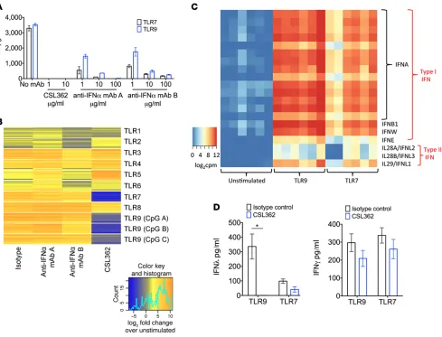
Using published DNA sequences and recombinant methods, we produced and purified 2 anti-IFN-α
mAbs for comparison to CSL362. Both anti-IFN-α mAbs neutralized TLR7- and TLR9-stimulated IFN-α, with >90% neutralization at doses ≥10 μg/ml. However, CSL362 reduced IFN-α more potently, with com-plete inhibition at equivalent doses (Figure 4A). Although the anti-IFN-α mAbs neutralized IFN-α, these had minimal impact on reducing TLR-induced IFN-upregulated gene expression in the IFN gene “signa-ture,” as compared with CSL362 (Figure 4B). This may be because activated pDCs produce more than one type of IFN that may contribute to the gene signature. Elevated levels of multiple type I IFN subtypes (IFNA, IFNB1, IFNW, IFNE) as well as type III IFN (IFNL1, IFNL2, IFNL3) levels were seen by RNA sequencing of isolated pDCs stimulated with TLR7 or TLR9 agonists (Figure 4C). Type II IFN (IFNG) levels were not differentially expressed. We then assessed the effect of CSL362 on TLR7- and TLR9- induced type III IFN (IFN-λ1, IFN-λ2 and IFN-λ3) levels in 9 healthy donors and 6 SLE donors. Three healthy donors and two SLE donors responded to TLR9 (CpG) stimulation with detectable (>50 pg/ml) type III IFN levels, as assessed by ELISA, which were reduced to negligible levels in all donors by CSL362 treatment (Figure 4D). In the 3 healthy donors and 3 SLE donors that responded to TLR7 (imiquimod) stimulation, CSL362 treatment led to a decreased production of IFN-λ in these donors (Figure 4D) that approached significance (P = 0.09). In contrast, there was no significant inhibition by CSL362 of TLR7- or TLR9-stimulated type II (IFN-γ) production in these same donors (Figure 4D).
R e s e a R c h a R t i c l e
TLR9 stimulation induced a greater plasmablast expansion than TLR7 and a higher concentration of CSL362 was required to suppress this TLR9-stimulated response (Figure 5D). Using CFSE staining, we showed that cellular proliferation in response to TLR7 and TLR9 agonists was inhibited by CSL362 in both memory B cells and plasmablasts (Figure 5E). This suggests that either pDCs or basophils provide a proliferative stimulus to B cells when activated by TLR7 or TLR9 agonists. Interestingly, the induction of naive B cell proliferation by TLR9 stimulation was not inhibited by CSL362, suggesting that TLR9-induced proliferation of naive B cells is a direct effect.
To determine the contribution of pDCs and basophils to plasmablast expansion, we reconstituted each cell type separately into CSL362-pretreated cultures. Reconstitution with pDCs, but not basophils, was able to restore TLR9-induced plasmablast expansion (Figure 6, A and B). To determine whether the reconstitution effect was direct or via secreted factors, conditioned media (CM) from pDCs was added to CSL362-pretreated cultures. CM from pDCs stimulated with TLR9 agonist (CpG) was able to restore plasmablast expansion (Figure 6C) and did so in a dose-dependent manner (Figure 6D), whereas CM from nonstimulated pDCs did not induce plasmablast expansion. These data suggested that a soluble factor or factors produced by CpG-activated pDCs promote plasmablast expansion. Similar experiments were per-formed using TLR7 agonist imiquimod as a stimulus; however, in these experiments, there was insufficient plasmablast expansion to allow a comparison of nonreconstituted and reconstituted cultures.
R e s e a R c h a R t i c l e
To identify the soluble factor or factors elaborated by stimulated pDCs that promote plasmablast expansion, Luminex and ELISA analysis of the CM was undertaken. Twenty-eight cytokines were ana-lyzed, and of these, only IFN-α, IL-6, and TNF-α were differentially elevated in TLR9-stimulated pDC CM (Supplemental Figure 5) when compared with CM from TLR9-stimulated basophils or unstimulated pDC CM. Furthermore, inhibition of IFN-α and IL-6, but not TNF-α, by neutralizing mAbs prevented restora-tion of plasmablast expansion with TLR9-stimulated pDC CM (Figure 6E). These data indicate that IFN-α
and IL-6 produced by CpG-activated pDCs stimulate plasmablast expansion.
R e s e a R c h a R t i c l e
vents TLR-induced plasmablast expansion than inhibiting IFN-α alone, possibly because activated pDCs produce additional cytokines, such as IL-6, that promote plasmablast expansion, as demonstrated above.
s.c. administration of CSL362 to cynomolgus macaques depletes pDCs and basophils in vivo and inhibits TLR9- induced IFN-α-inducible gene expression. We next sought to determine the in vivo effect of CSL362 on pDCs and subsequent IFN-α production when administered s.c., which is often the preferred route of delivery of biological therapeutics in chronic diseases such as SLE. CSL362 does not cross react with mouse CD123; however, there is a high degree of sequence homology between human and cynomolgus macaque CD123, which is also highly expressed on cynomolgus macaque pDCs and basophils. In addition, CSL362 demon-strated similar affinity between human and cynomolgus macaque CD123 and FcγRs and comparable activ-ity in cell-based assays (30). We treated naive cynomolgus monkeys with a single s.c. injection of CSL362 at varying doses (1, 10, 30 mg/kg). Maximal serum concentrations of CSL362 were detected at 48 hours (~12, 190, and 380 μg/ml at doses of 1, 10, and 30 mg/kg, respectively). Serum CSL362 was maintained Figure 5. CSL362 inhibits TLR7- and TLR9-stimulated plasmablast expansion and proliferation. Representative flow cytometric analysis from (A) healthy and (B) SLE donors of viable plasmablasts (Sytox Blue—, CD19+, CD27++, CD20–, CD38++) after stimulation with TLR9 (CpG C) or TLR7 (imiquimod) agonists,
R e s e a R c h a R t i c l e
Figure 6. Reconstitution of CSL362-treated peripheral blood mononuclear cells with plasmacytoid dendritic cells or plasmacytoid dendritic cell-con-ditioned media, but not basophils, restores TLR9-induced plasmablast expansion. (A) Representative flow cytometric analysis from a healthy donor of viable plasmablasts (Sytox Blue–, CD19+, CD27++, CD20–, CD38++)
R e s e a R c h a R t i c l e
above 1 μg/ml at day 7 for the 1 mg/kg dose and above 30 μg/ml for 14 days at the higher doses (Figure 7A). pDC depletion was achieved as early as 6 hours after administration for the highest dose and was maximal between days 5 and 15 for all doses (Figure 7, B and C). Similarly, basophils were also depleted by s.c. administration of CSL362 (Figure 7, B and D). These effects followed the peak serum drug levels and were maintained for 4 weeks at the 2 highest doses for pDCs and for 2 weeks for basophils (Figure 7, B–D). We assessed the effect of in vivo pDC depletion by CSL362 on IFN-induced gene expression by stimulating PBMCs, isolated at various time points after CSL362 administration, with CpG for 24 hours ex vivo. A decrease in IFN-inducible gene expression was observed following CSL362 administration (Figure 7E). The decrease in serum drug levels from day 22 onwards correlated with recovery of pDC numbers and an associated increase in CpG-stimulated IFN-α-inducible gene expression. Importantly, s.c. administration of CSL362 was well tolerated at all doses, and no overt toxicity was observed.
Discussion
We have shown that a humanized mAb targeting IL-3Rα (CSL362) has a unique activity profile, with effects against several key cell types and cytokines involved in SLE, thereby providing a rationale for CSL362’s evaluation as a therapeutic agent in this disease. CSL362 potently depletes pDCs, leading to selective abrogation of TLR7, TLR9, and SLE serum-induced IFN-α production and reduced expression of a panel of IFN-α-inducible genes. Of note, others have shown that depletion of pDCs ameliorates dis-ease in murine models of lupus (17, 18), suggesting that pDC depletion in human SLE may be an effective therapeutic strategy. In addition, we show that CSL362 also depletes basophils, inhibits TLR7- and TLR9-stimulated plasmablast expansion, neutralizes IL-3-mediated pDC survival, and has biological activity in vivo in nonhuman primates after s.c. administration. pDC and basophil depletion by CSL362 is dependent upon ADCC and NK cell activation. NK cells in SLE have an activated phenotype but normal ADCC capability, despite reduced CD16 expression (35). Importantly, we show that NK cells derived from patients with SLE can still be activated through the modified IgG1-Fc portion of CSL362, which is critical to cell depletion through enhanced ADCC.
Our data suggest that depletion of pDCs, rather than blockade of IFN-α alone, may be a more effective Figure 7. s.c. administration of CSL362 to cynomolgus macaques depletes plasmacytoid dendritic cells and basophils and inhibits TLR9-induced IFN-inducible gene expression. (A) Serum concentration of CSL362, determined by ELISA at various time points following s.c. administration of CSL362 (at doses of 1, 10, and 30 mg/kg) to cynomolgus macaques. (B) Representative flow cytometric analysis showing plasmacytoid dendritic cells (pDCs) (Lin1–,
HLA-DR+, BDCA2+, CD123+) and basophils (IgE+, CD123+) at baseline and at 6 hours and 36 days after single dose of CSL362. At various time points following
R e s e a R c h a R t i c l e
strategy to inhibit IFN-driven processes, such as gene expression and plasmablast expansion, in response to stimuli, such as immune complexes, that activate TLR7 and TLR9 in SLE. We have shown that by depleting pDCs, production of IFN types other than type I may be inhibited. Of particular interest is type III IFN, for which there is some evidence of aberrant regulation in SLE, with elevated serum levels compared with healthy controls (36). Increased levels of type III IFN were correlated with disease activity, anti-dsDNA antibody levels, glomerulonephritis, and arthritis (37). Although type III IFNs signal through a separate receptor complex than that of type I IFNs, they share common signaling pathways and, as a result, have been postulated to be responsible for ongoing disease activity, despite type I IFN blockade (38). Inhibition of multiple IFN types may therefore confer therapeutic advantage in these situations.
SLE is characterized by autoreactive antibodies, which reflect aberrant activation of B cells and dif-ferentiation into antibody-producing plasma cells. Our data suggest that pDC depletion as a therapeutic strategy may potentially combat two major pathogenic targets in SLE simultaneously, namely inhibiting IFN-α production and autoantibody production. We have shown that, by depleting pDCs with CSL362, plasmablast expansion and proliferation in response to TLR7 and TLR9 agonists is inhibited, although inhibition in SLE donors appears to be less robust than in healthy donors. Plasmablast expansion occurred in response to soluble factors released by activated pDCs, specifically, IFN-α and IL-6, confirming the importance of IFN-α and IL-6 in promoting plasmablast expansion found previously in a virally activated, CD40L-dependent system (34). Specific depletion of pDCs in murine models of SLE has not been found to alter peripheral plasma cell numbers; however, germinal centers in spleens were abolished, and there was a reduction in anti-dsDNA and anti-RNA antibodies (17, 18). It is possible that depleting pDCs may dif-ferentially effect short-lived compared with long-lived plasma cells in different compartments. Interestingly, we found that TLR9 agonist CpG C was a more robust ex vivo stimulus of plasmablast expansion than TLR7 agonist imiquimod and that the concentration of CSL362 required to inhibit imiquimod-induced plasmablast expansion closely followed that required to deplete pDCs and basophils. This suggests that the contribution of pDCs and soluble factors produced by pDCs to TLR7-stimulated plasmablast expansion may be greater than for TLR9.
The ability of CSL362 to deplete basophils and inhibit IL-3 signaling may be of additional benefit. Though basophils in SLE have been less studied compared with pDCs, they can be activated by IgE-con-taining immune complexes. Upon activation, basophils may augment autoantibody production by eliciting a Th2 response and production of the B cell survival factor BAFF (31, 39). Basophil depletion alleviated nephritis in a murine lupus model (31), and therapeutic targeting of basophils in SLE is currently being explored in a phase I trial of an anti-IgE mAb, omalizumab (clinical trial NCT01716312). IL-3 is a known maturation and survival factor for pDCs (32) and basophils (40). We found IL-3 blockade with higher doses of Fab′CSL362 depleted pDCs. Although IL-3 has not been extensively studied in SLE, elevated serum IL-3 levels have been reported in active SLE patients (41). More recently, administration of IL-3 in the MRL/lpr murine lupus model was found to exacerbate nephritis, and this was improved by IL-3 blockade, suggesting an important role for IL-3 in the progression of lupus nephritis (42). Therefore, the potential beneficial effects of CSL362 in SLE may extend to its ability to neutralize IL-3 in addition to depletion of pDCs and basophils.
A phase I trial in AML (clinical trial NCT01632852) using intravenously administered CSL362 in doses ranging from 0.3 to 12.0 mg/kg has recently been completed (43). In that study, there were no increased infections despite rapid (≤6 hours after dose) and complete pDC and basophil depletion at all doses for a fortnightly dosing frequency, which was sustained for ≥15 days for doses ≥3 mg/kg; however, a phase II trial will provide data regarding longer-term infection risk. In the current study, we report the use of s.c. administration in cynomolgus monkeys. This approach was taken because s.c. administration has a range of benefits in chronic diseases, such as SLE. Single s.c. administered doses of CSL362 given to cynomolgus macaques were well tolerated, and the biological effects were reversible, with recovery of depleted pDCs and basophils and of IFN-inducible gene expression occurring in step with waning serum drug levels.
In addition to SLE, we have shown that CSL362 was effective ex vivo in depleting pDCs and reducing IFN-α production in patients with a variety of type I IFN-associated autoimmune diseases, including pso-riasis, scleroderma, primary Sjogren’s syndrome, inflammatory myopathy (44), and rheumatoid arthritis (45). In a recent phase I trial in scleroderma, IFN receptor blockade showed promising results, with near complete inhibition of IFN-stimulated gene expression in peripheral blood and skin (46). In contrast, IFN-α
R e s e a R c h a R t i c l e
IFN-α and IFN-β signal) blockade may be more effective than IFN-α blockade alone. Our data suggest that CSL362 may be a new tool to dissect the role of pDCs, basophils, and IL-3 in various human diseases and highlight its potential role for use in the treatment of SLE and other IFN-dependent disorders.
Methods
Human subjects. SLE and autoimmune disease control (rheumatoid arthritis and psoriatic arthritis [n = 4 each], scleroderma [n = 3], psoriasis and ankylosing spondylitis [n = 2 each], seronegative inflammatory arthritis, primary Sjogren’s syndrome, polymyositis, granulomatosis with polyangiitis, and minimal change renal disease [n = 1 each]) blood samples were obtained from adult patients at The Royal Melbourne Hospital. SLE patients fulfilled SLICC classification criteria for SLE (48). Healthy controls were obtained from the Volunteer Blood Donor Registry at The Walter and Eliza Hall Institute of Medical Research. The SELENA-SLEDAI (Safety of Estrogens in Lupus Erythematosus National Assessment Systemic Lupus Erythematosus Disease Activity Index) score (49) was used to evaluate lupus disease activity.
Human sample collection. Whole blood was collected into lithium heparin tubes (BD, catalog 367526) for whole blood and PBMC assays. Serum was collected into SST II Advance tubes (BD, catalog 367958) for storage at –80°C after centrifugation. PAXgene Blood RNA tubes (BD, catalog 762165) were frozen at –20°C for later RNA extraction.
Tissue culture. PBMCs were isolated from whole blood by Ficoll density centrifugation (GE Health-care Life Sciences, catalog 17-1440-02). Culture of PBMCs was undertaken in RPMI 1640 media (Sigma- Aldrich, catalog R0083) supplemented with 10% heat-inactivated fetal calf serum (HyClone GE Health-care, catalog SH30084.03HI), 2 mM GlutaMAX (Gibco Life Technologies, catalog 35050-0610), and 0.5% penicillin/streptomycin (Gibco Life Technologies, catalog 15140-122), at 37°C with 5% CO2, unless otherwise stated.
Antibodies, reagents, and flow cytometry. Most flow cytometry antibodies and reagents used were com-mercially available and are listed in Supplemental Table 2. Only one antibody was not comcom-mercially available and was generated in-house. This was an anti-CD123-PE antibody (HU01C2) that was used in the pDC and basophil staining panels for the cynomolgus macaque study. Flow cytometry data were acquired with a MACSQuant Analyzer (Miltenyi Biotec) or a LSR Fortessa (BD Biosciences) and ana-lyzed with Flowjo software (Treestar). Cell sorting was performed with a FACSAria (BD Biosciences) or FACS Fusion (BD Biosciences).
Cell surface CD123 expression. Whole blood (50–200 μl) was stained with antibody cocktails, each of which included anti-CD123 PE. After red blood cell lysis with BD Lysing Solution (BD, catalog 349202), Quantibrite PE beads (BD, catalog 340495) were used to estimate the number of CD123 molecules on the surface of each cell type using the MACSQuant Analyzer. The cell types were defined by the following surface markers: pDCs (Lin1–, HLA-DR+, BDCA2++), basophils (Lin1–, CCR3+), mDCs (Lin1–, HLA-DR+, CD11c+, BDCA2–), CD56dim NK cells (NKp46+, CD3–, CD56dim), CD56+ NK cells (NKp46+, CD3, CD56+), naive B cells (CD19+, CD27–), memory B cells (CD19+, CD27+), plasmablasts (CD19+, CD27++, CD20–, CD38++), classical monocytes (CD3–, CD14++, CD16–), intermediate monocytes (CD3–, CD14++, CD16+), nonclassical monocytes (CD3–, CD14+, CD16++), neutrophils (CD16+, CD49d–), eosinophils (CD16–, CD49d+), CD4+ T cells (CD3+, CD4+, CD8–), and CD8+ T cells (CD3+, CD8+, CD4–).
R e s e a R c h a R t i c l e
Effect of CSL362 on NK cell activation ex vivo. PBMCs (0.5 × 10⁶) were cultured with 0.01 μM CSL362, Fab′CSL362, and two isotype controls for 18 to 21 hours. The first isotype (isotype control) contains the same Fc portion as CSL362, which has been modified to increase affinity for CD16. The second isotype (isotype 2) contains an unmodified IgG1-Fc. All cultures contained a 1:1,000 dilution of GolgiStop to promote intracellular recycling of CD107a. The percentage of viable (Sytox Blue–) CD107a+ NK cells (CD14–CD3–, CD56dim, and CD56+) was analyzed on a MACSQuant Analyzer.
Effect of CSL362 and anti-IFN-α mAbs on IFN-α production and IFN-α-upregulated gene expression ex vivo. PBMCs (0.5 × 10⁶ to 1.0 × 10⁶) were cultured with 0.01 μM CSL362, Fab′CSL362, isotype control, or one of two recombinantly produced and purified anti-IFN-α mAbs (KEGG DRUGS database numbers D09668 and D09662) for 6 to 24 hours, before stimulation with either TLR1–9 agonists (Human TLR1–9 Agonist kit, InvivoGen, catalog tlrl-kit1hw) or 50% sera from SLE patients with low (3.4–7.1 IU/ml), medium (91.8–104.3 IU/ml), or high (>470 IU/ml) anti-dsDNA antibody levels (as measured by radioim-munoassay; normal range 0–4 IU/ml) for 18 hours. The TLR agonists were used in the following concen-trations — TLR1 (pam3csk4) 1 μg/ml, TLR2 (HKLM) 1 × 10⁸ cells/ml, TLR3 (poly IC) 10 μg/ml, TLR4 (LPS) 10 μg/ml, TLR5 (flagellin) 2 μg/ml, TLR6 (FSL-1) 1 μg/ml, TLR7 (imiquimod) 2 μg/ml, TLR8 (ssRNA40) 2 g/ml, and TLR9 (CpG C ODN 2395) 0.5 μM. When SLE serum was used, fetal calf serum was omitted from culture medium. IFN-α levels in culture supernatants were measured by ELISA. Cell pellets were frozen at –80°C in Qiazol Lysis reagent (Qiagen, catalog 79306) for later RNA extraction.
Effect of depleting pDCs and basophils on IFN-α production ex vivo. PBMCs from healthy and SLE donors were depleted of pDCs (Lin1–, HLA-DR+, BDCA2++, CD123++) or basophils (Lin1–, CCR3+, CD123++) by sorting on a FACSAria. pDC-depleted and basophil-depleted PBMCs (1.0 × 10⁶) were cultured for 18 hours with 0.5 μM CpG C (Invivogen, catalog tlrl-2395-1) or 2 μg/ml imiquimod (Invivogen, catalog tlrl-imqs). IFN-α levels in culture supernatant were analyzed by ELISA.
Extraction of RNA from PBMCs and whole blood for assessment of IFN-α inducible gene expression. RNA extrac-tion from PBMCs, and from whole blood stored in PAXgene tubes, was performed with the miRNeasy Mini Kit (Qiagen, catalog 217004). The Ambion TURBO DNA-free kit (Invitrogen, catalog AM1907) was used to remove contaminating DNA before RNA was converted to cDNA using the SuperScript III First-Strand Synthesis SuperMix for qRT-PCR kit (Invitrogen, catalog 11752-050). All kits were utilized as per the manufacturers’ instructions. Expression of a panel of 11 IFN-α-upregulated genes (IFI44L, IFIT1, IFIT3, IRF7, ISG15, MX1, MX2, OAS1, OAS2, SERPING1, XAF1) was determined using TaqMan custom-ized gene arrays (Applied Biosystems, catalog 4342247) analyzed on the 7900HT Fast RT-PCR System (Applied Biosystems).
RNA sequencing of isolated pDCs. Healthy donor PBMCs were cultured for 18 hours with 0.5 M CpG C, 0.5 μg/ml imiquimod, or media alone for 18 hours. Following culture, cells were stained with Lin1 FITC and magnetically sorted using an Easysep FITC kit (Stemcell Technologies, catalog 18552), keeping only the negative unlabeled fraction. This Lin1– enriched fraction was then stained with Lin1, BDCA1, BDCA2, HLA-DR, CD11c, CD123, and propidium iodide (PI) and sorted using a BD FACS Fusion. pDCs were sorted as PI–, Lin1–, HLADR+, CD11c–, BDCA2+, and CD123+. Sorted pDCs were immediately stored in RNAprotect Cell Reagent (Qiagen, catalog 76526) and frozen at –80°C until RNA extraction using an RNeasy plus micro kit (Qiagen, catalog 74034). RNA was then submitted to the Australian Genome Research Facility for next-generation sequencing on the Illumina HiSeq platform. The data discussed in this publication have been deposited in NCBI’s Gene Expression Omnibus (50) and are accessible through GEO Series accession number GSE79272 (http://www.ncbi.nlm.nih.gov/geo/query/acc.cgi?acc=GSE79272).
Effect of CSL362 and anti-IFN-α mAbs on TLR7 and TLR9 agonist and CD40L-induced plasmablast expansion and proliferation ex vivo. PBMCs (0.5 × 10⁶) were cultured with 1 μg/ml CSL362, one of two recombinantly produced anti-IFN-α mAbs, or isotype control for 24 hours. 0.5 μM CpG C, CpG B/ODN2006 (Invi-voGen, catalog tlrl-2006), CpG A/ODN 2216 (Invi(Invi-voGen, catalog tlrl-2216), imiquimod, or 0.5 μg/ml CD40L (R&D Systems, catalog 6245-CL-050) was added for 6 days to stimulate plasmablast expansion. The percentage of viable (Sytox Blue–) naive B cells (CD19+, CD27–), memory B cells (CD19+, CD27+), or plasmablasts (CD19+, CD27++, CD20–, CD38++) was analyzed on a LSR Fortessa. To assess proliferation of the B cell subsets, cells were labeled with CFSE prior to culture for 7 days, as described above.
R e s e a R c h a R t i c l e
Human Basophil Kit II (Miltenyi Biotec, catalog 130-092-662), respectively, on an AutoMACS ProSepara-tor (Miltenyi Biotec). Nondepleted PBMCs (0.5 × 10⁵) were treated with 1 μg/ml CSL362 or isotype for 24 hours and then washed 3 times to ensure drug removal. Cultures were reconstituted with isolated pDCs or basophils, at varying concentrations, and stimulated with CpG C, or imiquimod, for 6 days. The percentage of viable plasmablasts was analyzed on a LSR Fortessa.
To produce CM, isolated pDCs and basophils (1.5 × 10⁵) were cultured with 0.5 μM CpG C, imiqui-mod, or media alone for 24 hours. Supernatants were then added to PBMCs (0.5 × 10⁵) that had been pretreated for 24 hours with 1 μg/ml of CSL362 or isotype control. CpG C or imiquimod or media alone was added to the culture for 6 days. The percentage of viable plasmablasts was analyzed on a LSR Fortessa.
ELISAs. IFN-α levels in supernatant and CM were quantified with the VeriKine Human IFN-α Multi-subtype ELISA kit (PBL Assay Science, catalog 41105). BAFF and IL-3 levels in CM were determined by the BAFF Quantikine ELISA kit (R&D Systems, catalog SBLYS0B) and the IL-3 Duo Set (R&D Systems, catalog DY203), respectively. Type III IFN levels in supernatant were assessed with the DIY Human IFN lambda 3/1/2 (IL-28B/29/28A) ELISA kit (PBL Assay Science, catalog 61840). Type II IFN levels were determined using the VeriKine Human Interferon Gamma ELISA Kit (PBL Assay Science, catalog 41500-1). All kits were used as per the manufacturers’ protocols.
Luminex assays. Levels of 25 cytokines in CM were analyzed using Milliplex Multiplex Assays (Merck Millipore, catalog HT17MG-14K-PX25) on a Luminex 200 analyser. Cytokines analyzed were GM-CSF, IFN-γ, MIP-3α, TNF-α, TNF-β, IL-1β, IL-2, IL-4, IL-5, IL-6, IL-9, IL-10, IL-12p70, IL-13, IL-15, IL-17A, IL-17E/IL25, IL-17F, IL-21, IL-22, IL-23, IL-27, IL-28A, IL-31, and IL-33.
Effect of blockade of IFN-α, IL-6, and TNF-α by neutralizing mAbs on plasmablast expansion restored by acti-vated pDC CM. PBMCs (0.5 × 10⁶) were cultured with 0.5 μM CpG C for 6 days, following pretreatment with 1 μg/ml of CSL362 for 24 hours. CM from healthy donor pDCs that had been stimulated with CpG C was added to restore plasmablast expansion at the same time as the CpG, in addition to 50 μg/ml of a recombinantly produced anti-IFN-α mAb (KEGG Drugs database D09668), an anti-IL-6 mAb (R&D Systems, catalog MAB2061), or an anti-TNF-α mAb (Etanercept, Pfizer). The percentage of viable plasma-blasts was analyzed on a LSR Fortessa.
Effect of s.c. administered CSL362 on pDCs and IFN-α-inducible gene expression in cynomolgus macaques. Naive cynomolgus monkeys were administered a single dose (1 mg/kg, 10 mg/kg, or 30 mg/kg) of CSL362 s.c. Peripheral blood was collected at various time points for analysis of CSL362 serum levels by ELISA, and pDC (Lin1–, HLA–DR+, BDCA2+, CD123+) and basophil (IgE+, CD123+) numbers were assessed by flow cytometry on a LSR Fortessa. PBMCs were cultured with CpG C for 24 hours, after which expression of a panel of IFN-α-inducible genes (IFI35, IFIT1, IRF7, MX1, MX2, OAS1) was determined by quantitative PCR.
Statistics. Comparisons between two groups were analyzed with the Mann Whitney U test. A P value of less than 0.05 was considered statistically significant. Statistical analyses were performed with GraphPad Prism Software (version 6.0).
Study approval. Animal studies were conducted at Maccine Pte Ltd, Singapore, in accordance with standard operating procedures and were approved by the Institutional Animal Care and Use Commit-tee of Maccine (259-2012, amendment 34). The human research ethics commitCommit-tees of Melbourne Health (2012.039) and The Walter and Eliza Hall Institute of Medical Research (12/05) in Parkville, approved the human studies. All human subjects signed a written consent form prior to participation in the study.
Author contributions
SO, GV, EM, ADN, IPW, and NJW designed the research. SO, HH, TYT, MN, MB, and KM performed the experiments. SO, MN, KM, MB, IPW, and NJW analyzed the data. SO, IPW, and NJW wrote the man-uscript. All authors assisted in revising the manman-uscript.
Acknowledgments
R e s e a R c h a R t i c l e
Practitioner Fellowship [1023407], NHMRC Program Grant ([1016647], S. Oon Postgraduate Scholarship [1039026]), operational infrastructure grants through the Australian government’s Institute for Research and Innovation in Social Services, the Victorian State Government, Arthritis Australia (South Australia Lupus, Scleroderma, and Sjogren’s Support Group grant), CSL Limited, and Janssen Research and Development.
Address correspondence to: Ian P. Wicks, Division of Inflammation, The Walter and Eliza Hall Institute of Medical Research, 1G Royal Parade, Parkville, Victoria, Australia, 3052. Phone: 61.3.9345.2466; E-mail: wicks@wehi.edu.au. Or to: Nicholas Wilson, CSL Limited, Bio21 Institute, 30 Flemington Road, Park-ville, Victoria, Australia, 3052. Phone: 61.3.9389.2153; E-mail: nick.wilson@csl.com.au.
Tsin Yee Tai’s present address is: Division of Inflammation, The Walter and Eliza Hall Institute of Medical Research, Parkville, Victoria, Australia.
1. Bernatsky S, et al. Mortality in systemic lupus erythematosus. Arthritis Rheum. 2006;54(8):2550–2557.
2. Borchers AT, Keen CL, Shoenfeld Y, Gershwin ME. Surviving the butterfly and the wolf: mortality trends in systemic lupus erythematosus. Autoimmun Rev. 2004;3(6):423–453.
3. Bennett L, et al. Interferon and granulopoiesis signatures in systemic lupus erythematosus blood. J Exp Med. 2003;197(6):711–723. 4. Bauer JW, et al. Elevated serum levels of interferon-regulated chemokines are biomarkers for active human systemic lupus
ery-thematosus. PLoS Med. 2006;3(12):2274–2284.
5. Petri M, et al. Sifalimumab, a human anti-interferon-alpha monoclonal antibody, in systemic lupus erythematosus: a phase I randomized, controlled, dose-escalation study. Arthritis Rheum. 2013;65(4):1011–1021.
6. McBride JM, et al. Safety and pharmacodynamics of rontalizumab in patients with systemic lupus erythematosus: results of a phase I, placebo-controlled, double-blind, dose-escalation study. Arthritis Rheum. 2012;64(11):3666–3676.
7. Kalunian K, et al. A phase II study of the efficacy and safety of rontalizumab (rhuMAb inteferon-α) in patients with systemic lupus erythematosus (ROSE). Ann Rheum Dis. 2016;75(1):196–202.
8. Furie R, et al. Anifrolumab, an anti-interferon alpha receptor monoclonal antibody, in moderate to severe systemic lupus erythe-matosus (SLE). Arthritis Rheumatol. 2015; 67(suppl 10):3223.
9. Liu YJ. IPC: professional type 1 interferon-producing cells and plasmacytoid dendritic cell precursors. Annu Rev Immunol. 2005;23:275–306.
10. Llanos C, Mackern-Oberti JP, Vega F, Jacobelli SH, Kalergis AM. Tolerogenic dendritic cells as a therapy for treating lupus. Clin
Immunol. 2013;148(2):237–245.
11. Banchereau J, Pascual V. Type I interferon in systemic lupus erythematosus and other autoimmune diseases. Immunity. 2006;25(3):383–392.
12. Båve U, Magnusson M, Eloranta ML, Perers A, Alm GV, Rönnblom L. Fcγ RIIa is expressed on natural IFN-α-producing cells (plasmacytoid dendritic cells) and is required for the IFN-α production induced by apoptotic cells combined with lupus IgG.
J Immunol. 2003;171(6):3296–3302.
13. Boule MW, Broughton C, Mackay F, Akira S, Marshak-Rothstein A, Rifkin IR. Toll-like receptor 9-dependent and -independent dendritic cell activation by chromatin-immunoglobulin G complexes. J Exp Med. 2004;199(12):1631–1640.
14. Honda K, et al. IRF-7 is the master regulator of type-I inteferon-dependent immune responses. Nature. 2005;434(7034):772–777. 15. Means TK, Latz E, Hayashi F, Murali MR, Golenbock DT, Luster AD. Human lupus autoantibody–DNA complexes activate
DCs through cooperation of CD32 and TLR9. J Clin Invest. 2005;115(2):407–417.
16. Chan VS, Nie YJ, Shen N, Yan S, Mok MY, Lau CS. Distinct roles of myeloid and plasmacytoid dendritic cells in systemic lupus erythematosus. Autoimmun Rev. 2012;11(12):890–897.
17. Sisirak V, et al. Genetic evidence for the role of plasmacytoid dendritic cells in systemic lupus erythematosus. J Exp Med. 2014;211(10):1969–1976.
18. Rowland SL, et al. Early, transient depletion of plasmacytoid dendritic cells ameliorates autoimmunity in a lupus model. J Exp
Med. 2014;211(10):1977–1991.
19. Farkas L, Beiske K, Lund-Johansen F, Brandtzaeg P, Jahnsen F. Plasmactyoid dendritic cells (natural interferon-α/β-producing cells) accumulate in cutaneous lupus erythematous lesions. Am J Pathol. 2001;159(1):237–243.
20. Cederblad B, Blomberg S, Vallin H, Perers A, Alm GV, Rönnblom L. Patients with systemic lupus erythematosus have reduced numbers of circulating natural interferon-α producing cells. J Autoimmun. 1998;11(5):465–470.
21. Blomberg S, Eloranta ML, Magnusson M, Alm GV, Rönnblom L. Expression of the markers BDCA-2 and BDCA-4 and pro-duction of interferon-α by plasmacytoid dendritic cells in systemic lupus erythematosus. Arthritis Rheum. 2003;48(9):2524–2532. 22. Jin O, et al. Systemic lupus erythematosus patients have increased number of circulating plasmacytoid dendritic cells, but
decreased myeloid dendritic cells with deficient CD83 expression. Lupus. 2008;17(7):654–662.
23. Miyashita A, et al. Proportion of lymphocytic inflammation with CD123 positive cells in lupus erythematous profundus predict a clinical response to treatment. Acta Derm Venereol. 2014;94(5):563–567.
24. Lande R, et al. 2011. Neutrophils activate plasmacytoid dendritic cells by releasing self-DNA-peptide complexes in systemic lupus erythematosus. Sci Transl Med. 2011;3(73):73ra19.
25. Dzionek A, et al. BDCA-2, a novel plasmacytoid dendritic cell-specific type II C-type lectin, mediates antigen capture and is a potent inhibitor of interferon α/β induction. J Exp Med. 2001;194(12):1823–1834.
R e s e a R c h a R t i c l e
27. Zhan Y, et al. Bcl-2 antagonists kill plasmacytoid dendritic cells from lupus-prone mice and dampen interferon-alpha produc-tion. Arthritis Rheumatol. 2015;67(3):797–808.
28. Olweus J, et al. Dendritic cell ontogeny: a human dendritic cell lineage of myeloid origin. Proc Natl Acad Sci U S A. 1997;94(23):12551–12556.
29. Broughton SE, et al. Dual mechanism of interleukin-3 receptor blockade by an anti-cancer antibody. Cell Rep. 2014;8(2):410–419. 30. Busfield SJ, et al. Targeting of acute myeloid leukemia in vitro and in vivo with an anti-CD123 mAb engineered for optimal
ADCC. Leukemia. 2014;28(3):2213–2221.
31. Charles N, Hardwick D, Daugas E, Illei GG, Rivera J. Basophils and the T helper 2 environment can promote the development of lupus nephritis. Nat Med. 2010;16(6):701–707.
32. Grouard G, Rissoan M, Filgueira L, Durand I, Banchereau J, Liu YJ. The enigmatic plasmacytoid T cells develop into dendritic cells with interleukin (IL)-3 and CD40-ligand. J Exp Med. 1997;185(6):1101–1111.
33. Kadowaki N, et al. Subsets of human dendritic cell precursors express different toll-like receptors and respond to different microbial antigens. J Exp Med. 2001;194(6):863–869.
34. Jego G, Palucka AK, Blanck JP, Chalouni C, Pascual V, Banchereau J. Plasmacytoid dendritic cells induce plasma cell differen-tiation through type I interferon and interleukin 6. Immunity. 2003;19(2):225–234.
35. Hervier B, et al. Phenotype and function of natural killer cells in systemic lupus erythematosus: excess interferon-gamma pro-duction in patients with active disease. Arthritis Rheum. 2011;63(6):1698–1706.
36. Lin SC, Kuo CC, Tsao JT, Lin LJ. Profiling the expression of interleukin (IL)-28 and IL-28 receptor α in systemic lupus erythe-matosus patients. Eur J Clin Invest. 2012;42(1):61–69.
37. Wu Q, Yang Q, Lourenco E, Sun H, Zhang Y. Interferon-lambda1 induces peripheral blood mononuclear cell-derived chemokines secretion in patients with systemic lupus erythematosus: its correlation with disease activity. Arthritis Res Ther. 2011;13(3):R88.
38. Amezcua-Guerra LM, Ferrusquia-Toriz D, Castillo-Martínez D, Márquez-Velasco R, Chávez-Rueda AK, Bojalil R. Limited effectiveness for the therapeutic blockade of interferon α in systemic lupus erythematosus: a possible role for type III interfer-ons. Rheumatology (Oxford). 2015;54(2):203–205.
39. Davidson A, Diamond B. Activated basophils give lupus a booster shot. Nat Med. 2010;16(6):635–636. 40. Voehringer D. Basophil modulation by cytokine instruction. Eur J Immunol. 2012;42(10):2544–2550.
41. Fishman P, et al. Interleukin-3 immunoassay in systemic lupus erythematosus: preliminary data. Int Arch Allergy Immunol. 1993;100(3):215–218.
42. Renner K, et al. IL-3 contributes to development of lupus nephritis in MRL/lpr mice. Kidney Int. 2015;88(5):1088–1098. 43. Smith BD, et al. 120 first-in man, phase I study of CSL362 (anti-IL3Rα/anti-CD123 monoclonal antibody) in patients with
CD123+ acute myeloid leukemia (AML) in CR at high risk for early relapse. Blood. 2014;124(21):120.
44. Cao W. Pivotal functions of plasmacytoid dendritic cells in systemic autoimmune pathogenesis. J Clin Cell Immunol. 2014;5(2):212–234.
45. Rodríguez-Carrio J, López P, Suárez A. Type I IFNs as biomarkers in rheumatoid arthritis: towards disease profiling and per-sonalized medicine. Clin Sci (Lond). 2015;128(8):449–464.
46. Goldberg A, et al. Dose-escalation of human anti-interferon-alpha receptor monoclonal antibody MEDI-546 in subjects with systemic sclerosis: a phase 1, multicenter, open label study. Arthritis Res Ther. 2014;16(1):R57.
47. Bissonnette R, et al. A randomized, double-blind, placebo-controlled, phase I study of MEDI-545, an anti-interferon-alpha monoclonal antibody, in subjects with chronic psoriasis. J Am Acad Dermatol. 2010;62(3):427–436.
48. American College of Rheumatology Ad Hoc Committee on Systemic Lupus Erythematosus Response Criteria. The American College of Rheumatology response criteria for systemic lupus erythematosus clinical trials: measures of overall disease activity.
Arthritis Rheum. 2004;50(11):3418–3426.
49. Petri M, et al. Derivation and validation of the Systemic Lupus International Collaborating Clinics classification criteria for sys-temic lupus erythematosus. Arthritis Rheum. 2012;64(8):2677–2686.
50. Edgar R, Domrachev M, Lasha AE. Gene expression omnibus: NCBI gene expression and hybridization array data repository.