Role of Phosphoramidate Antisense in Specific
Inhibition of K562 Leukemic Cell Line
N. Naghsh,
1A. Haeri Ruhani,
1M. Rabbani,
2M. Salehi,
3H. Mirmohammad
Sadeghi,
2and M.R. Noori Daloii
4,*1
Department of Physiology, Research and Science Centre, Islamic Azad University, Tehran Branch, Tehran, Islamic Republic of Iran
2
Department of Biotechnology, School of Pharmacy, Isfahan University of Medical Sciences, Isfahan, Islamic Republic of Iran
3
Department of Genetics and Molecular Biology, Faculty of Medicine, Isfahan University of Medical Sciences, Isfahan, Islamic Republic of Iran
4
Department of Medical Genetics, Faculty of Medicine, Tehran University of Medical Sciences, Tehran, Islamic Republic of Iran
Abstract
Telomeres are specialized heterochromatin structures that act as protective caps at
the ends of the chromosomes. Telomere maintenance in the majority of tumor cells is
achieved by telomerase, a reverse transcriptase enzyme that catalyzes the synthesis of
further telomeric DNA. Telomerase is detected in the majority of malignant cells.
Thus, telomerase may represent a very good candidate for targeted therapy in cancer.
To inhibit telomere maintenance by telomerase, approaches that directly target either
telomerase and telomeres or the telomerase regulatory mechanisms have been used.
Moreover, strategies targeting telomerase-positive cells as a means to directly kill the
tumor cells have been tested. The aim of this study was to evaluate effects of
antisense against the template region in the, telomerase RNA (hTR). The K562 cells
were cultured and to inhibit telomerase activity, a 19-mer antisense oligonucleotide,
complementary to hTR was designed. The modification of this sequence was
N3
′→
P5
′
phosphoramidate and FuGENE6 was used as transfection reagent.
Telomerase activity was measured using TRAP assay. Telomerase activity decreased
using both sense and antisense hTR after 3 days with 1 µM comparing to the
scramble oligonucleotides. There was morphological changes observed in K562 cells
and MTT assay demonstrated that treatment of these cells efficiently reduced cell
viability. Our results clearly demonstrated that antisense phosphoramidate is a very
good candidate for targeted therapy in K562 leukemic cell line. The applied antisense
inhibits transcription of hTR efficiently and can be considered as a potential
therapeutic approach for leukemia treatment.
Keywords: Telomerase; Antisense; Oligonucleotides; FuGENE6
*E-mail: nooridaloii@ut.ac.ir
Introduction
Telomeres are specialized heterochromatin structures that act as protective caps at the ends of chromosomes.
division and, when they reach a critically short length, cells exit from the cell cycle and undergo a replicative senescence [1]. By contrast, immortal cells as well as germline cells adopt some mechanisms to bypass the senescence checkpoint. Telomere maintenance in 80-95% of tumor cells is achieved by telomerase, a reverse transcriptase enzyme that catalyzes the synthesis of further telomeric DNA repeats. The telomerase holoenzyme consists of the catalytic subunit reverse transcriptase protein hTERT the telomerase RNA template subunit, hTR and other associated proteins. Telomeric DNA and the core telomerase components of telomerase, hTR and hTERT, are definitely required for telomerase function and therefore they are good targets for anti-telomerase strategy. Telomerase activity is typically absent from most normal human cells, but is expressed in nearly all human cancer cells. All these findings suggest that telomerase might represent a very good candidate for targeted therapy in leukemia. To inhibit telomere maintenance by telomerase, approaches that directly target either telomerase and telomeres or the telomerase-associated regulatory has been applied [2,3].
N3′→P5′ phosphoramidate oligonucleotides have been designed, synthesized and evaluated as telomerase inhibitors. The oligonucleotides were targeted at a segment of hTR downstream from the hTR template region. It behaves like classical active-site enzyme inhibitors, i.e. ‘template antagonists’. The oligo-nucleotides were employed because they form very stable duplexes with single-stranded RNA, are resistant to nucleases and display a high affinity for nucleic acids, but not proteins. The same group then tested the anti-telomerase effect of N3′→P5′ phosphoramidate oligonucleotides. Telomerase inhibition occurred 24 h after transfection of an immortalized human breast epithelial cell line at submolar concentrations [4,5]. Long-term treatment with NP oligonucleotides resulted in gradual telomere shortening followed by cellular senescence and apoptosis, while the scramble control compound had no effect on cell proliferation [6,7]. Optimization of the sequence, length and bioavailability resulted in the selection of the novel telomerase template antagonist [5,8]. Although antisense against hTERT has widely been studied, but there are only a few study on antisense against hTR, application of antisense against hTR in cell line with high telomerase activity has not been studied. The aim of this study was to evaluate a relatively long antisense (19-mer) on a cell line with high telomerase activity (K562). To the best of our knowledge it is the first study, using the mentioned settings.
Material and Methods
Cell Culture Conditions
Cells were maintained in RPMI-1640 supplemented with 10% fetal bovine serum (FBS), streptomycin and penicillin at 37°C in a humidified atmosphere containing 5% CO2 as previously described[1,5].
Antisense Oligonucleotides
The sense, antisense and scramble oligonucleotides were synthesized by Faza Pajoh Company and were purified by HPLC. They were designed to have 19-mer lengths and to be complementary to the template (5′ -CUAACCCUAA-3′) region of the telomerase RNA. To increase instability, these oligonucleotides were modified with phosphoramidate groups. The sequence of antisense oligonucleotide was (5′-CTCAGTTAGGG TTAGACAA-3′).
Oligonucleotide Transfection Protocol
Before transfection, cells were plated on 6-well plates and were washed with FCS-free medium RPMI-1640. Then 900 µl FCS-free RPMI-1640 were added in to each well prior to transfection, for the preparation of the complex of antisense oligomers/transfector, the FuGENE6 transfection reagent (Roche, Germany) was diluted with serum-free medium and incubated for 5 min at room temperature. Then, the desired amount of oligomer was added into the FuGENE6 for 15 min at room temperature. The final volume of the complex in each sample was 100 µl. These complexes were added drop wise to the cell culture containing 900 µl FCS-free medium. A control containing only FuGENE6 reagent was also included. The cultures were incubated for 4 h at 37°C; 1 ml of 20% FCS in RPMI-1640 was added into each well, to a final FCS concentration of 10%.
Effect of Oligonucleotides on Number and Viability of K562 Cells
K562 cells were suspended at a concentration of 5×10⁴/ml. Then 200 µl of the cell suspension was placed in each well of a triplicate 96-well microtiter plate. Different concentrations (1 µM and 8 µM) of
phosphoramidate oligonucleotides together with
performed three times and the average results were calculated [3,4].
Telomerase Activity with Telomerase Repeat Amplification Protocol (TRAP) Assay
Cultured cells in logarithmic growth were suspended at a concentration of 2×10⁴/ml, and then 5 ml was placed into a cell culture flask. Cells were harvested after 12 h, 24 h, and 36 h, washed once with wash buffer and homogenized in 150 µl cell lysis buffer, on ice for 30 min. The homogenates were then centrifuged at 12000×g for 20 min at 4°C. The supernatants were snap-frozen in liquid nitrogen and stored at −80°C. Telomerase activity was then measured on samples using TRAP-PCR-ELISA assay. In brief, 2 µl of tissue extract and 48 µl TRAP reaction mixture were placed into tubes. PCR was performed at 94°C for 120 s (1 cycle) followed by 35 cycles of 94°C for 30 s, 48°C for 30 s, and 72°C for 90 s. The PCR products (20 µl) were hybridized to a digoxigenin (DIG)-labelled telomeric repeat specific detection probe. The PCR products immobilized via the biotin-labelled primer to a streptavidin-coated microtiter plate. The immobilized PCR products detected with a peroxidase-conjugated anti-DIG antibody and visualized following addition of the stop reagent. Microtitre plate was assessed on an enzyme-linked immunosorbent assay (ELISA) plate reader at 960 and 540 nm wavelength [8,10].
Effect of Oligonucleotides on K562 Cell Morphological Changes
Morphology of the cells were observed using light microscopy and compared to the control cells in different time intervals (24 h, 48 h and 72 h) after transfection.
Results
Inhibitory Effect of N3′→P5′ Phosphoramidate Oligonucleotides on Telomerase Activity
Differences between absorbance in 540 nm and 960 nm were used as indication of telomerase activity. Using 1 µM oligonucleotides, it was observed that telomerase activity was 1.8 in control group, 1.3 in scramble (non-significant decrease), 0.6 in antisense and 0.7 in sense oligonucleotides (significant decrease in both). In summary, telomerase activity decreased (p<0.05) with N3′→P5′ phosphoramidate hTR sense and antisense after 3 day with 1 µM compared to scramble oligonucleotides and control group (Fig. 1).
Effects of Oligonucleotides on Cell Proliferation and Cell Number
K562 cells were treated with different concentrations ofsense and antisense oligonucleotides for 3 days. The applied antisense and sense oligonucleotides reduced the cell number (Fig. 2). The result of MTT assay, which was used to measure proliferation and viability in K562 cells, is shown in Figure 3. The applied nucleotides significantly (p<0.05) inhibited these leukemic cell proliferations in 8 µM concentration 3 days after transfection (Fig. 3).
Morphological Changes in K562 Cells
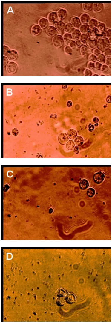
The results of light microscopy demonstrated that the cells became round after treated with antisense for 24 h. After 48 h, the cells were crimpled and floated. The cells also had big nucleoli and aberrant nuclei and the cell membranes were also disrupted (Fig. 4).
Discussion
In this investigation we applied antisense and sense oligonucleotides against hTR of a leukemic cell line, K562. We applied a long, 19-mer, N3′→P5′ phosphor-ramidated oligonucleotide, on a leukemic cell line with a very high telomerase activity. To the best of our knowledge it is the first report of application of antisense technology with modification, length, cell line, time and concentration of treatment that we used. Our results clearly demonstrated that telomerase activity in K562 cells was efficiently decreased with N3′→P5′
Figure 1. ELISA-TRAP assay demonstrating acquisition of telomerase activity in K562 cells transfected after 3 days with phosphoramidate oligonucleotides (formulated with FuGENE6). The concentrations of all nucleotides were 1 µM. Telomerase activity decreased significantly in both antisense and sense, but not in scramble compared with control group.
Figure 2. Effect of antisense, sense and scramble
phospho-ramidate Oligonucleotides (formulated with FuGENE6) cell number 24 h post transfection. The concentrations of all oligonucleotides were 1 µM. There were no significant differences between groups.
Figure 3. Effect of antisense, sense and scramble
phosphoramidate oligonucleotides on K562 cells ELISA-based MTT assay demonstrating number of the cell in 8 µM concentration. Optical density decreased significantly in both antisense and sense, but not in scramble compared with control group.
Figure 4. The morphological changes in the K562 cell line
microscopy experiments on our cells demonstrated that the morphology of the cells changed greatly as effect of both antisense and sense oligonucleotides treatment, so that cells were very similar to pre-apoptotic cells. These results are in accordance with the findings of others that telomere shortening and senescence can promote apoptosis [13] and the cells are probably arrested in G2/M. Interestingly it is demonstrated that induced apoptosis is related to decreased telomerase activity [14]. The role of Bcl-2 inhibition in cell apoptosis is shown by other investigators and it is also shown that telomeres shortening and Bcl-2 reduced expression are related to apoptosis. Therefore the effects of our treatment might be through Bcl-2 inhibition. Although there are some evidences to support this idea, but more investigations is necessary to prove this idea [13,14].
Our results also demonstrated that Phosphoramidate oligonucleotides can be considered as an efficient modification in antisense technology. Other modifications have also been used by others [13], but our results demonstrated that application of Phosphoramidate modified oligonucleotides is a very powerful approach in antisense technology [15].
In conclusion, our results as well as others in different settings clearly demonstrated that these modified oligonucleotides could effectively cause telomerase inhibition in human malignant cell lines. Therefore it may provide important insights into therapeutic approach against leukemia. We suggest that this technology could be considered as a strong approach in treatment of leukemic cells in human.
References
1. Arai J., Yasukawa M., Ohminami H., Kakimoto M., Hasegawa A., and Fujita S. Identification of human telomerase reverse transcriptase-derived peptides that induce HLA-A24-restricted antileukemia cytotoxic T lymphocytes. Blood, 97: 2903-2907 (2001).
2. Bodnar A.G., Kim N.W., Effros R.B., and Chiu C.P. Mechanism of telomerase induction during T cell acti-vation. Experimental Cell Research, 228: 58-64 (1996). 3. Bodnar A.G., Ouellette M., Frolkis M., Holt S.E., Chiu
C.P., Morin G.B., Harley C.B., Shay J.W., Lichtsteiner S., and Wright W.E. Extension of life-span by introduction of telomerase into normal human cells.
Science, 279: 349-352 (1998).
4. Bernard D., Pourtier-Manzanedo A., Gil J. and Beach D.H. Myc confers androgen-independent prostate cancer cell growth. Journal of Clinical Investigation, 112:
1724-1731 (2003).
5. Biroccio A., Amodei S., Benassi B., Scarsella M., Cianciulli A., Mottolese M., Del Bufalo D., Leonetti C., and Zupi G. Reconstitution of hTERT restores tumorigenicity in melanoma-derived c-Myc low-expressing clones. Oncogene, 21: 3011-3019 (2002a). 6. Asai A., Oshima Y., Yamamoto Y., Uochi T.A., Kusaka
H., Akinaga S., Yamashita Y., Pongracz K., Pruzan R., Wunder E., Piatyszek M., Li S., Chin A.C., Harley C.B., and Gryaznov S. A novel telomerase template antagonist (GRN163) as apotential anticancer agent. Cancer Research, 63: 3931-3939 (2003).
7. Biroccio A., Amodei S., Antonelli A., Benassi B., and Zupi G. Inhibition of c-Myc oncoprotein limits the growth of human melanoma cells by inducing cellular crisis. Journal of Biological Chemistry, 278: 35693-35701 (2003a).
8. Biroccio A., Gabellini C., Amodei S., Benassi B., Del Bufalo D., Elli R., Antonelli A., D’Incalci M., and Zupi G. Telomere dysfunction increases cisplatin and ecteinascidin-743 sensitivity of melanoma cells.
Molecular Pharmacology, 63: 632-638 (2003b).
9. Kondo Y., Koga S., Komata T., and Kondo S. Treatment of prostate cancer in vitro and in vivo with 2-5A-anti-telomerase RNA component. Oncogene, 19(18): 2205-11 (2000).
10. Dalla-Favera R., Bregni M., Erikson J., Patherson D., Gallo R.C., and Croce C.M. Human c-myc oncogene is located on the region of chromosome 8 that is translocated in Burkitt lymphoma cells. PNAS, 79: 7824-7827 (1982).
11. Culig Z., Hoffmann J., Erdel M., Eder I.E., Hobisch A., Hittmair A., Bartsch G., Utermann G., Schneider M.R., Parczyk K., and Klocker H. Switch from antagonist to agonist of the androgen receptor bicalutamide is associated with prostate tumour progression in a new model system. British Journal of Cancer, 81: 242-251 (1999).
12. Caprio V., Guyen B., Opoku-Boahen Y., Mann J., Gowan S.M., Kelland L.M., Read M.A., and Neidle S. A novel inhibitor of human telomerase derived from 10H-indolo[3,2-b]quinoline. Bioorg. Med. Chem. Lett., 10(18): 2063-6 (2000).
13. Sun L. and Wang X. Effects of allicin on both telomerase activity and apoptosis in gastric cancer SGC-7901 cell. ISSN 1007-9327 CN 14-1219/R, World J. Gastroenterol.,
9(9): 1930-1934 (2003).
14. Biroccio A., Benassi B., Filomeni G., Amodei S., Marchini S., Chiorino G., Rotilio G., Zupi G., and Ciriolo M.R. Glutathione in.uences c-Myc-induced apoptosis in M14 human melanoma cells. Journal of Biological Chemistry, 277: 43763-43770 (2002b).